
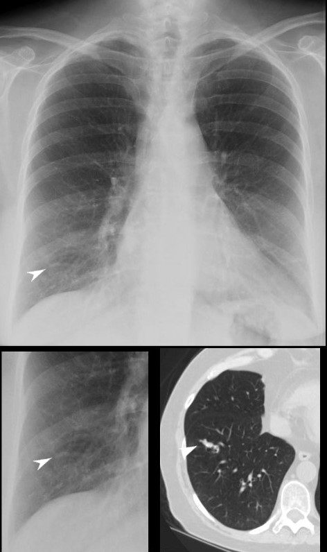
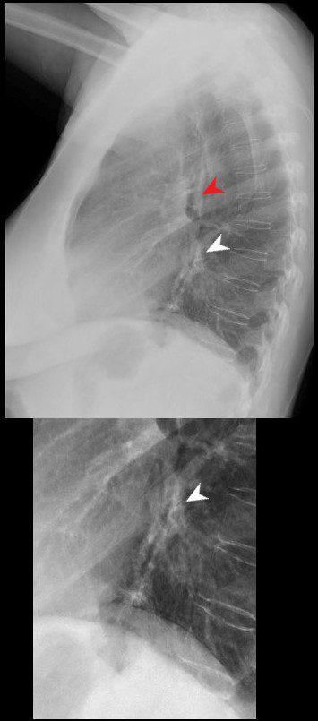
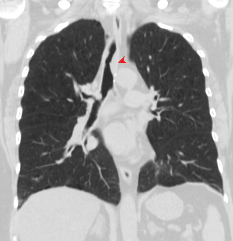
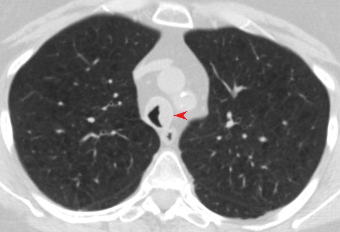
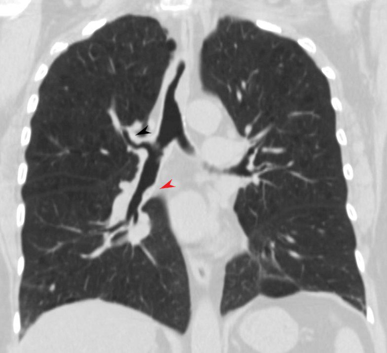
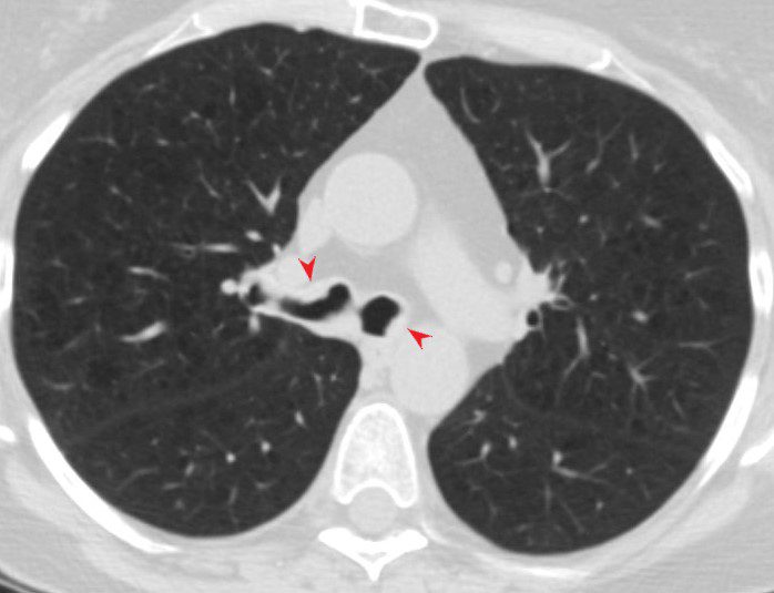
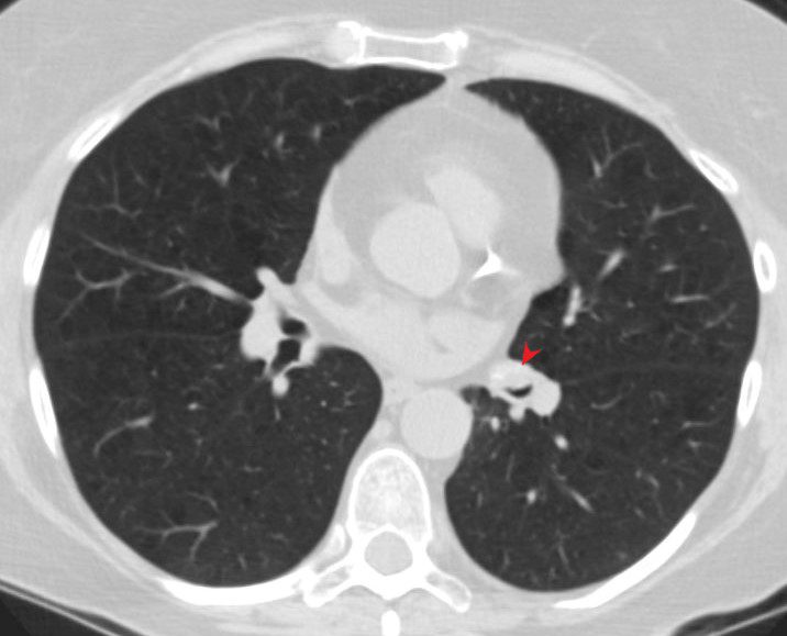
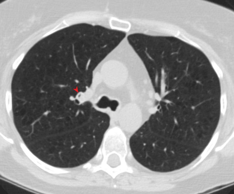
56-year-old female with stable tracheobronchial amyloidosis.
The CXR shows in the AP projection shows a small tubular density which on CT reflect as a small bronchus which is thickened and contains calcification. The lateral exam shows bronchial wall thickening
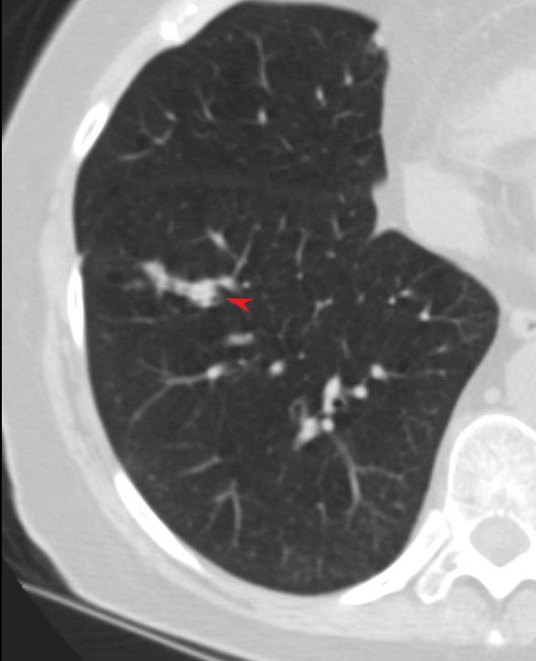
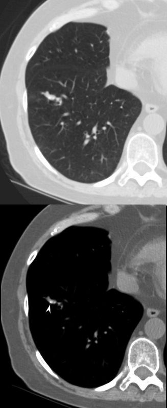
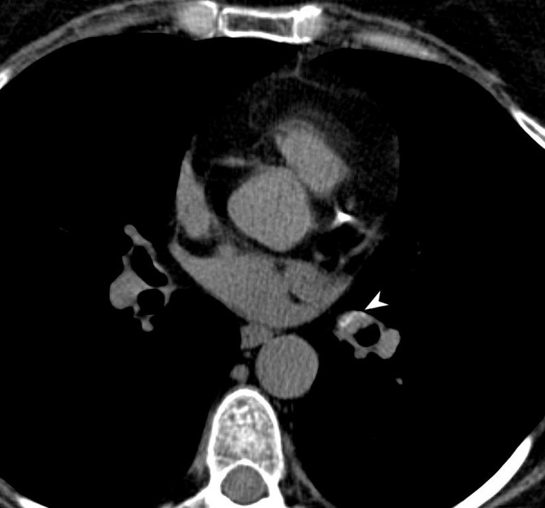
The CT shows multicentric thickening of the trachea and bronchi.
The bronchi to the left lower lobe and a middle lobe bronchus are thickened and contain calcium
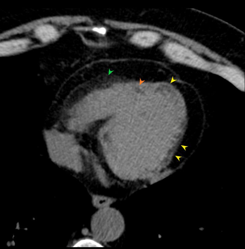
There is nodular fat accumulation along the lateral wall and apex of the LV and within the RV. There is also linear fat accumulation in the mid septal region and prominent pericardial fat around the anterior wall of the RV
AMYLOIDOSIS TRACHEA BRONCHI BRONCHIOLES
Ashley Davidoff MD
AMYLOIDOSIS TRACHEA BRONCHI BRONCHIOLES
Ashley Davidoff MD
AMYLOIDOSIS TRACHEA BRONCHI BRONCHIOLES
Ashley Davidoff MD
Ashley Davidoff MD
Ashley Davidoff MD
Ashley Davidoff MD
Ashley Davidoff MD
Ashley Davidoff MD
Ashley Davidoff MD
Ashley Davidoff MD
Ashley Davidoff MD
Ashley Davidoff MD