

CALCIFIED NODES OF AMYLOIDOSIS
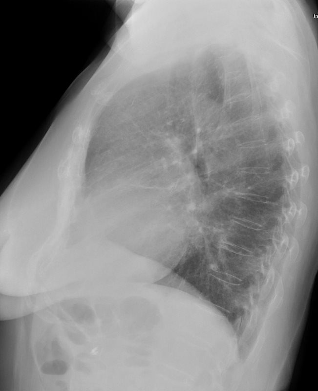
72-year-old male with history of an amyloidoma removed with right middle lobectomy
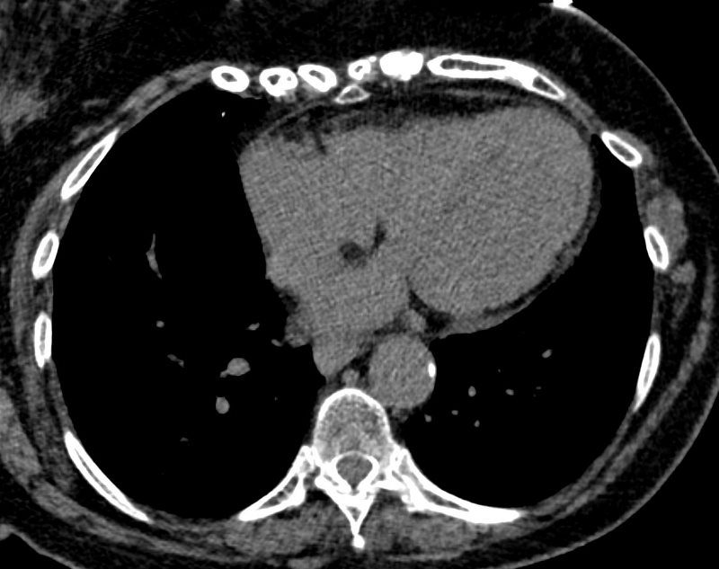
The CXR shows left ventricular enlargement
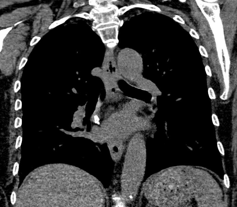
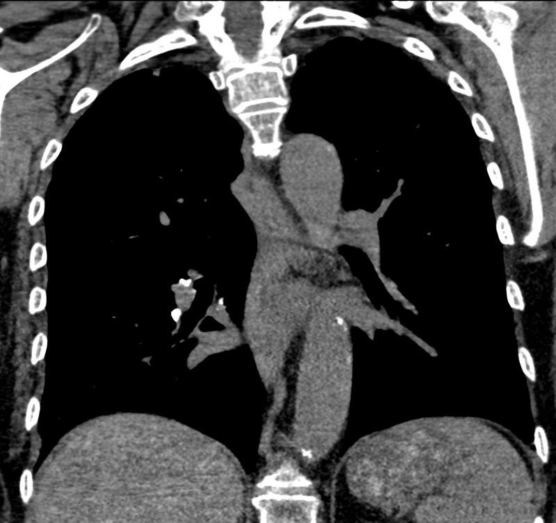
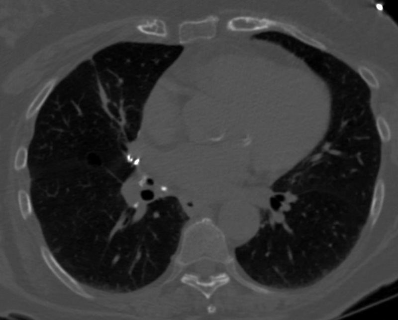
The current CT is characterized by stable small hilar nodal calcifications that likely represent amyloidosis
There are calcifications on right side of the left atrium and by the left heart border with associated focal regions of pericardial thickening. Involvement of the pericardium may be due to amyloidosis. However the LS calcification could also be post op and the calcification along on the left heart border could also be branch of circumflex with unusually chunky appearance which would be out of proportion to the degree of calcification elsewhere in the coronaries.
Fat in the LV apex indicates previous LAD territory infarction and likely account for the LVE noted on CXR
Ashley Davidoff MD
– AMYLOIDOSIS
72-year-old male with history of an amyloidoma removed with right middle lobectomy
The CXR shows left ventricular enlargement
The current CT is characterized by stable small hilar nodal calcifications that likely represent amyloidosis
There are calcifications on right side of the left atrium and by the left heart border with associated focal regions of pericardial thickening. Involvement of the pericardium may be due to amyloidosis. However the LS calcification could also be post op and the calcification along on the left heart border could also be branch of circumflex with unusually chunky appearance which would be out of proportion to the degree of calcification elsewhere in the coronaries.
Fat in the LV apex indicates previous LAD territory infarction and likely account for the LVE noted on CXR
Ashley Davidoff MD
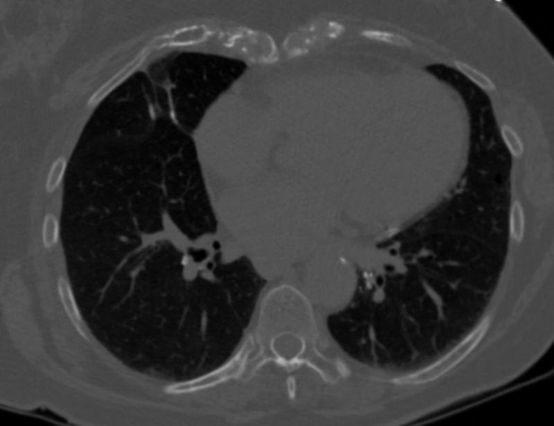
Hilar Adenopathy
72-year-old male with history of an amyloidoma removed with right middle lobectomy
The CXR shows left ventricular enlargement
The current CT is characterized by stable small hilar nodal calcifications that likely represent amyloidosis
There are calcifications on right side of the left atrium and by the left heart border with associated focal regions of pericardial thickening. Involvement of the pericardium may be due to amyloidosis. However the LS calcification could also be post op and the calcification along on the left heart border could also be branch of circumflex with unusually chunky appearance which would be out of proportion to the degree of calcification elsewhere in the coronaries.
Fat in the LV apex indicates previous LAD territory infarction and likely account for the LVE noted on CXR
Ashley Davidoff MD
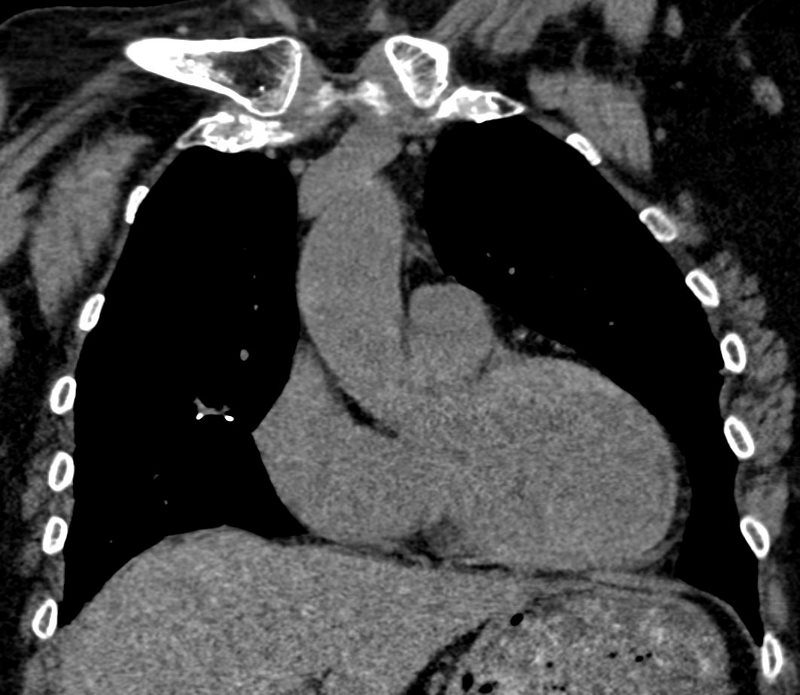
Pericardial Disease
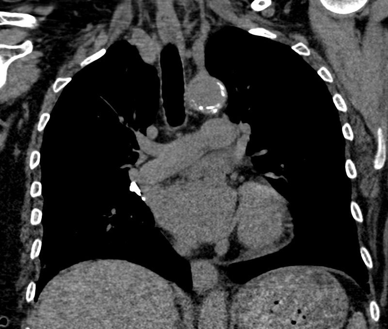
72-year-old male with history of an amyloidoma removed with right middle lobectomy
The CXR shows left ventricular enlargement
The current CT is characterized by stable small hilar nodal calcifications that likely represent amyloidosis
There are calcifications on right side of the left atrium and by the left heart border with associated focal regions of pericardial thickening. Involvement of the pericardium may be due to amyloidosis. However the LS calcification could also be post op and the calcification along on the left heart border could also be branch of circumflex with unusually chunky appearance which would be out of proportion to the degree of calcification elsewhere in the coronaries.
Fat in the LV apex indicates previous LAD territory infarction and likely account for the LVE noted on CXR
Ashley Davidoff MD
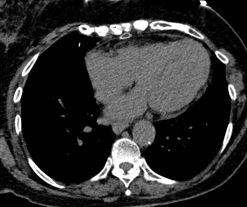

AMYLOIDOSIS
72-year-old male with history of an amyloidoma removed with right middle lobectomy
The CXR shows left ventricular enlargement
The current CT is characterized by stable small hilar nodal calcifications that likely represent amyloidosis
There are calcifications on right side of the left atrium and by the left heart border with associated focal regions of pericardial thickening. Involvement of the pericardium may be due to amyloidosis. However the LS calcification could also be post op and the calcification along on the left heart border could also be branch of circumflex with unusually chunky appearance which would be out of proportion to the degree of calcification elsewhere in the coronaries.
Fat in the LV apex indicates previous LAD territory infarction and likely account for the LVE noted on CXR
Ashley Davidoff MD
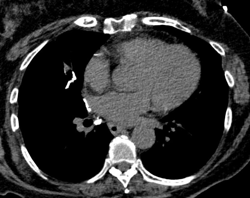
Fat in the LV Post MI and LVE
72-year-old male with history of an amyloidoma removed with right middle lobectomy
The CXR shows left ventricular enlargement
The current CT is characterized by stable small hilar nodal calcifications that likely represent amyloidosis
There are calcifications on right side of the left atrium and by the left heart border with associated focal regions of pericardial thickening. Involvement of the pericardium may be due to amyloidosis. However the LS calcification could also be post op and the calcification along on the left heart border could also be branch of circumflex with unusually chunky appearance which would be out of proportion to the degree of calcification elsewhere in the coronaries.
Fat in the LV apex indicates previous LAD territory infarction and likely account for the LVE noted on CXR
Ashley Davidoff MD