
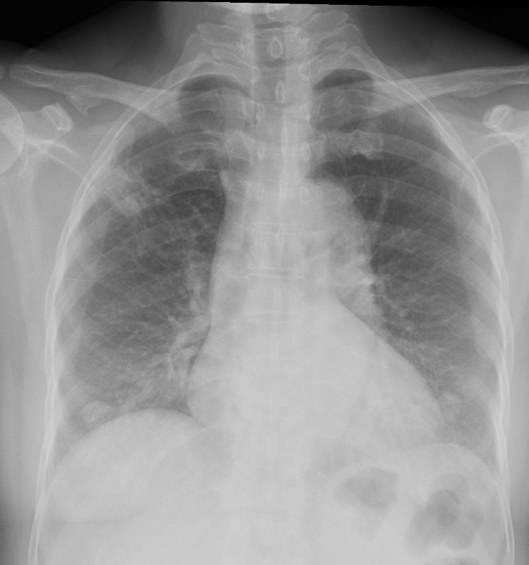
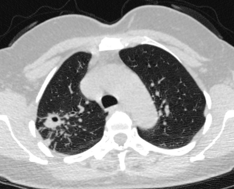
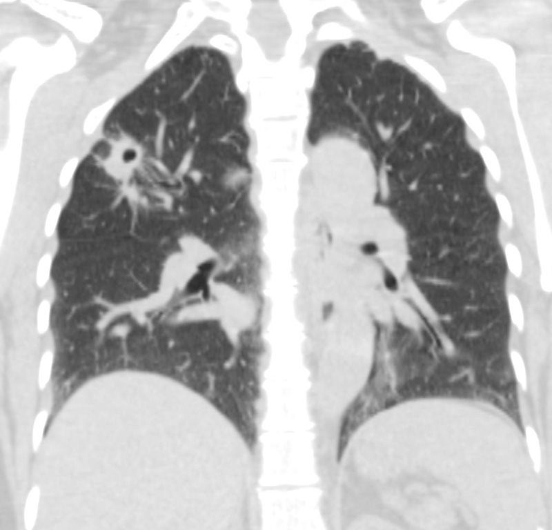
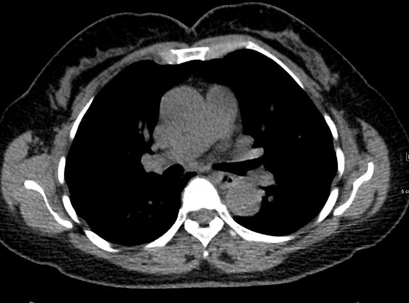
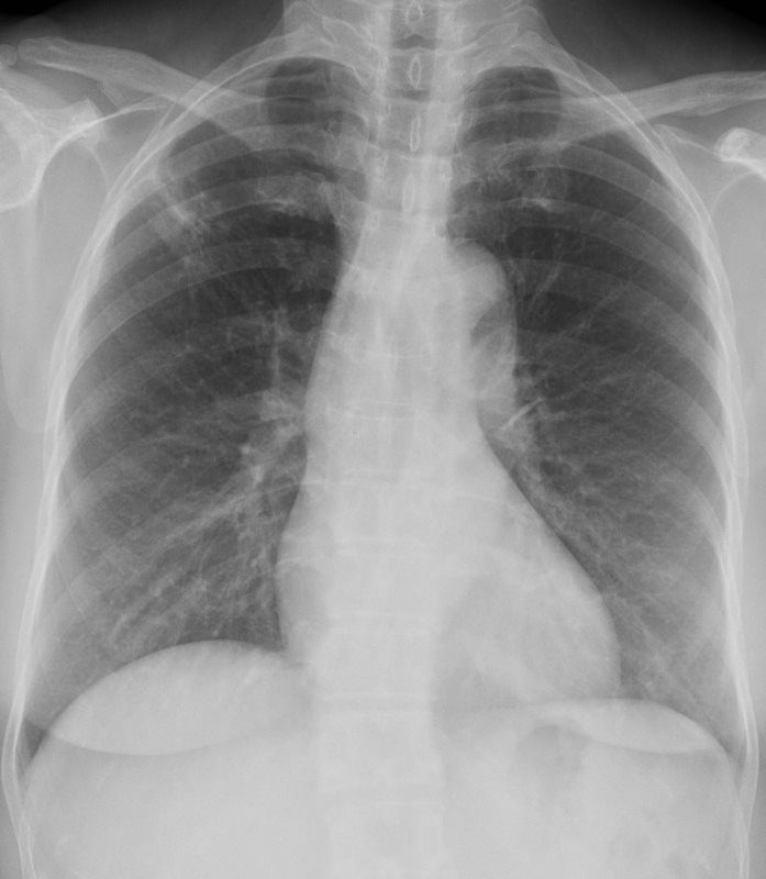
50-year-old Female with history of TB presents with a focal ground glass infiltrate in the RUL.
CT shows a cavitating nodule in the right upper lobe with thickened interlobular septa. No obvious mediastinal nor hilar adenopathy
CXR shows 3 months later after treatment shows persistent but improved subsegmental infiltrate
Ashley Davidoff MD
TB – BEFORE TREATMENT
50-year-old Female with history of TB presents with a focal ground glass infiltrate in the RUL.
CT shows a cavitating nodule in the right upper lobe with thickened interlobular septa. No obvious mediastinal nor hilar adenopathy
CXR shows 3 months later after treatment shows persistent but improved subsegmental infiltrate
Ashley Davidoff MD