
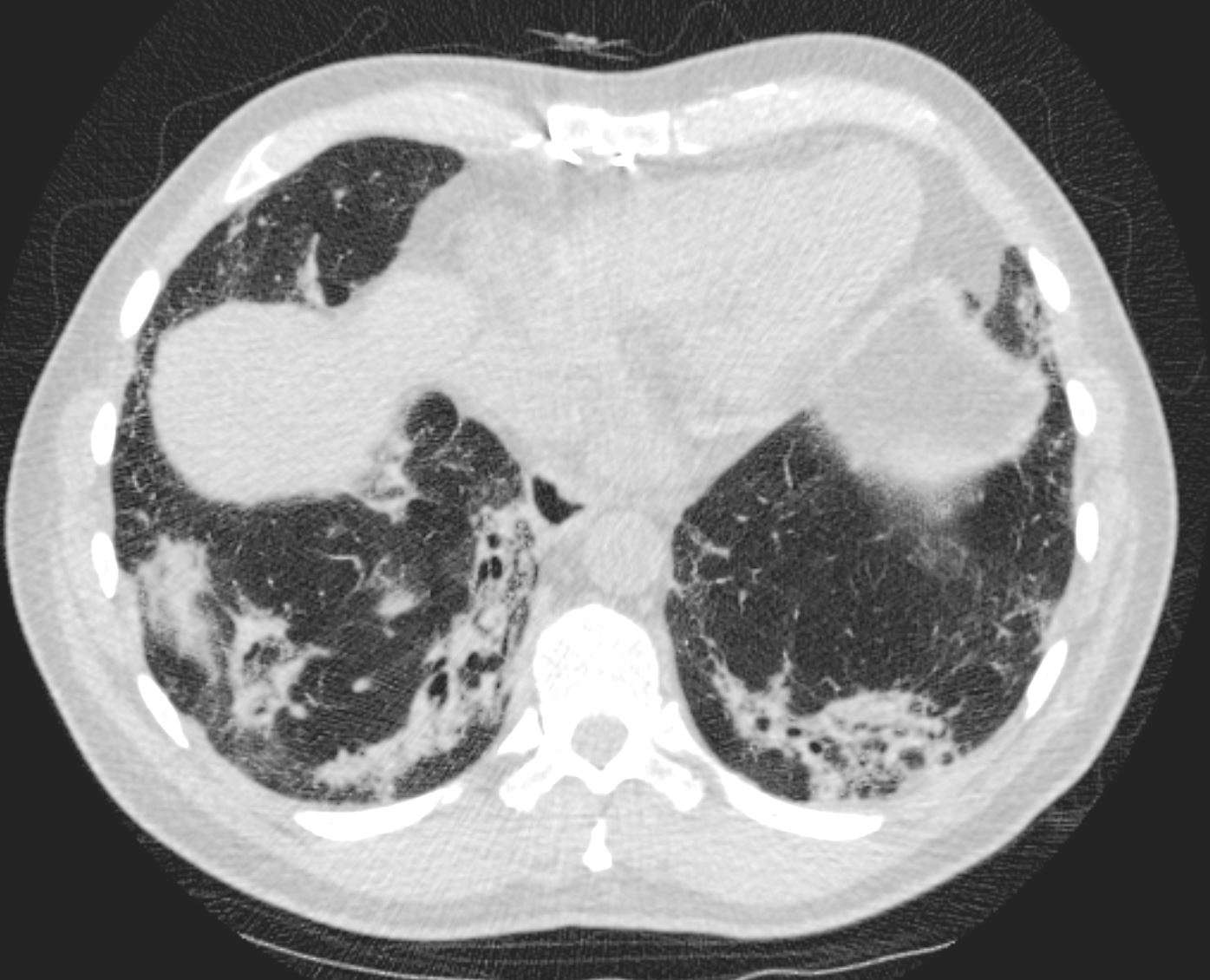
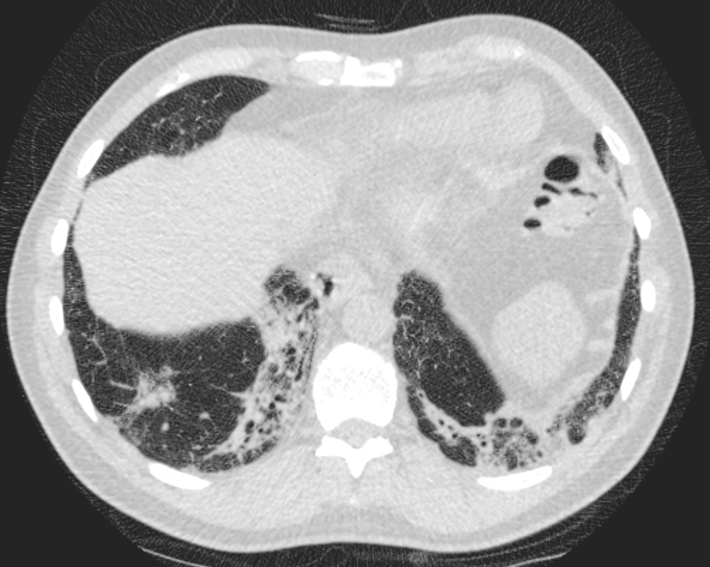
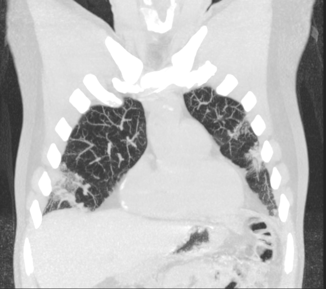
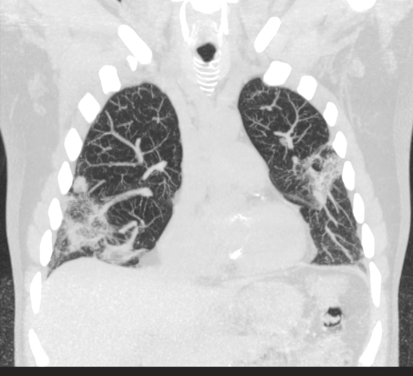
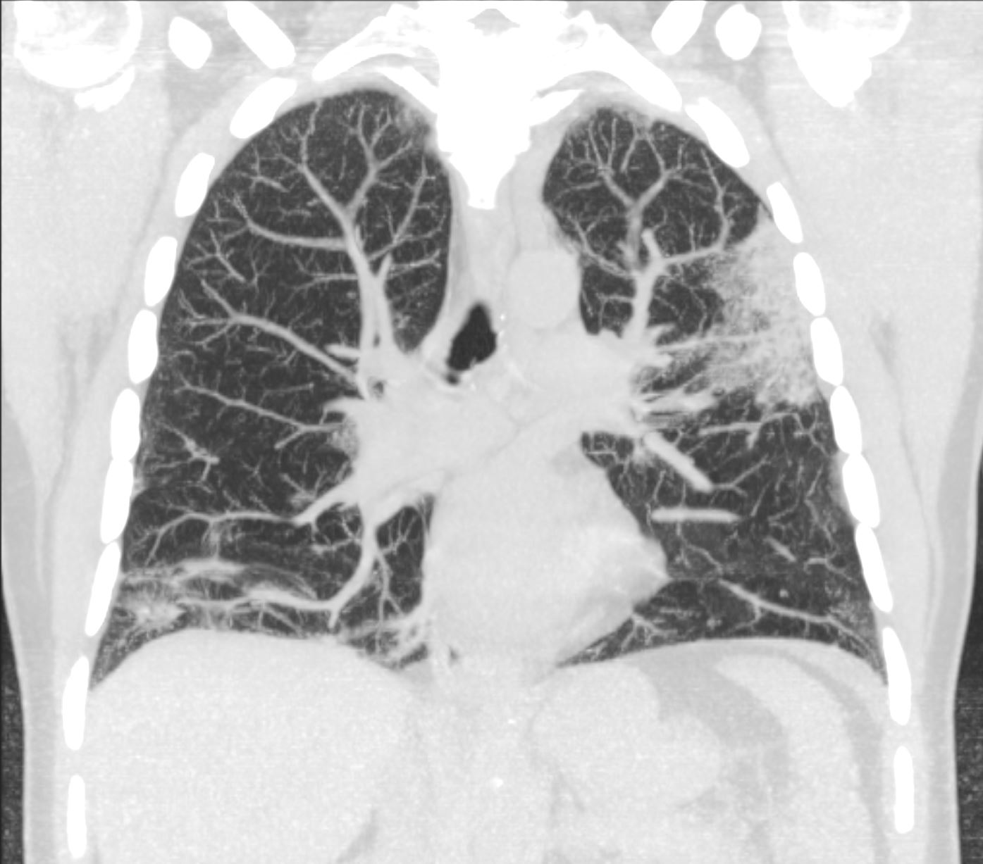
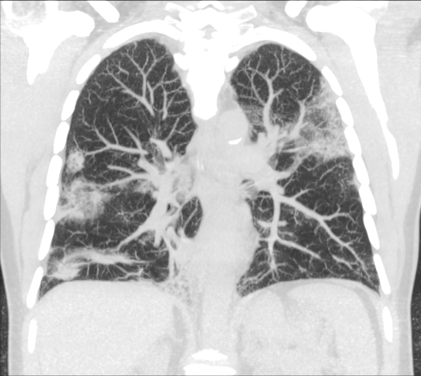
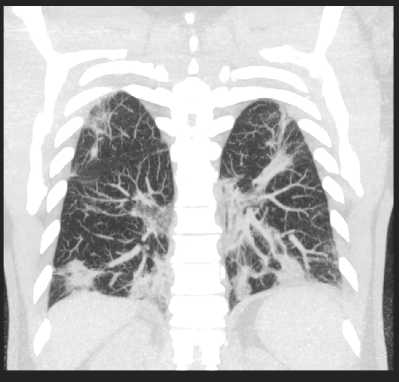
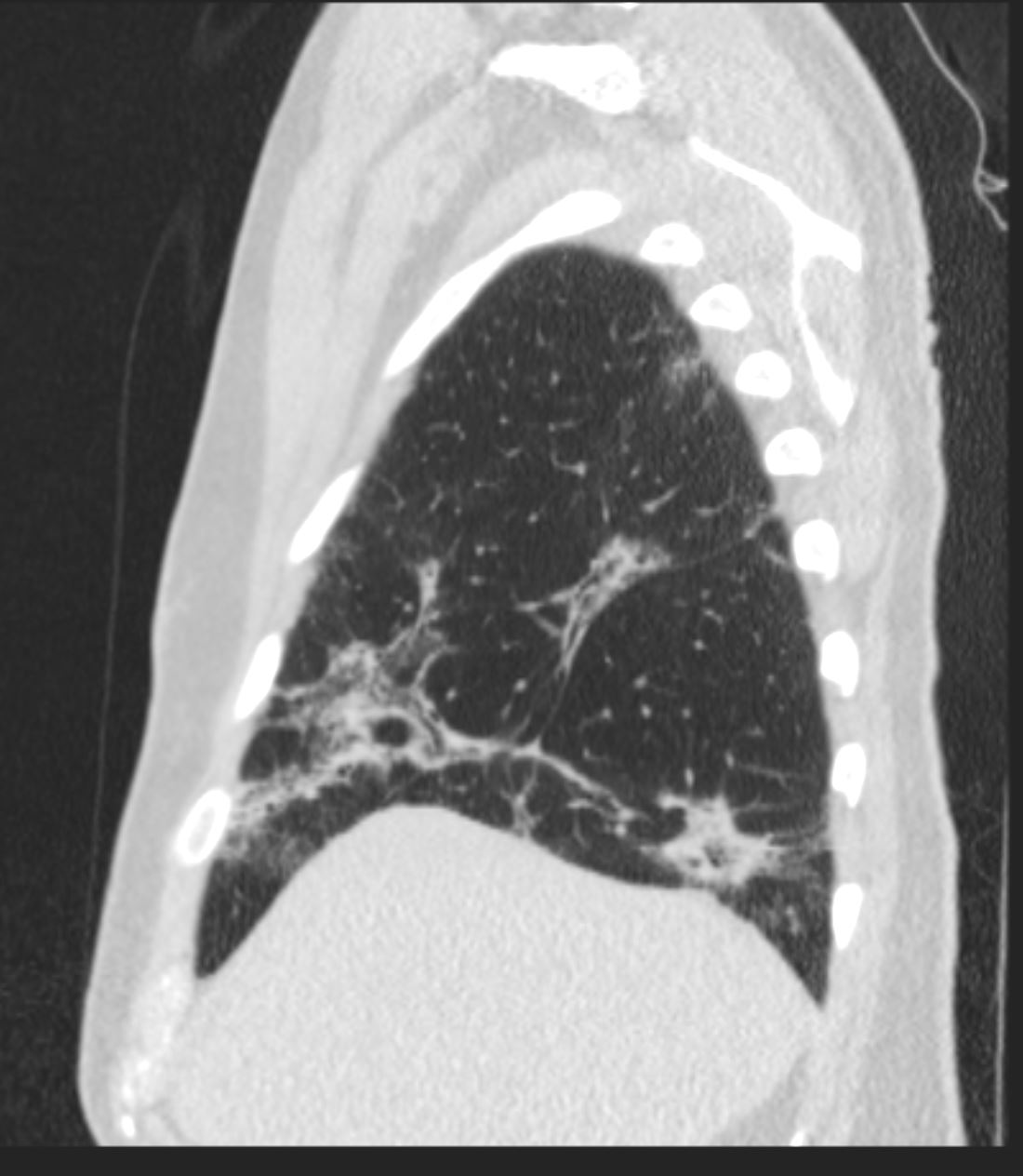
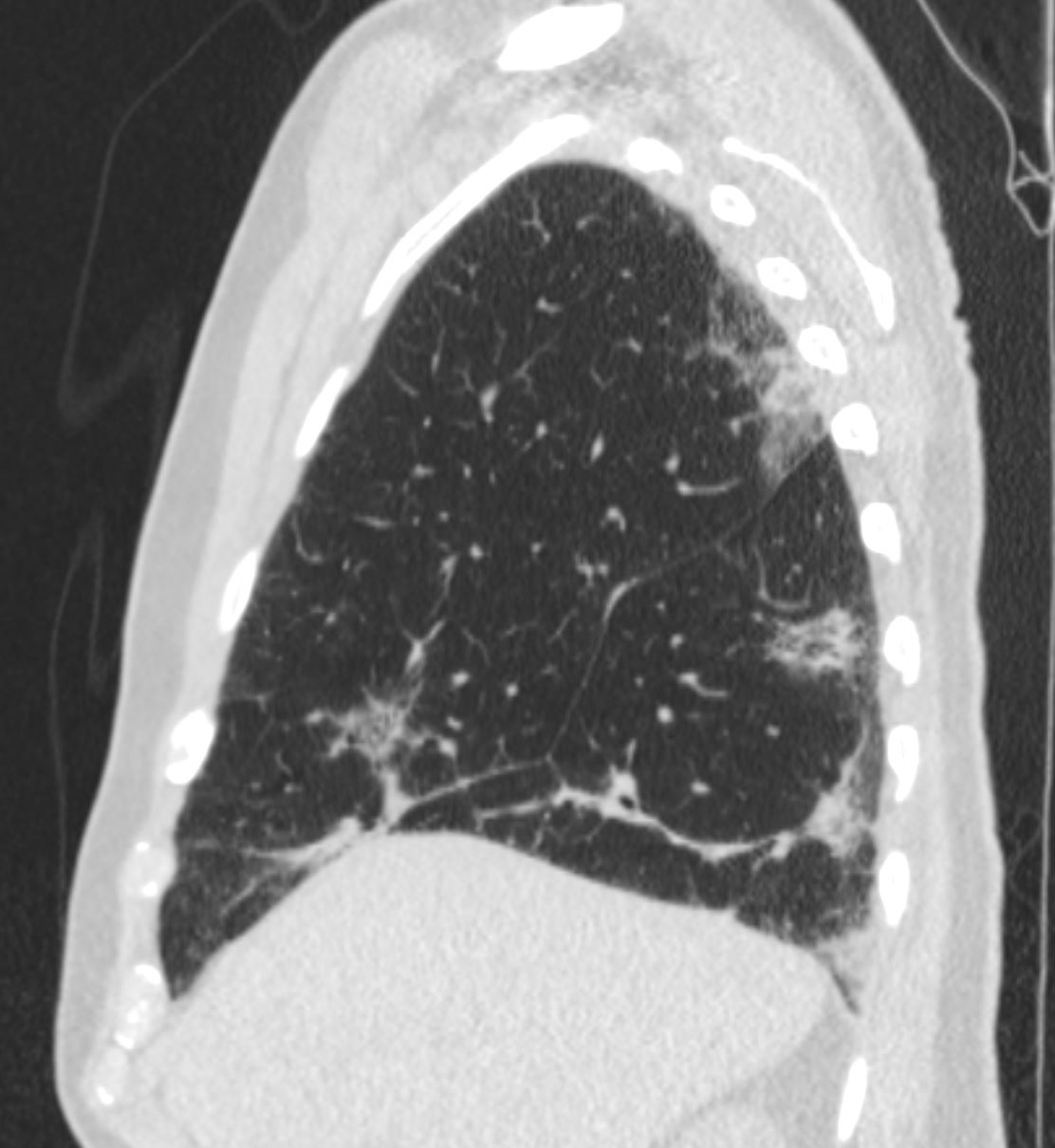
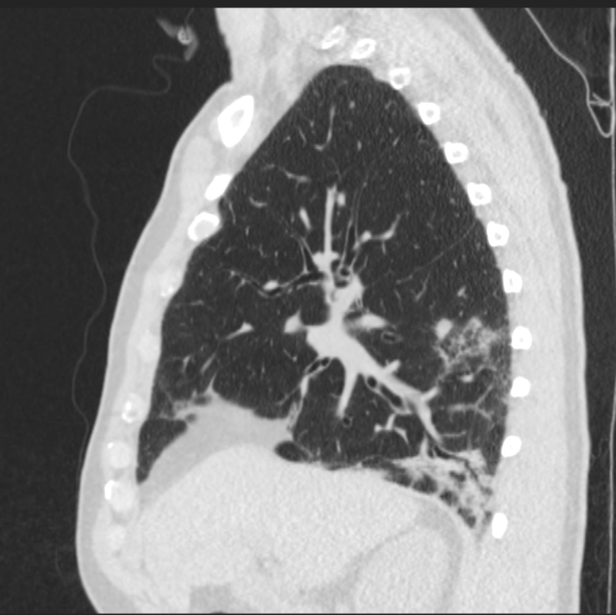
Had COVID19 1month ago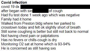 cxr from 1 weeks ago
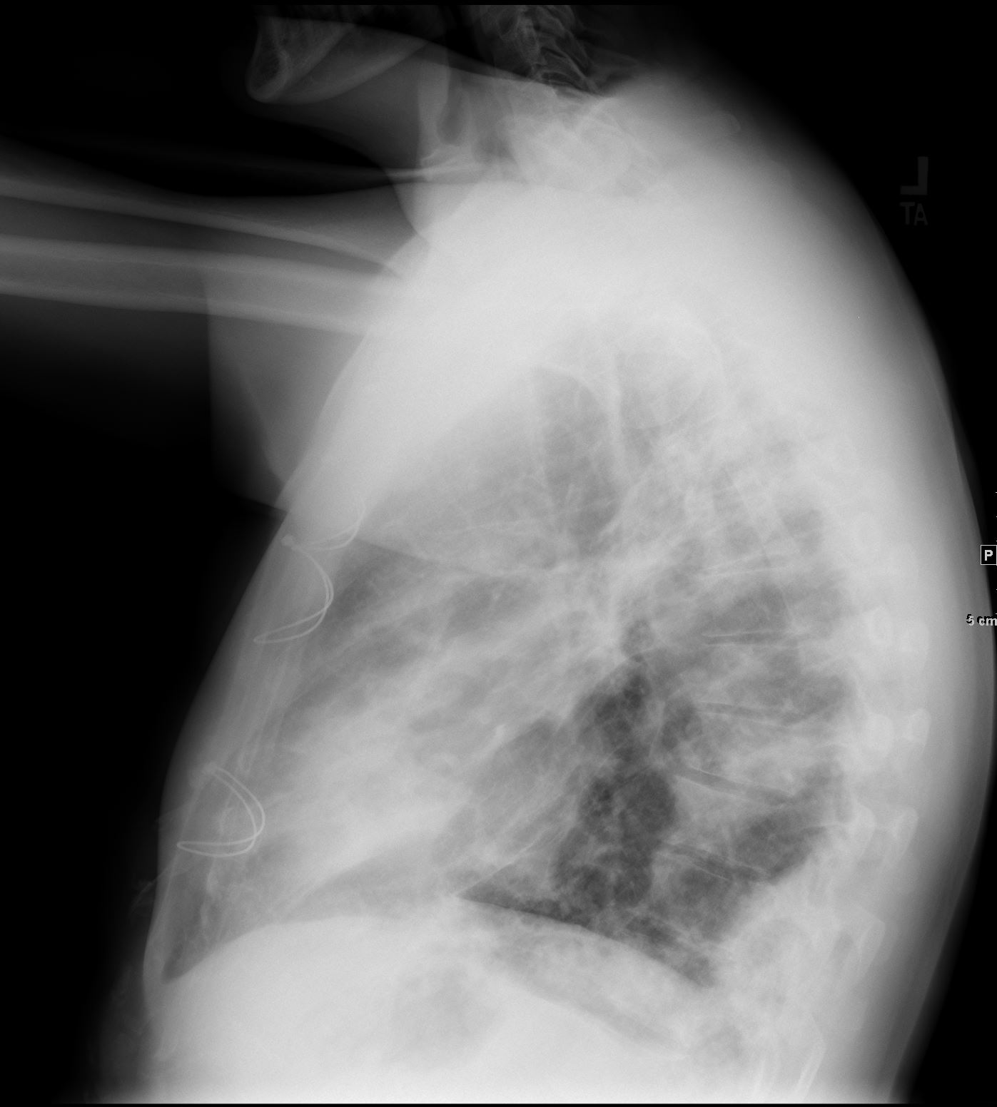
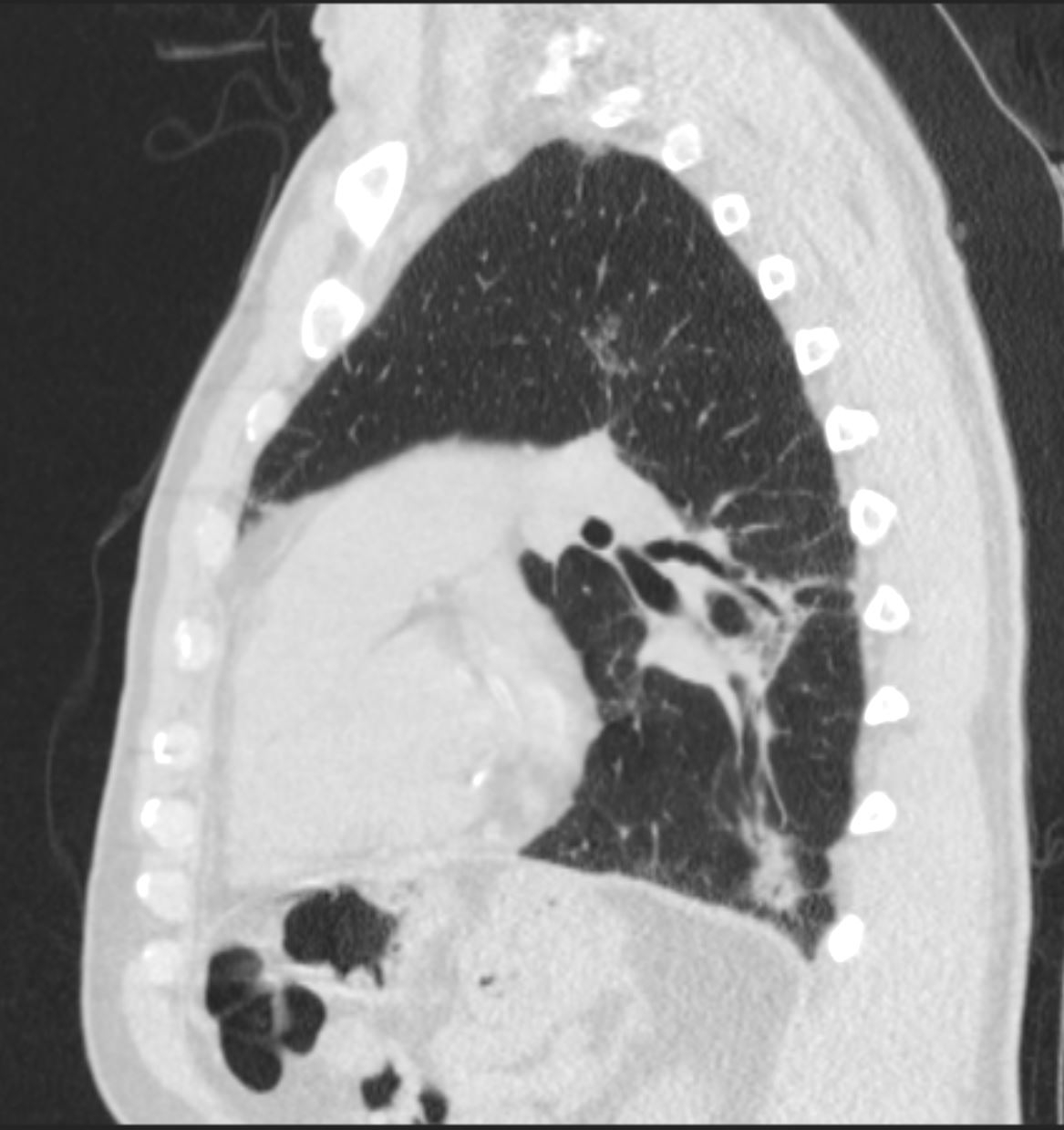
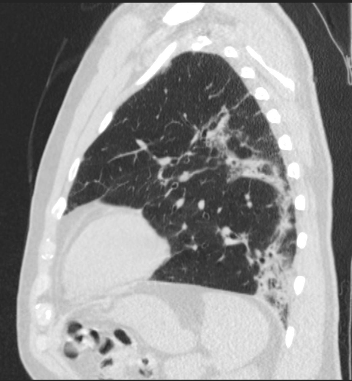
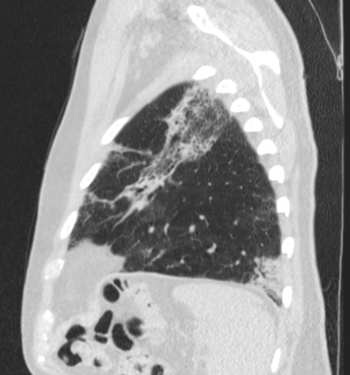
 arcade-like sign ?
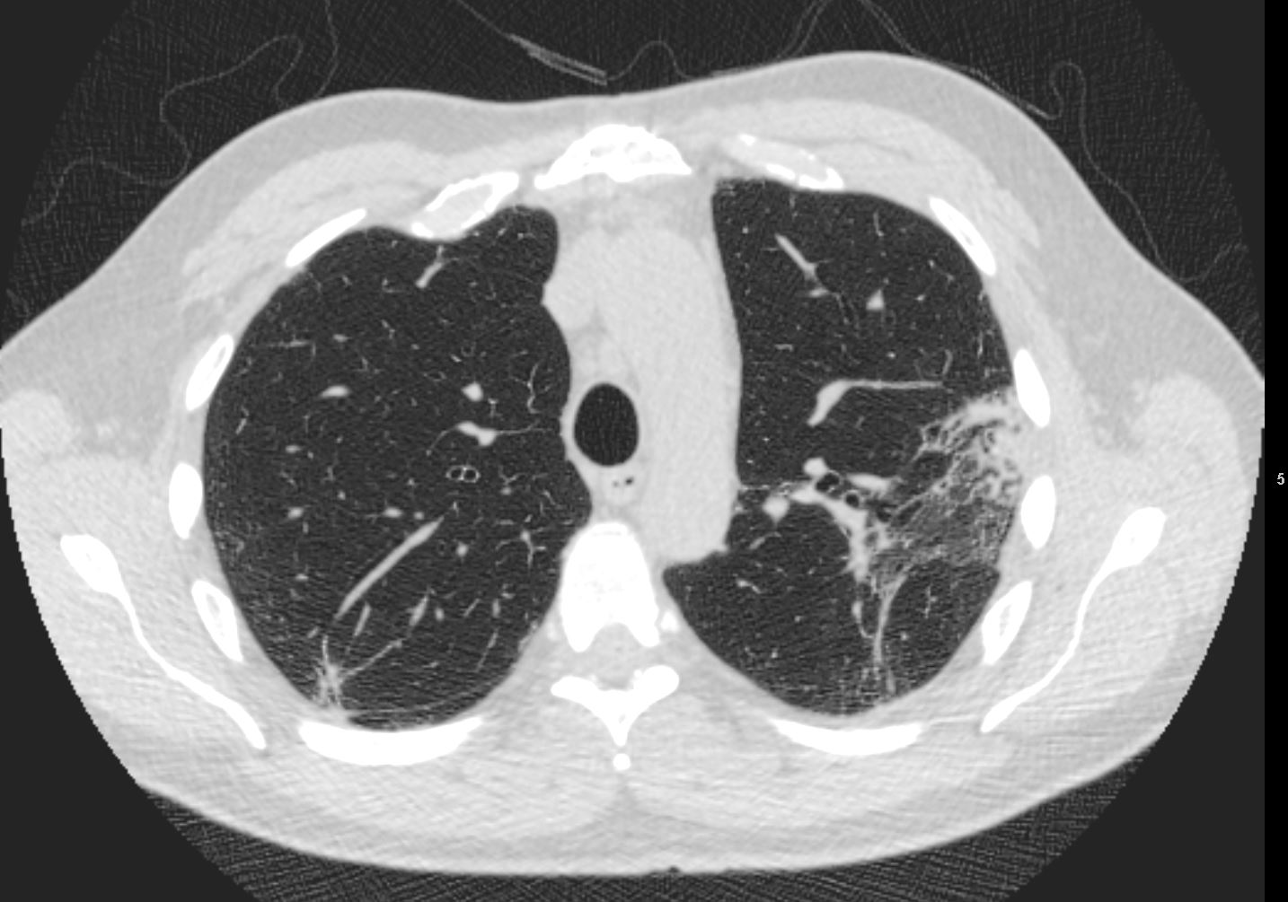
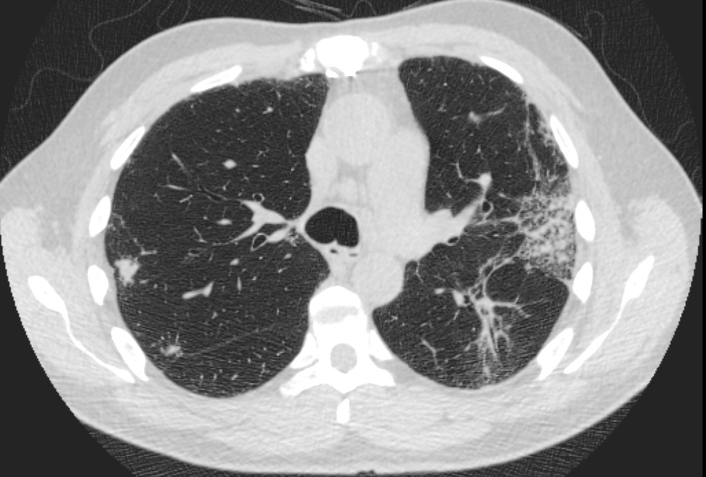
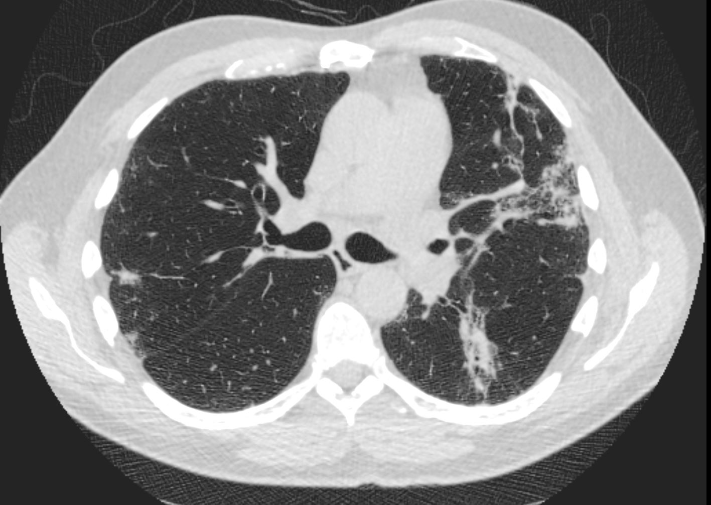
?arcade-like sign 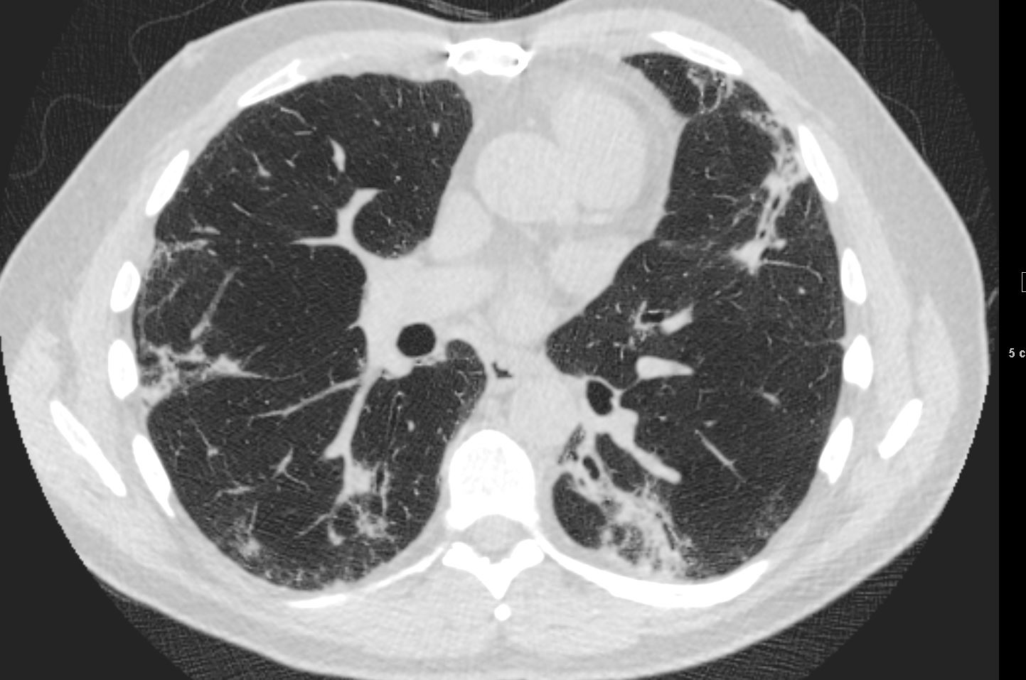
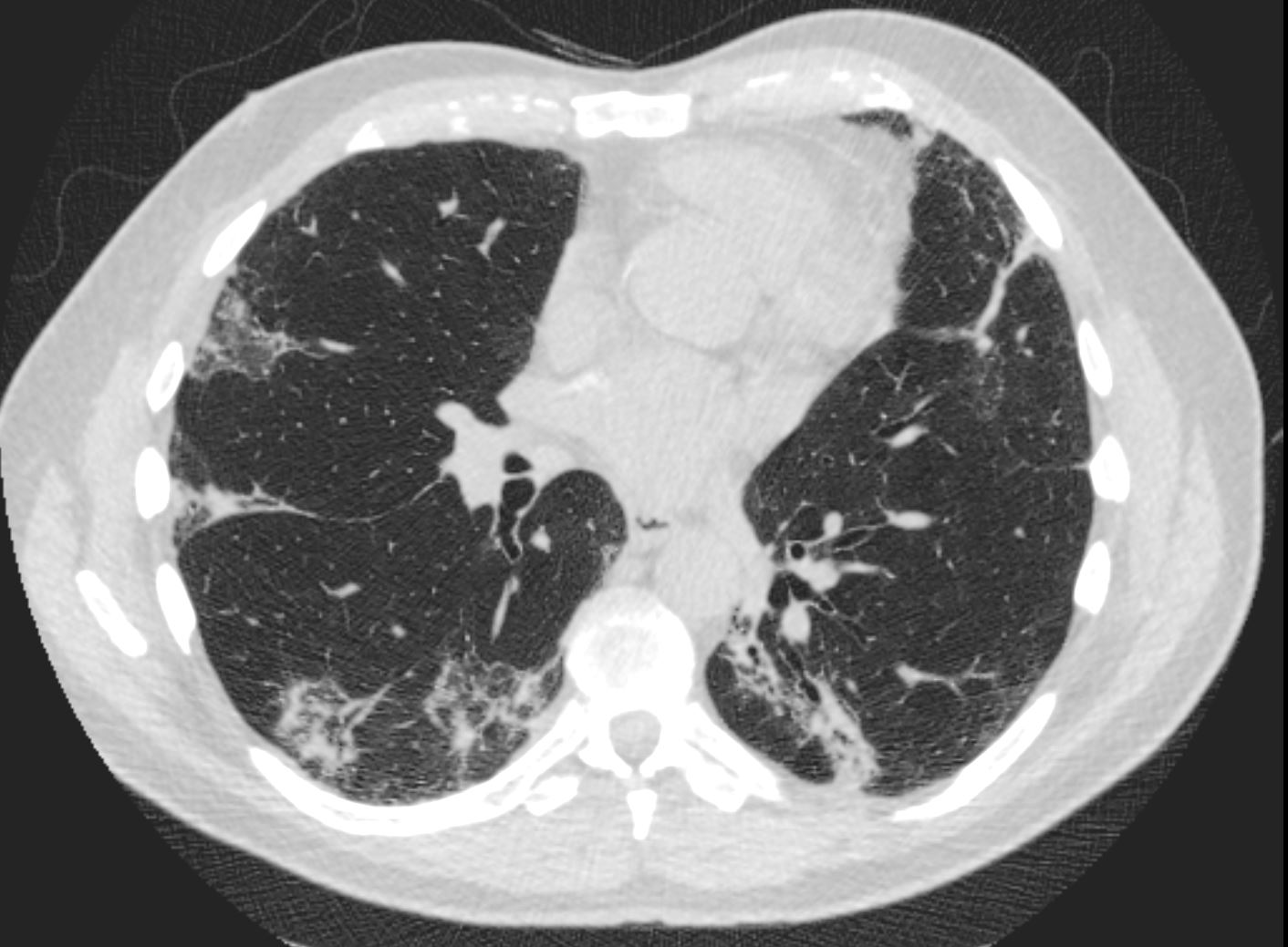
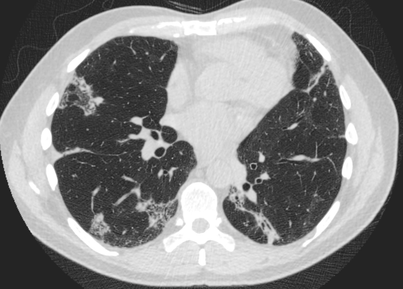
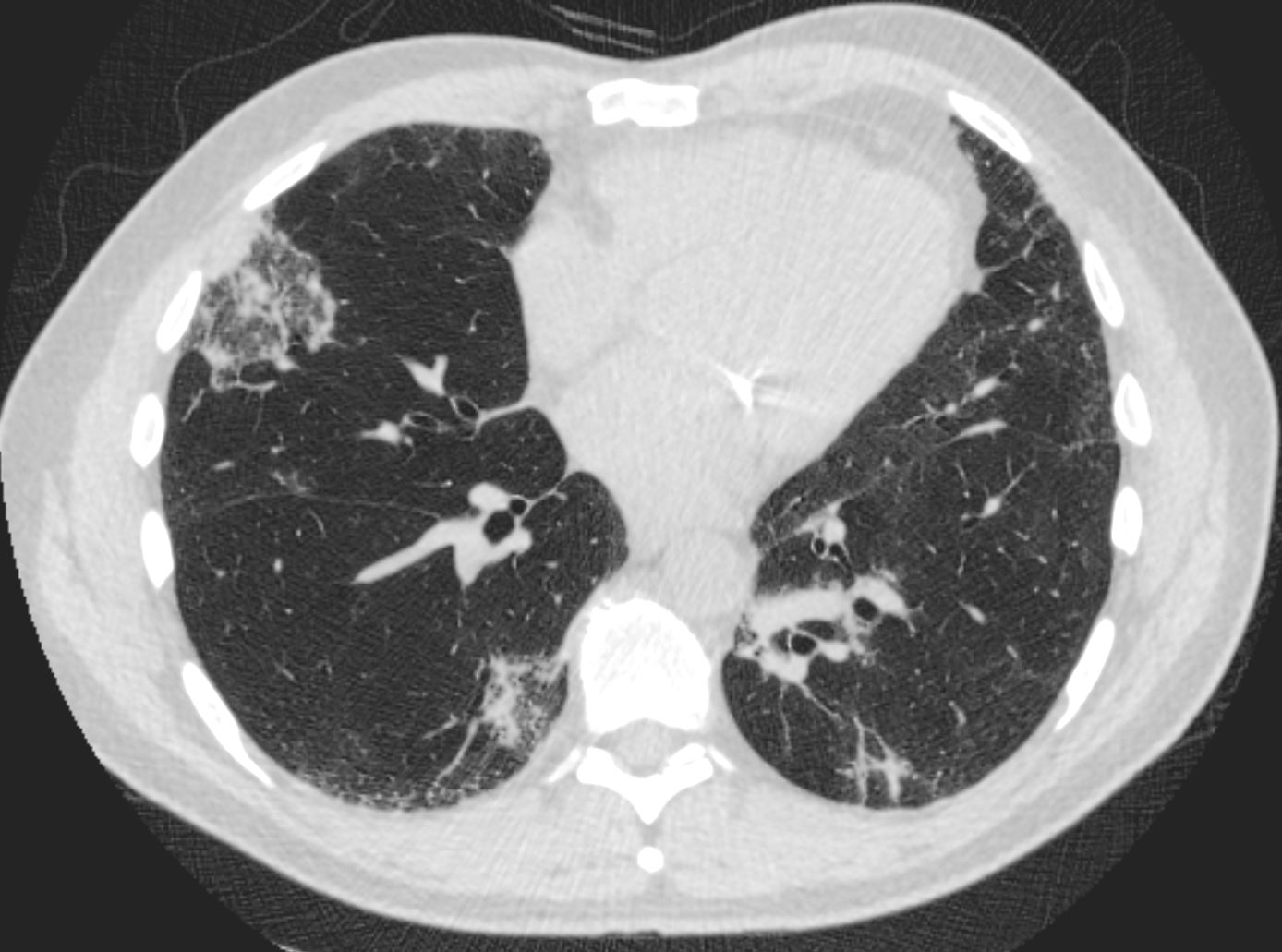
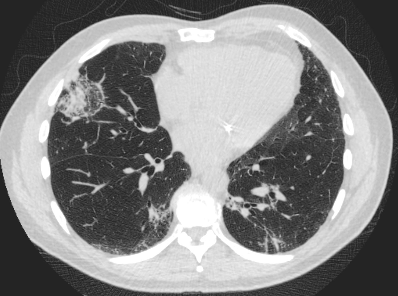
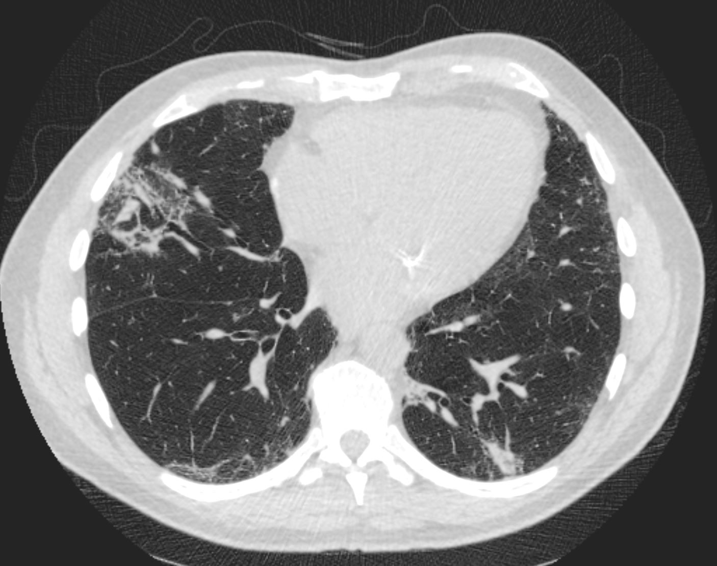 arcade-like sign
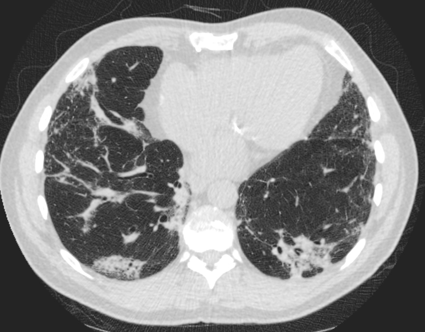
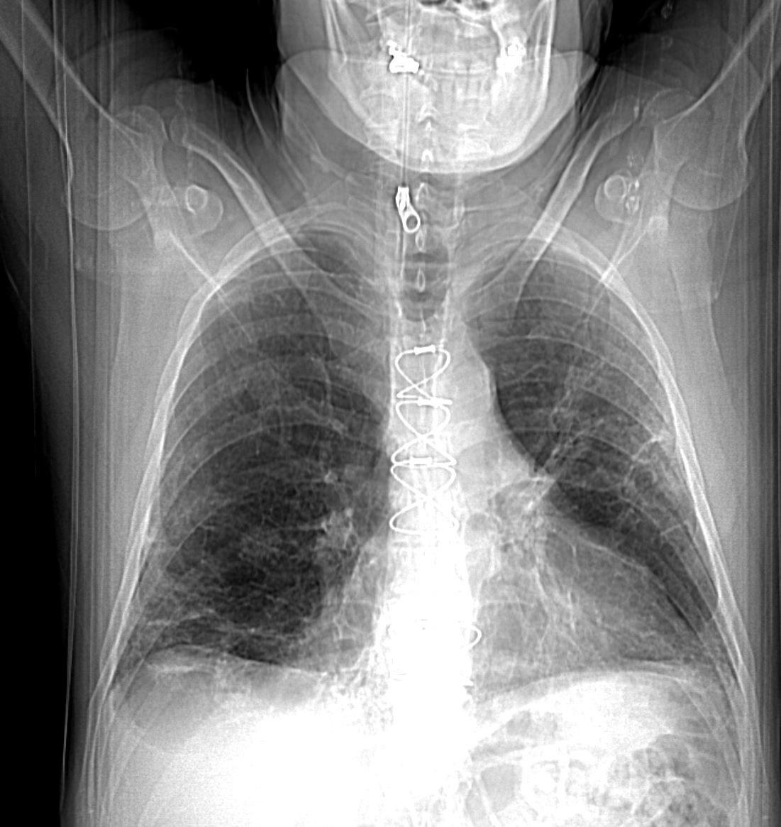
“Organising pneumonia (OP), a form a DPLD, is a distinct clinicopathological entity that may occur as a pulmonary reaction to various injuries, including ARDS. OP is characterised by the patchy filling of alveoli and bronchioles by loose plugs of connective tissue with concomitant diffuse alveolar damage, the hallmark of ARDS. Radiological findings typically include peripheral consolidation, ground-glass infiltrates and/or solitary nodules. The definitive diagnosis of OP requires histological assessment and the primary treatment of OP is, apart from treating the underlying disease, corticosteroid administration over several months, initially at relatively high doses.5″ ( Vadász)
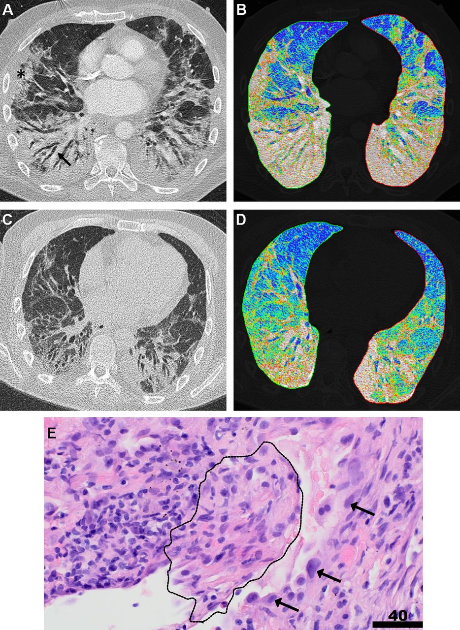
“Organising pneumonia complicating severe COVID-19. (A) Thoracic CT of a patient with COVID-19 on clinical deterioration and markedly decreasing pulmonary compliance 16 days after the initiation of mechanical ventilation, revealing extensive subpleural patchy consolidation (black asterisk), fibrotic bands and traction bronchiectasis (black arrow) of the middle lobe, the lingula and both lower lobes, compatible with organising pneumonia. (B) A colour-coded lung density map (based on Hounsfield units) at the same level as (A). Blue areas represent normal lung tissue with normal lung density values, whereas green areas represent lung tissue with slightly increased density values, consistent with ground-glass opacification and red/white areas characterise lung tissue with markedly increased density values consistent with consolidations/fibrotic changes. (C) Follow-up CT approximately 8 weeks after the initiation of corticosteroid therapy, showing partially reversed peribronchovascular consolidation, fibrotic bands and bronchiectasis. (D) The corresponding colour-coded lung density map confirms the decreased density values of the affected areas. (E) Histology of a transbronchial biopsy from the same patient with H&E staining at a magnification of ×400 is illustrated. The organisation of an actin+ fibrous plug (immunostainings not shown) within an alveolus, intermingled with lymphocytes (dotted line), corresponding to organising pneumonia with prominent hyperplasia of surrounding alveolar pneumocytes (arrows). Scale bar corresponds to 40 µm.” ( Vadász)
OP is a process of pulmonary tissue repair that can be either:
- •
-
cryptogenic;
- •
-
secondary to a lung injury such as infection, drug toxicity, inhalation of a pathogen (cocaine), inhalation of toxic gas, gastroesophageal reflux, collagenosis, organ transplant, or radiotherapy [5], [6], [7], [8];
- •
-
it can be histologically associated with pulmonary lesions of another nature such as vasculitis, lymphoma, lung cancer [9], hypersensitivity pneumonitis, eosinophilic pneumonia, acute interstitial pneumonia, non-specific interstitial pneumonia, or usual interstitial pneumonia [5] (Table 1).