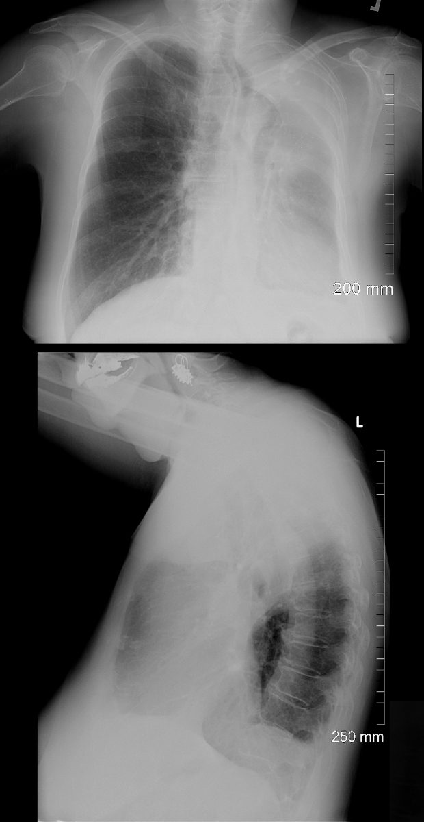
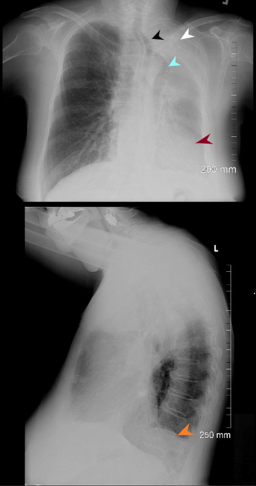
76-year-old female presents with dyspnea. PA CXR shows airless consolidation of the left upper lobe, volume loss of the left hemithorax, with leftward shift of the trachea, and cardio mediastinal structures. The right lung is hyperinflated with herniation across the midline. The lateral examination shows an anterior airless consolidation, and elevation of the left hemidiaphragm. Granulomatous calcifications are noted in the left upper lobe atelectatic segment Ashley Davidoff MD TheCommonVein.net76-year old female presents with dyspnea. PA CXR shows airless consolidation of the left upper lobe (white arrowhead), volume loss of the left hemithorax, with leftward shift of the trachea (black arrowhead) and cardio mediastinal structures (maroon arrowhead). The right lung is hyperinflated with herniation across the midline (light blue arrowhead). The lateral examination shows an anterior airless consolidation, and elevation of the left hemidiaphragm orange arrowhead). Granulomatous calcifications are noted in the left upper lobe atelectatic segment Ashley Davidoff MD TheCommonVein.net76-year-old female presents with dyspnea CT scan shows findings of left upper lobe atelectasis characterized by airless consolidation, with leftward shift of the cardio mediastinal structures. There is evidence of encasement of the pulmonary artery with a soft tissue mass surrounding it. There is occlusion of the left upper lobe bronchus. Granulomatous calcifications are noted in the left upper lobe atelectatic segment Ashley Davidoff MD TheCommonVein.net76-year-old female presents with dyspnea CT scan shows findings of left upper lobe atelectasis characterized by airless consolidation (blue arrowheads a,b,and c), with leftward shift of the cardio mediastinal structures (orange arrowheads e,f). There is evidence of encasement of the pulmonary artery (maroon arrowhead ) with a soft tissue mass surrounding it (white asterisks a,b). There is occlusion of the left upper lobe bronchus (yellow arrowhead, b). Granulomatous calcifications are noted in the left upper lobe atelectatic segment (b) Ashley Davidoff MD TheCommonVein.net