Diffuse scleroderma diagnosed 30 years ago
History of scleroderma renal crisis 24 years ago
Severe Raynaud’s and recurrent digital ischemic ulcers s/p amputations
Diffuse skin disease that has improved since time of diagnosis
Severe GI disease with chronic diarrhea and weight loss. Has required multiple courses of antibiotics and fecal transplants and disease complicated by multiple episodes of c.diff.
Recurrent pericardial effusions
Interstitial lung disease stable on most recent chest CT
Musculoskeletal disease with flexion contractures in both hands and elbows. +inflammatory arthritis and history of tendon friction rubs
surgery
PERICARDIUM
Final Diagnosis
PERICARDIUM:
FIBROCONNECTIVE TISSUE WITH FIBROSIS AND ASSOCIATED REACTIVE MESOTHELIUM WITH MILD CHRONIC INFLAMMATION AND FIBRINOUS EXUDATE.
NO TUMOR IDENTIFIED.
Still have to upload all pictures from pic library on work (home computer in folder called acroosteolysis
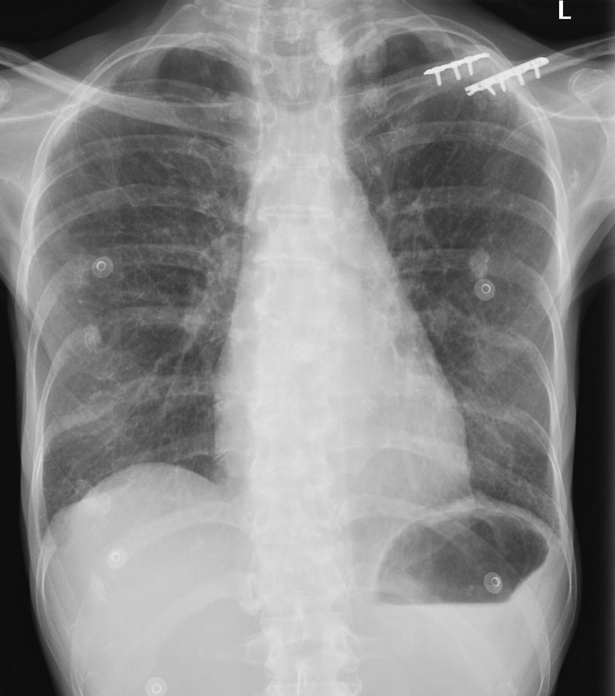
CXR
Heart and mediastinum: The cardiomediastinal silhouette is stable
without significant cardiomegaly.
Lungs and pleura: Lungs are hyperinflated. Scarring at the bilateral
lung apices. Lungs are otherwise clear. No pneumothorax. No pleural
effusions
Ashley Davidoff MD
The CommonVein.net
4 years ago
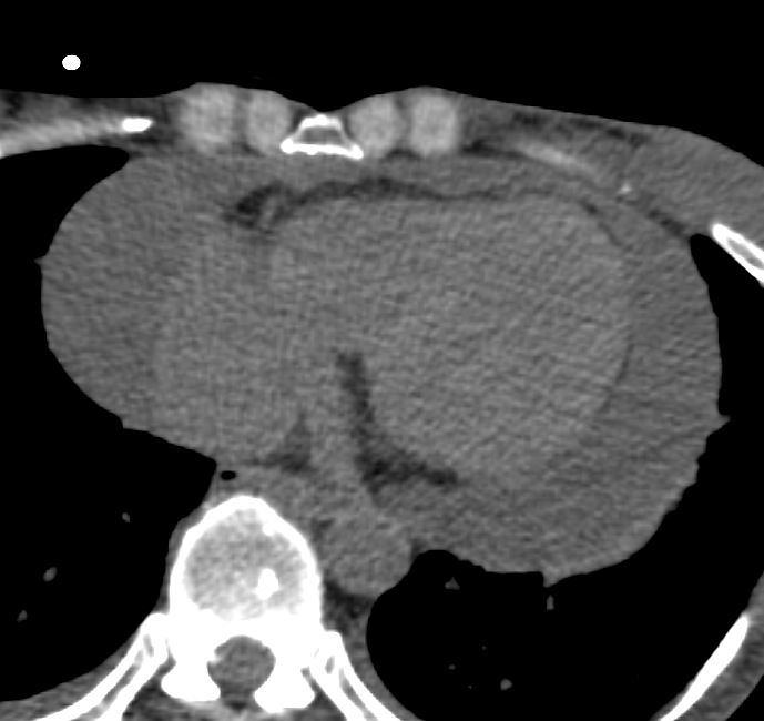
Presented with a large pericardial effusion, significantly increase in volume since J1 month prior, of uncertain
most likely chronic given the placement of a
pericardial drainage in the past therefore likely associated with
connective tissue disease. Prior pericardial drain has been removed.

Ashley Davidoff MD
The CommonVein.net
Echocardiogram
Showed a large circumferential pericardial effusion with
significant fibrinous exudate adherent to the epicardial surface.
Delayed diastolic expansion of the distal RV suggested raised
intrapericardial pressure.
IVC size was normal with blunted respirophasic variation suggestive of high normal RA pressure (5-10 mm Hg).
Pericardial window followed
Path report
Pericardium:
Fibroconnective tissue with fibrosis and associated reactive mesothelium with mild chronic inflammation and fibrinous exudate.
No tumor identified.
CT of the Chest
- Mild groundglass opacities predominating within the peripheral /subpleural lung apices and lower lobes bilaterally.
- multiple bilateralpulmonary nodules.Pleura: No pleural effusion.Heart and pericardium: No pericardial effusion. There is a
pericardial drain with its tip terminating adjacent to the pulmonary
trunk. There is trace air within the peritoneum where the pericardial
drain is exiting from the body inferior to the sternum.
Ashley Davidoff MD
The CommonVein.net
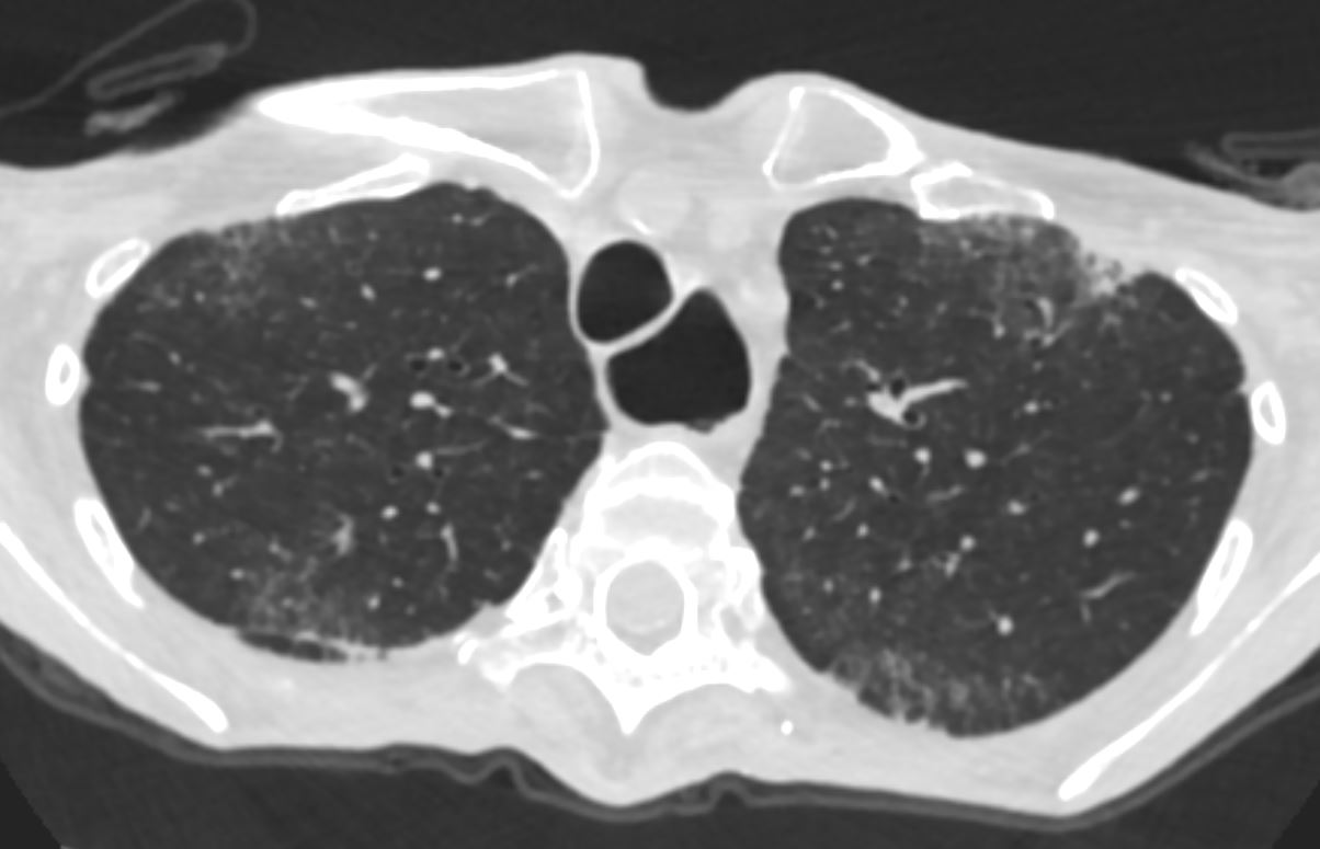
Mild Interstitial Lungh Disease
Ashley Davidoff MD
The CommonVein.net
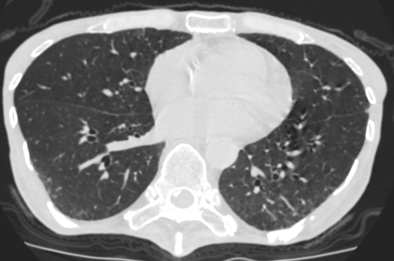
Mild Interstitial Lungh Disease
Ashley Davidoff MD
The CommonVein.net
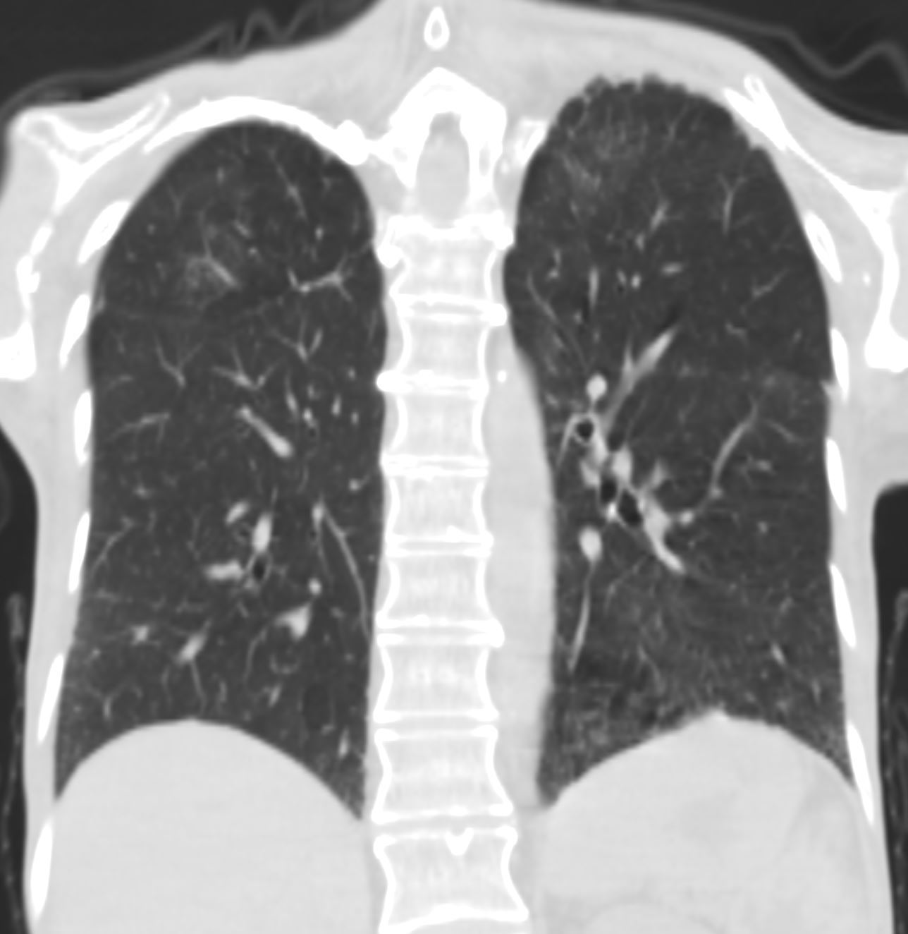
Mild Interstitial Lungh Disease
Ashley Davidoff MD
The CommonVein.net
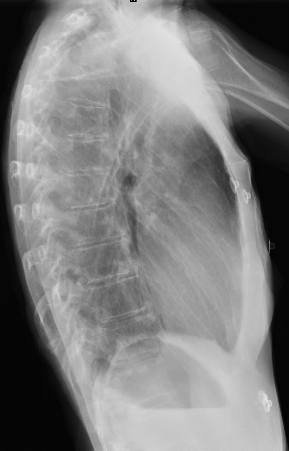
Dilated Esophagus

Dilated Esophagus
Ashley Davidoff MD
The CommonVein.net
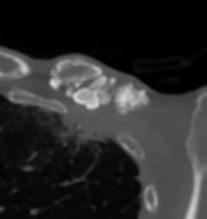
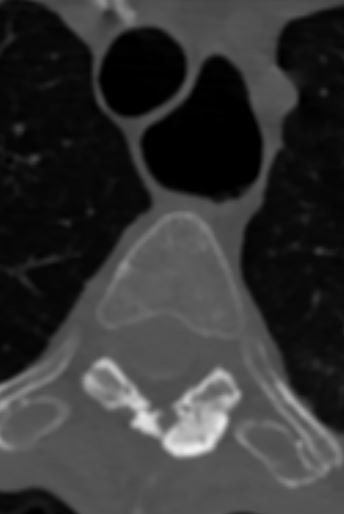
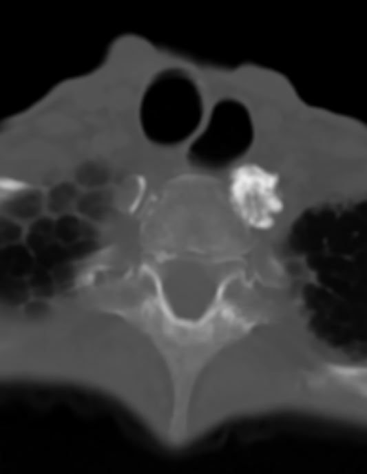
Soft Tissue Calcification and Ossification
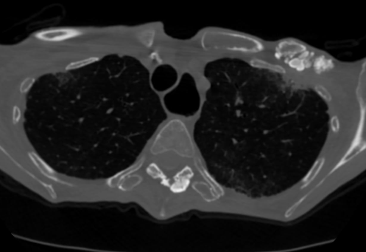
Soft Tissue calcification and ossification
Ashley Davidoff MD
The CommonVein.net
Soft Tissue calcification and ossification
Ashley Davidoff MD
The CommonVein.net
Soft Tissue calcification and ossification
Ashley Davidoff MD
The CommonVein.net
Soft Tissue calcification and ossification
Ashley Davidoff MD
The CommonVein.net
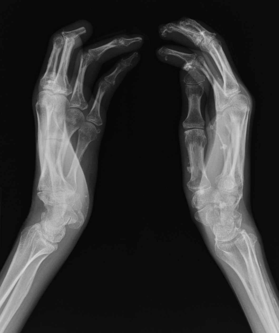
Acro-osteolysis, Contraxctures and Soft Tissue Calcification
Right HAnd
- soft tissue swelling about the 3rd digit.
- dDystrophic calcifications about the
distal phalanges along the medial aspect of the 5th metacarpal. - joint space narrowing involving the 2nd through 5th DIPs. contracture deformities
are compatible with a history of scleroderma.
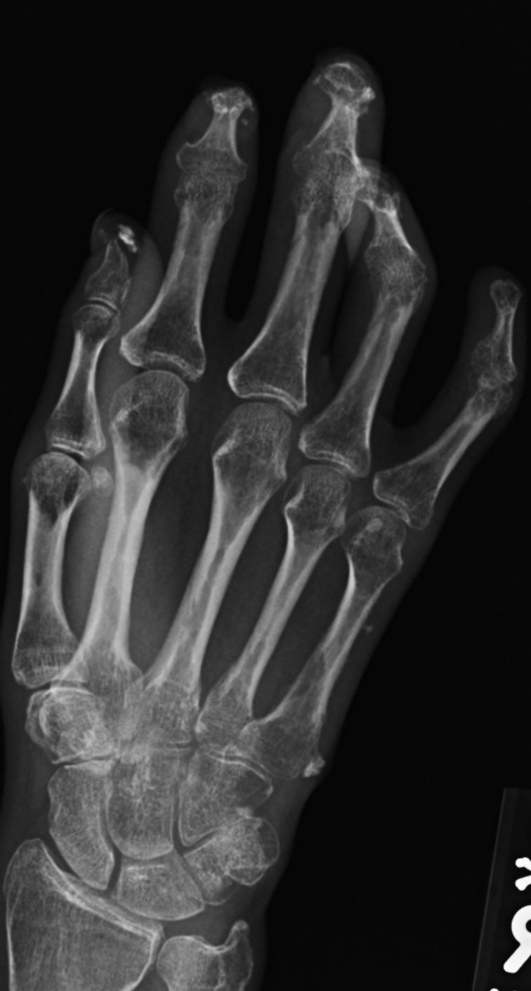
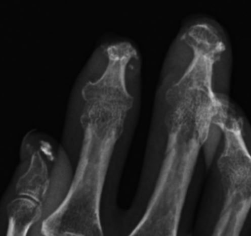
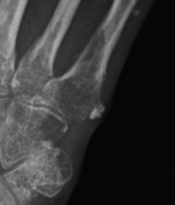
Hands show distal acro- osteolysis contractures and soft tissue calcification
Ashley Davidoff MD
The CommonVein.net
Hands show distal acro- osteolysis contractures and soft tissue calcification
Ashley Davidoff MD
The CommonVein.net
Hands show distal acro- osteolysis contractures and soft tissue calcification
Ashley Davidoff MD
The CommonVein.net
Hands show distal acro- osteolysis contractures and soft tissue calcification
Ashley Davidoff MD
The CommonVein.net
Hands show distal acro- osteolysis contractures and soft tissue calcification
Ashley Davidoff MD
The CommonVein.net
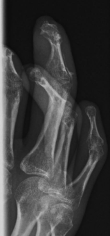
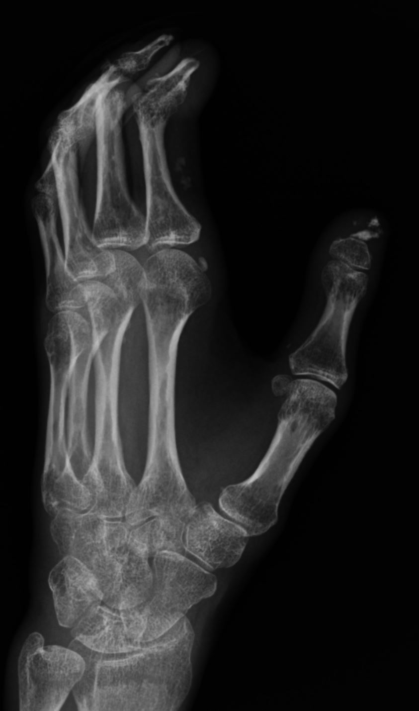
Left Hand
Severe acroosteolysis of the third digit to the level of the distal
metaphysis of the proximal phalanx. There is also acroosteolysis of
the thumb and index fingers. Scattered regions of soft tissue
calcification in the tuft of the thumb and in the proximal soft
tissues of the index finger. Narrowing of the remaining IP joints
with periarticular osteopenia. No acute fracture. Alignment is
anatomic.
Findings suggestive of scleroderma

hands show distal acro- osteolysis contractures and soft tissue calcification
Ashley Davidoff MD
The CommonVein.net
hands show distal acro- osteolysis contractures and soft tissue calcification
Ashley Davidoff MD
The CommonVein.net
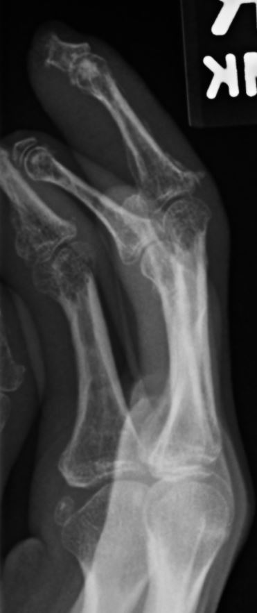
Ballcatchers view
severe acroosteolysis of the middle finger to the
level of the proximal phalanx distal metaphysis, consistent with
known history of scleroderma. There is also acroosteolysis of the
thumb distal phalanx and of the index finger to the level of the
middle phalanx. Scattered areas of soft tissue calcification in the
tuft of the thumb and in the proximal soft tissues of the index
finger. Mild to moderate narrowing of the remaining IP joints. No
acute fracture. No marginal erosions.