
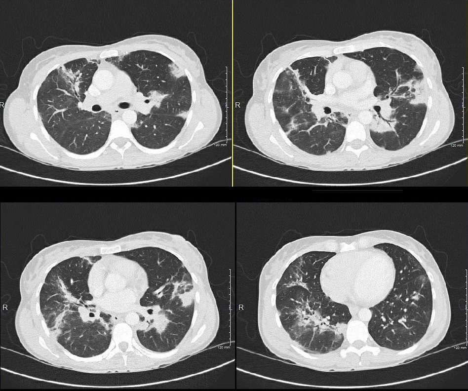
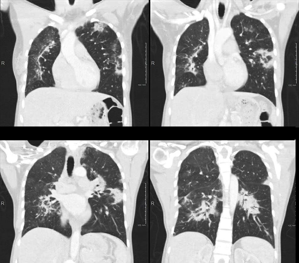
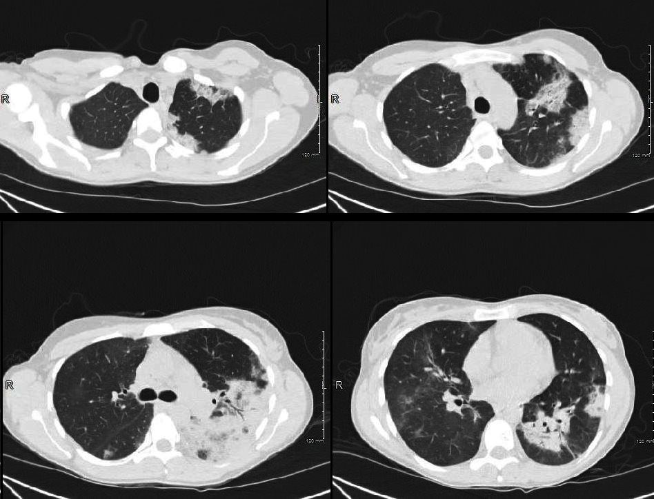
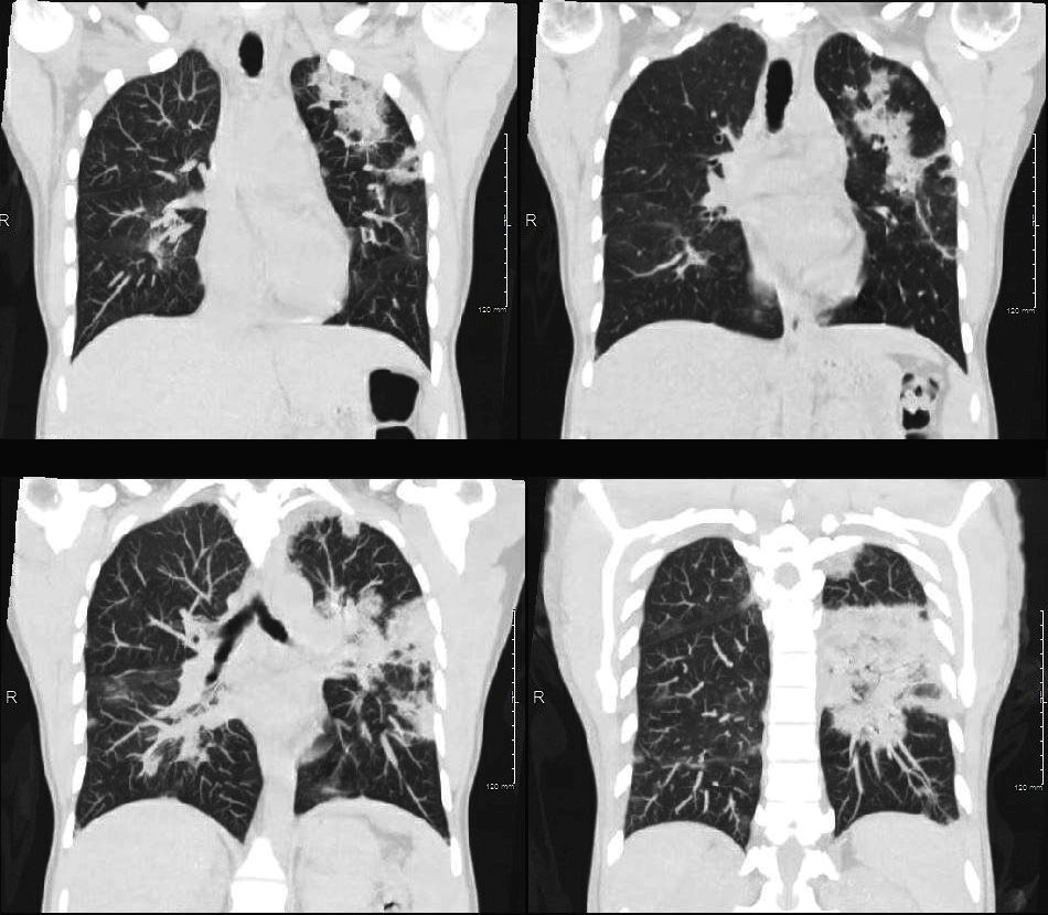
- bilateral ground-glass areas: common
- interlobular septal thickening: common
- pleural effusions: can be present in ~80% (range 60-100%) of cases
- thickening of bronchovascular bundles: present in around two-thirds of cases
- air-space consolidation: present in around half of the cases
- ill-defined centrilobular nodules: present in around one-third of cases
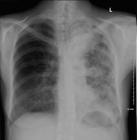
36 year old female who presented with dyspnea
CXR revealed
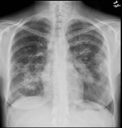
CXR shows bibasilar patchy infiltrates. She was treated with antibiotics
Ashley Davidoff MD
TheCommonVein.net
Ashley Davidoff MD
TheCommonVein.net
Ashley Davidoff MD
TheCommonVein.net
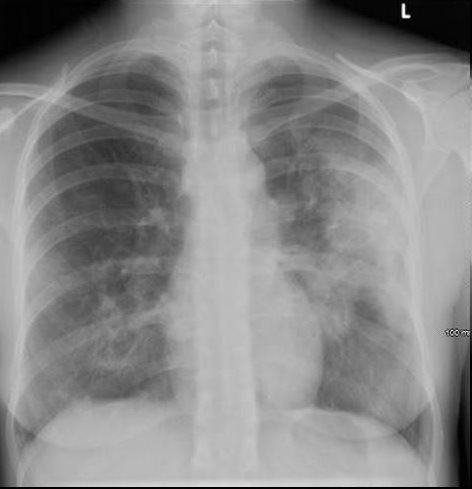
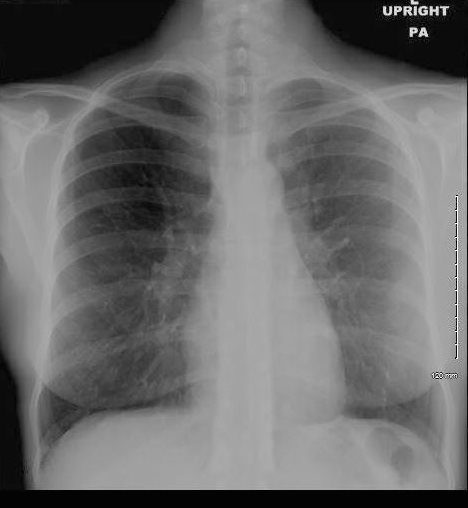
1 Week Later
Ashley Davidoff MD
TheCommonVein.net
dx acute eosinophillic pneumonia
A Few Days LAter
Ashley Davidoff MD
TheCommonVein.net
dx eosinophillic pneumonia
Ashley Davidoff MD
TheCommonVein.net
dx eosinophillic pneumonia
Patient was placed on streroids
10 days Later
Ashley Davidoff MD
TheCommonVein.net
dx eosinophillic pneumonia
1 Month Later
Ashley Davidoff MD
TheCommonVein.net
dx eosinophillic pneumonia
dx eosinophillic pneumonia