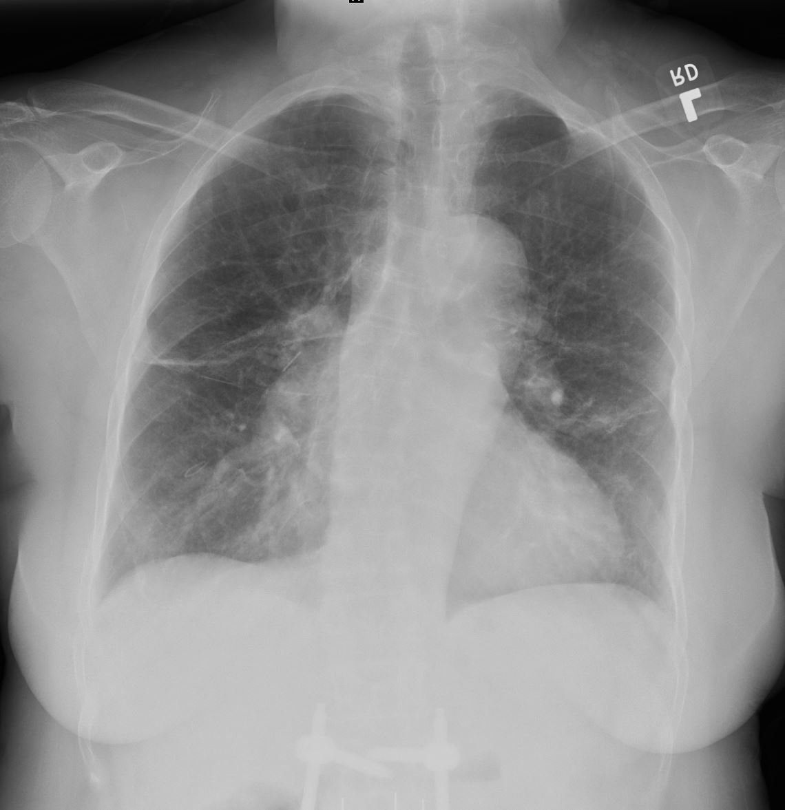
60year old female cigarette smoker with COPD presents with dyspnea and new lung nodules on CXR
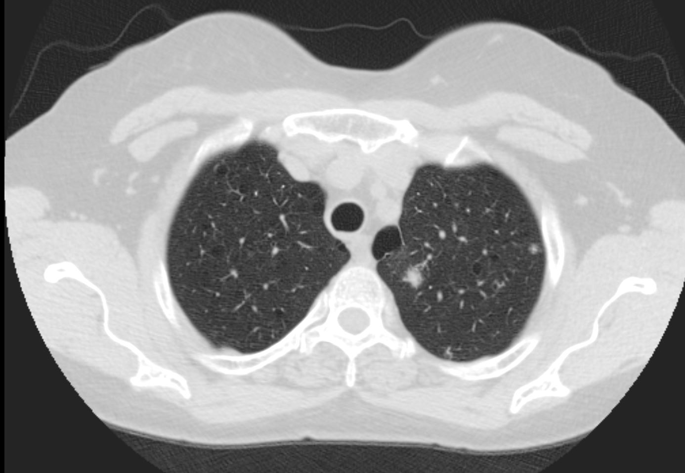
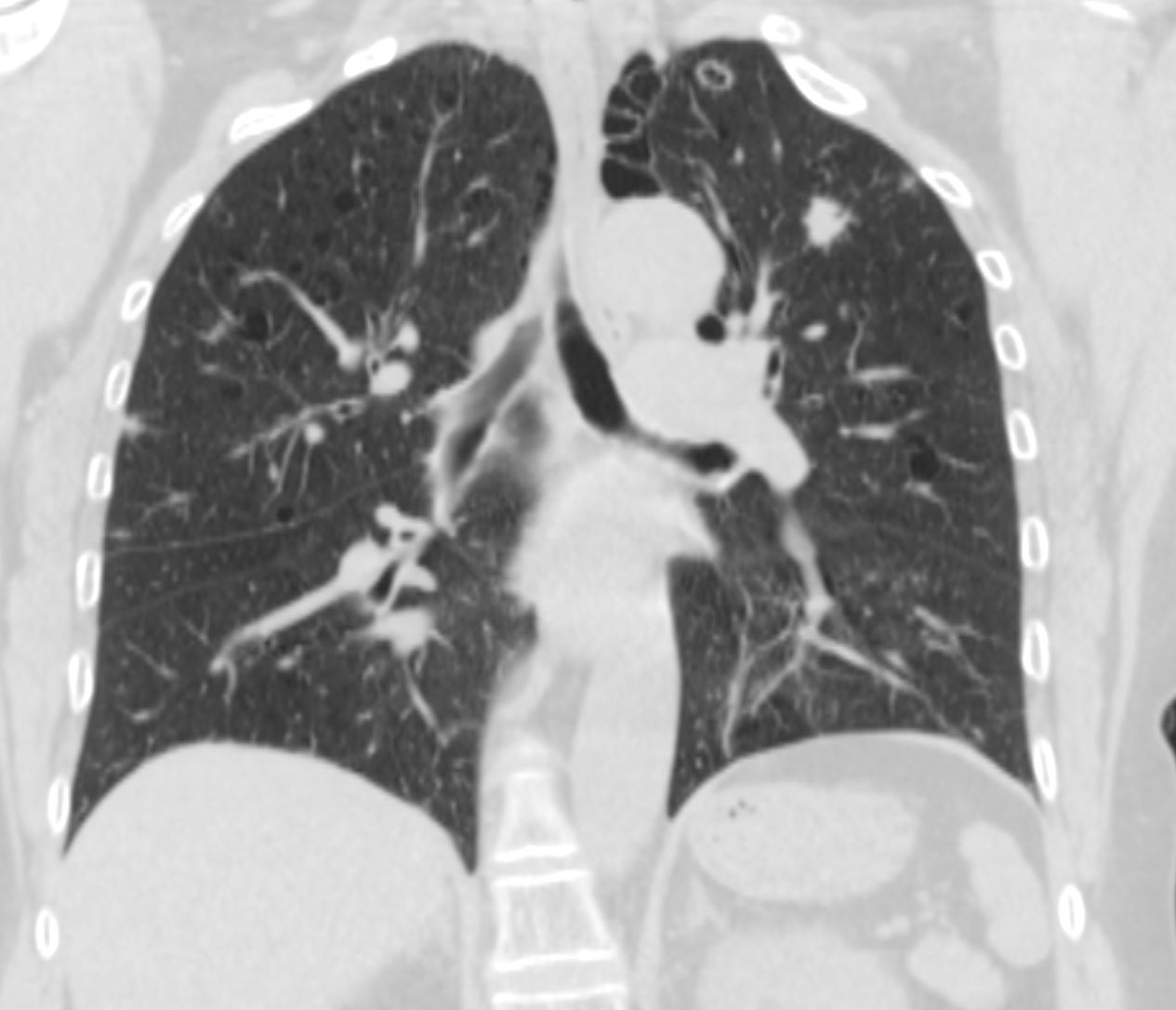
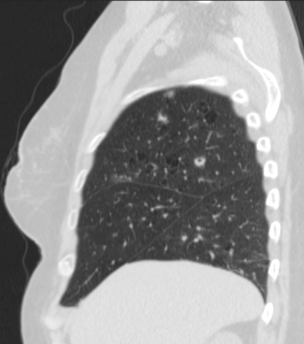
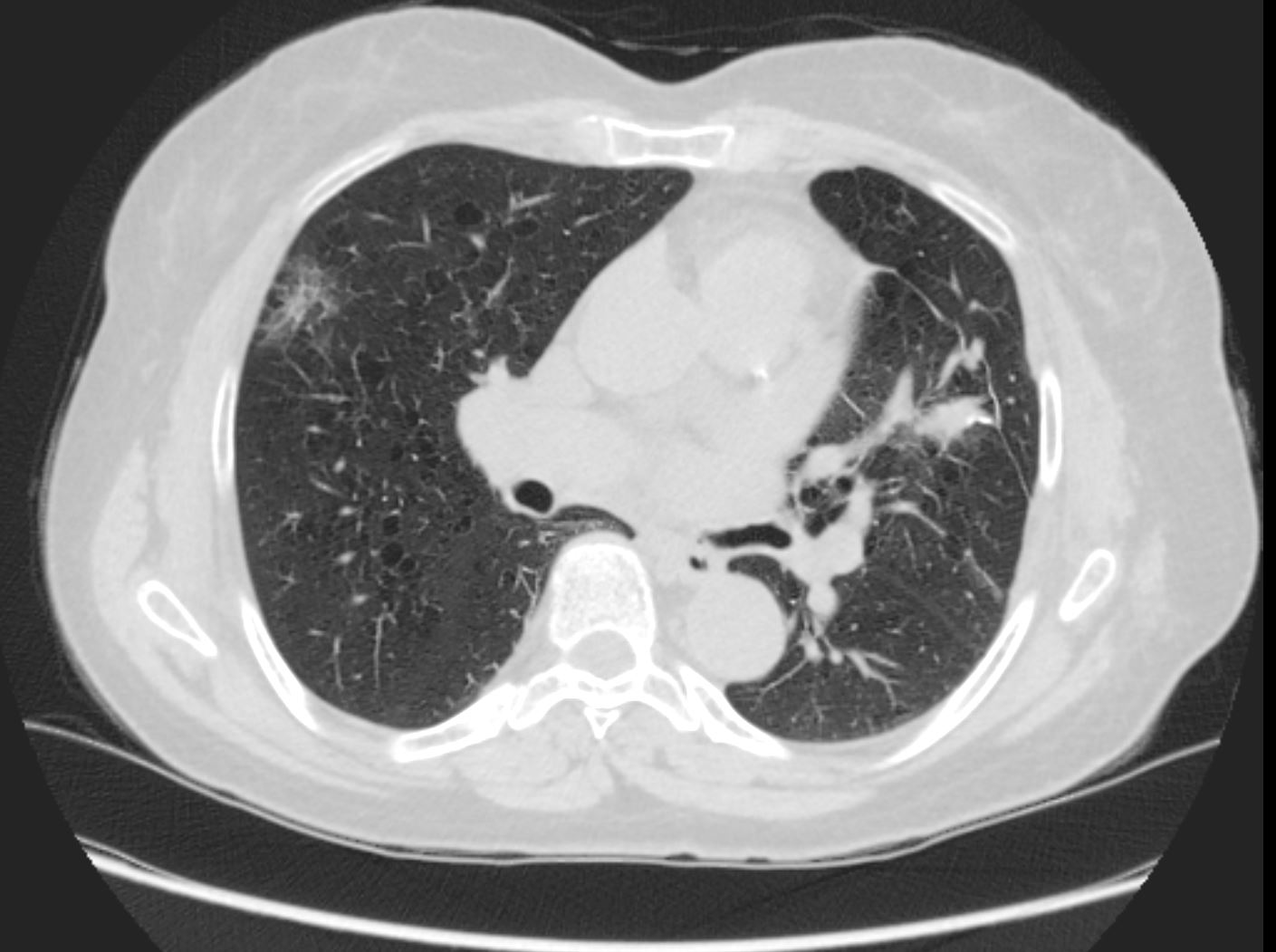
CT shows a mixture of thick walled cysts, nodules and cavitating nodules which reflects an evolving process of Langehans histiocytosis disease in the lungs
Thick walled cyst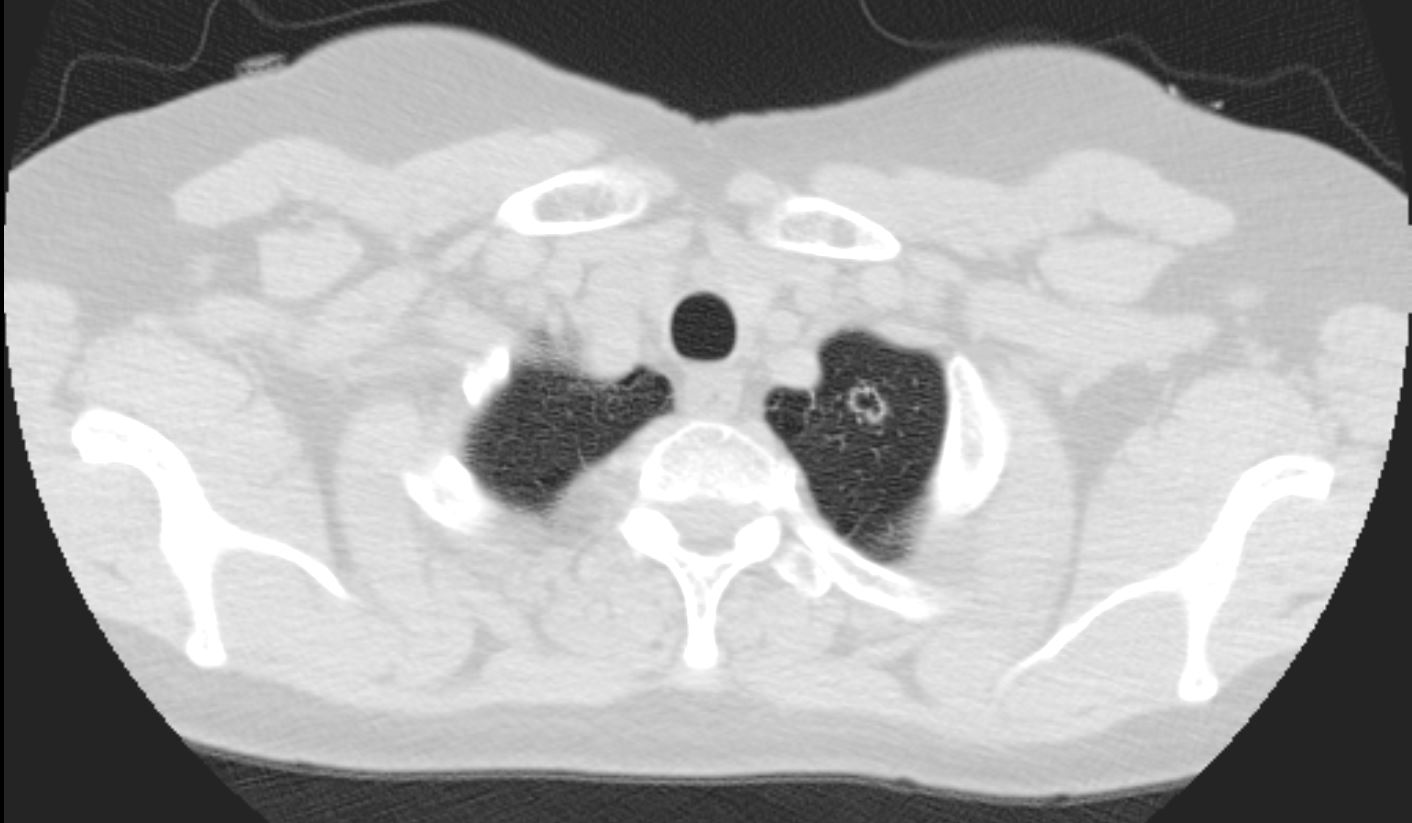
Nodules and evidence of centrilobular emphysema and paraseptal emphysema
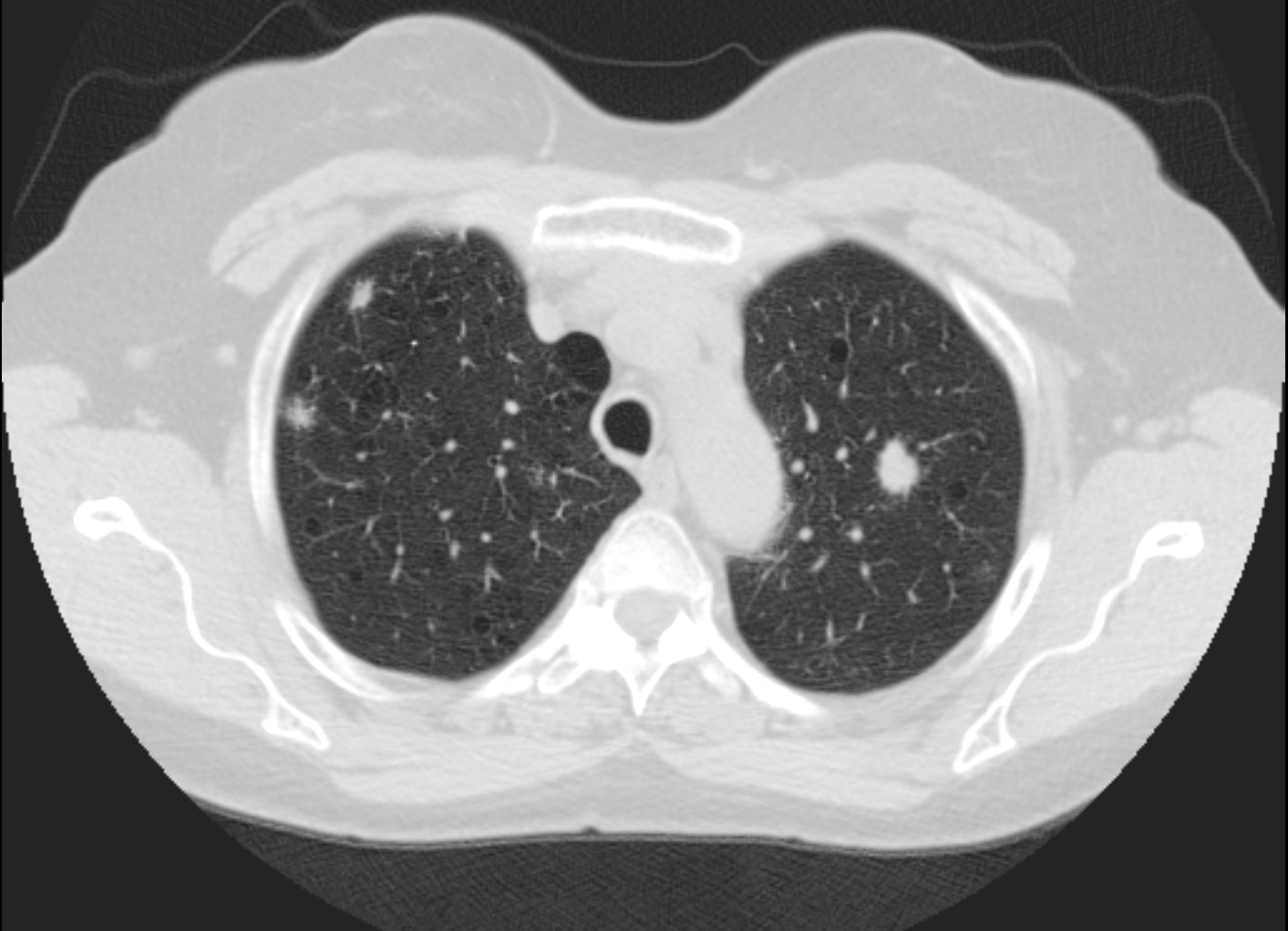
Cavitating Spiculated Nodule
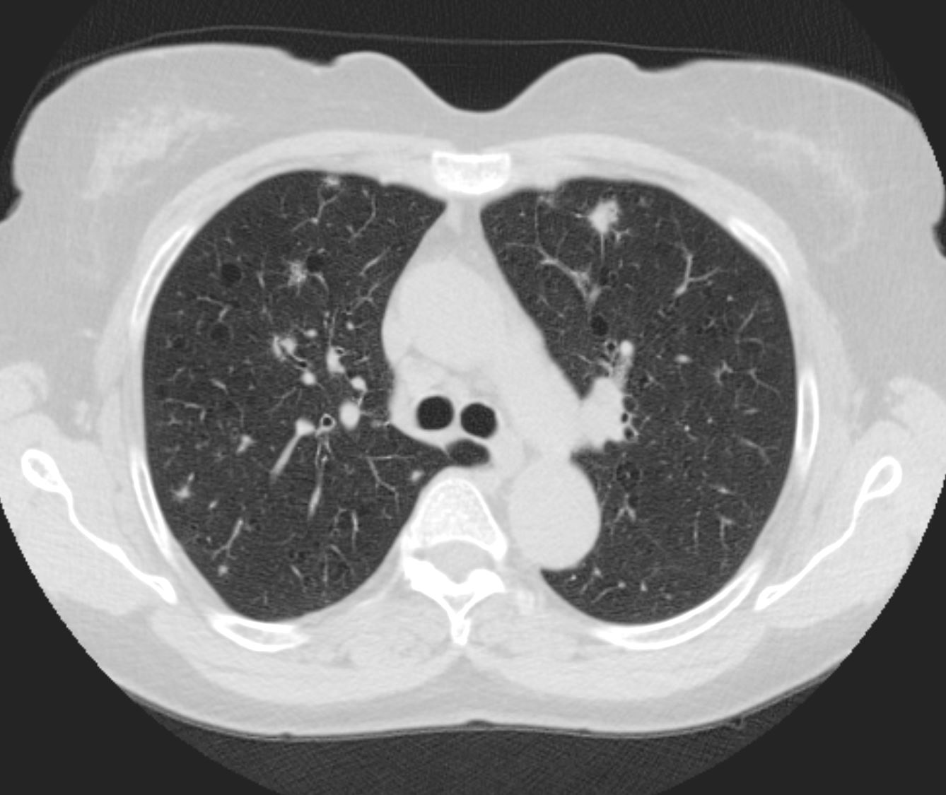
10 Years Ago
By this time she had had surgical biopsy in the LUL and LLL and the nodules were improving
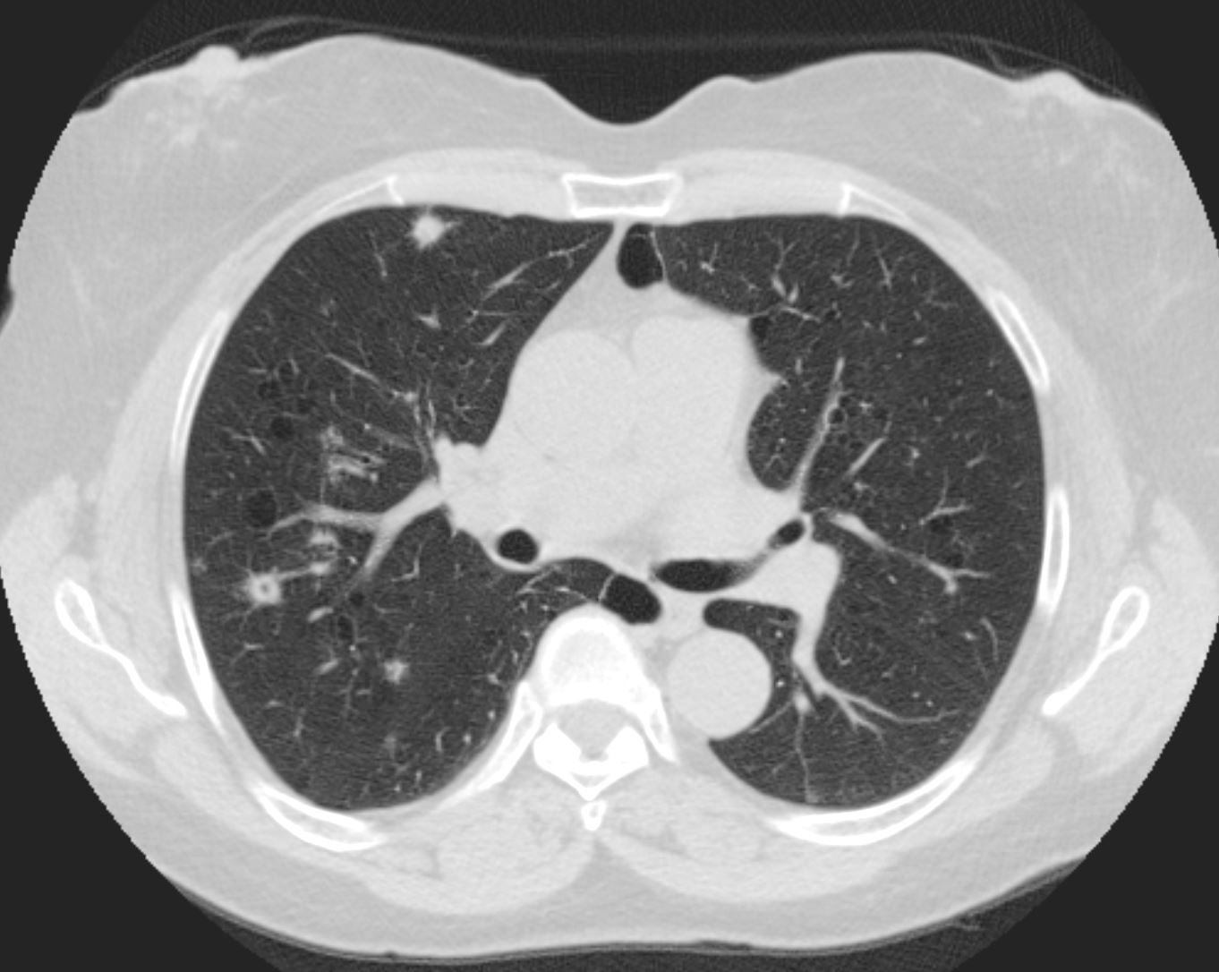
9 Years ago
5 Years ago
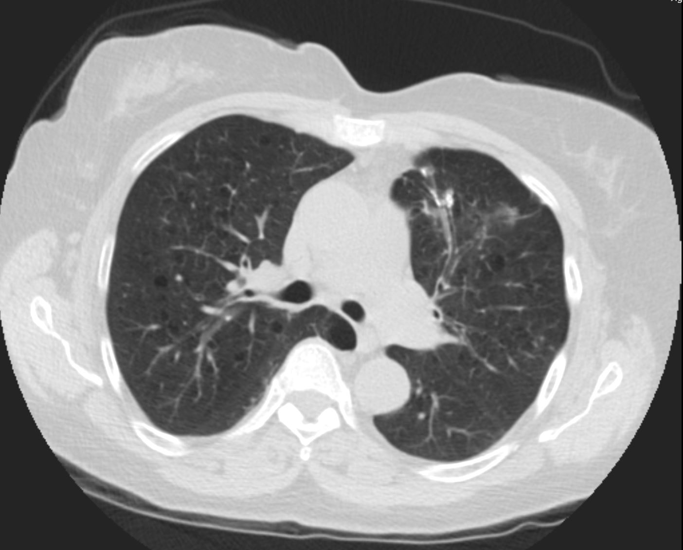
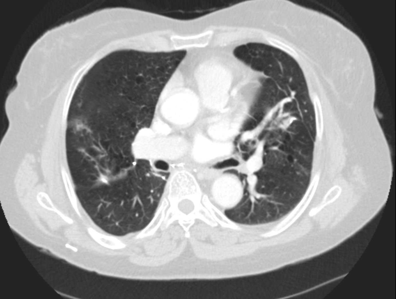
She developed a new spiculated nodule in the RUL


Proved to be malignant – – large cell carcinoma
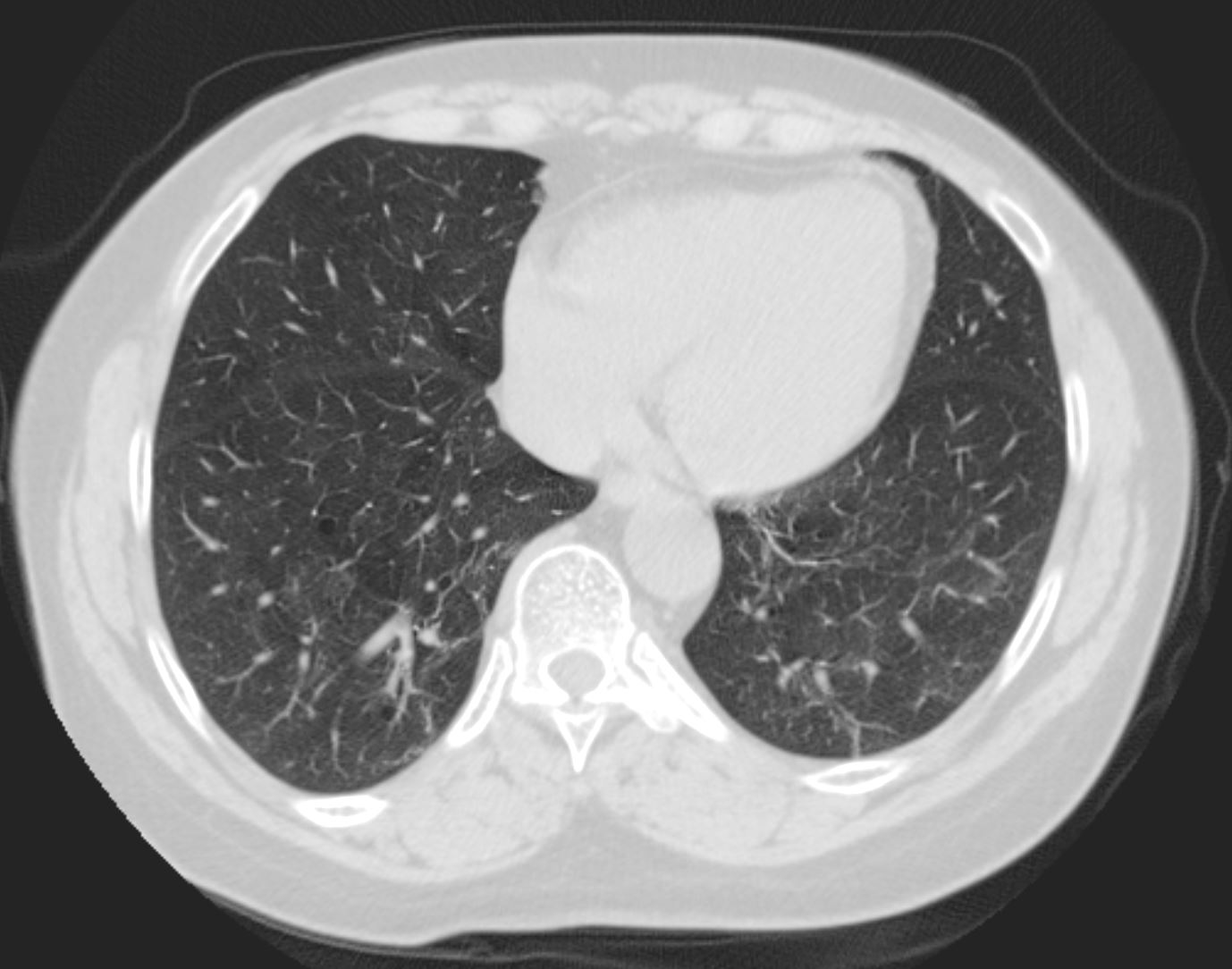
4 Years ago
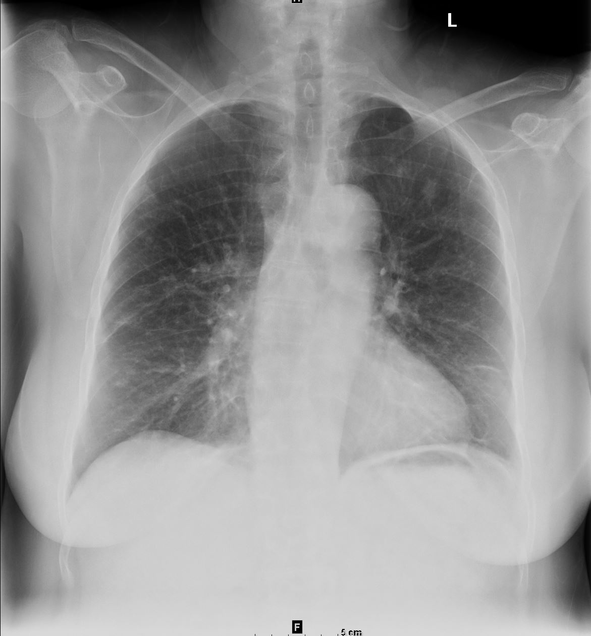
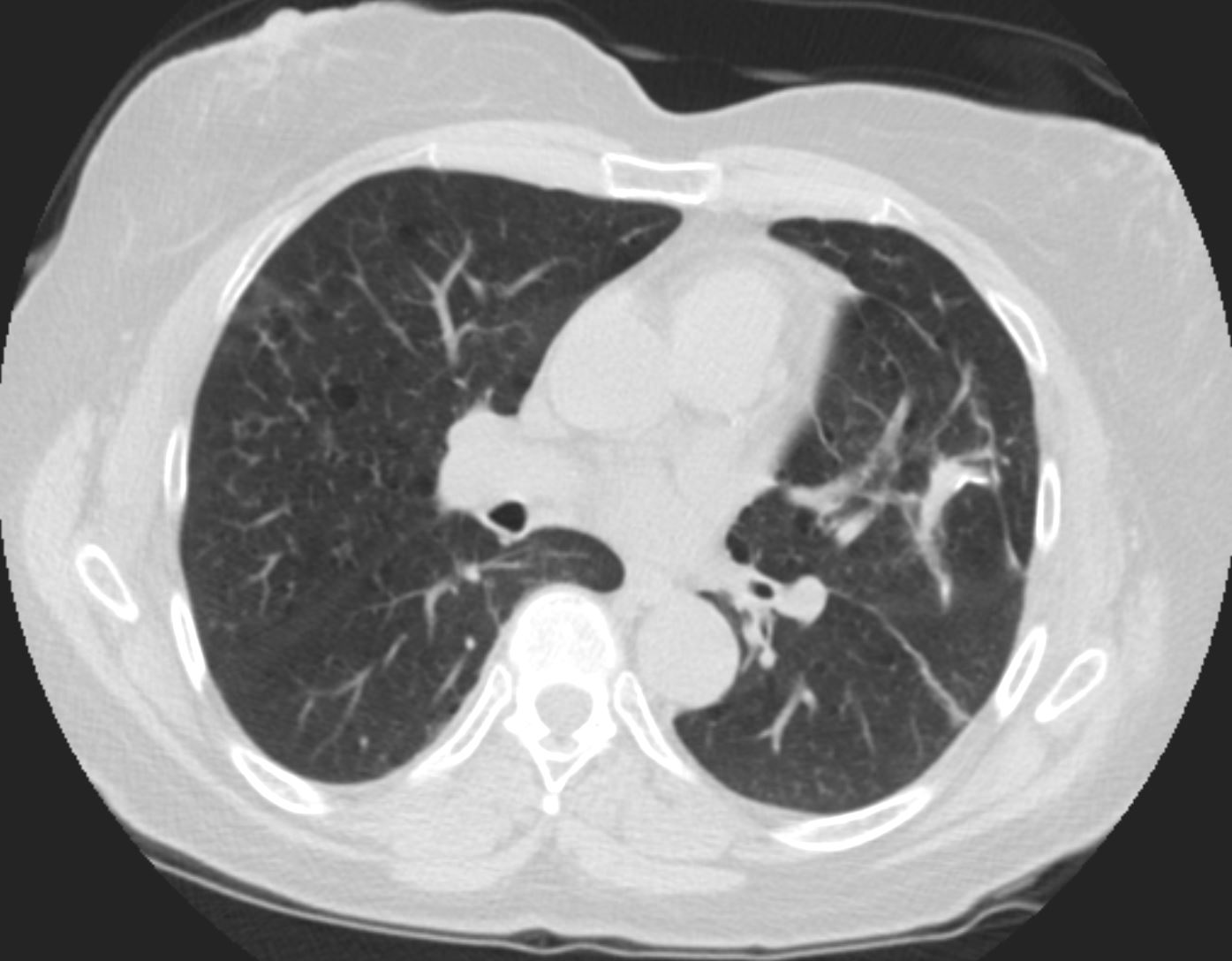
Shows post resection of the RUL nodule and soft tissue changes in the scar in the LUL
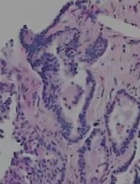
LUL lesion resected showing an adenocarcinoma
Emphysema s/p bilateral adenocarcinoma