
Adenocarcinoma Left Lung with Pneumonic Consolidation
Bilateral Lymphangitis Carcinomatosis

50 year old female with primary adenocarcinoma of the left lung with diffuse bilateral lymphangitic spread of disease characterized by lymphovascular distribution.
The nodularity on the fissures characterize the lymphatic distribution and the nodules are likely of a mixed nature, some being in the interlobular septa, and some in a centrilobular distribution .
Ashley Davidoff MD 158Lu 131022
CXR CT Correlation
Lymphangitic Spread and Fissural Involvement
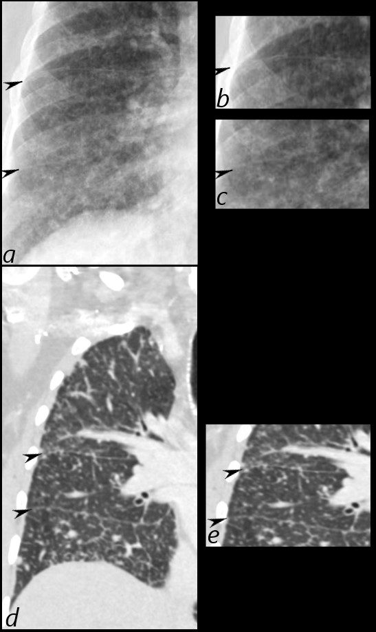
50 year old female with primary adenocarcinoma of the left lung presenting with pneumonic consolidation. And diffuse reticulonodular changes bilaterally
Images from the CXR (a, and magnified in b and c) show thickening of the fissures of the right lung (black arrowheads) with diffuse reticulonodular changes.
The coronal CT scan (d, magnified in e) confirm nodular thickening of the fissures (black arrowheads). In addition there are nodular changes with centrilobular location and along the interlobular septa, reminiscent of lymphovascular spread. These findings are consistent with the diagnosis of lymphangitis carcinomatosa
Ashley Davidoff MD TheCommonVein.net 158Lu 131022c
CT Adenocarcinoma of Left Lung in the form of Consolidation with Bilateral Lymphangitic Spread
50 year old female with primary adenocarcinoma of the left lung presenting with pneumonic consolidation, and diffuse reticulonodular changes bilaterally
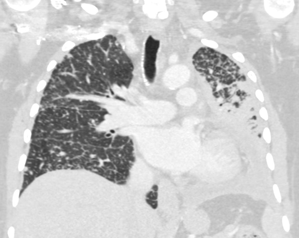
CT in a coronal plane shows intralobular nodules, nodular changes along the interlobular septa,
nodular changes along the minor fissure, centrilobular nodules and diffuse nodular thickening of the interlobular septa. These findings represent lymphatic spread along the lymphovascular bundles and are consistent with the diagnosis of lymphangitis carcinomatosa
Ashley Davidoff MD TheCommonVein.net 158Lu 131023
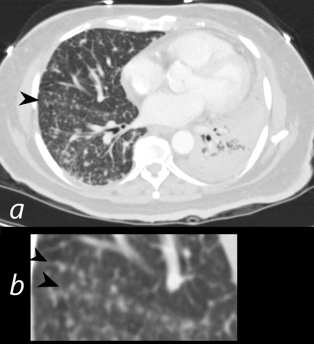
50-year-old female with primary adenocarcinoma of the left lung presenting with pneumonic consolidation of the left lower lobe, and diffuse reticulonodular changes bilaterally
The axial CT through the mid chest shows nodular changes along the major fissure (a and b, black arrowheads). These findings are consistent with the diagnosis of lymphangitis carcinomatosa.
The left lung shows consolidation in the posterior aspect of the left lower lobe
Ashley Davidoff MD TheCommonVein.net 158Lu 131029c01L
Coarsened Interlobular Septa
Bilateral Lymphangitic Spread
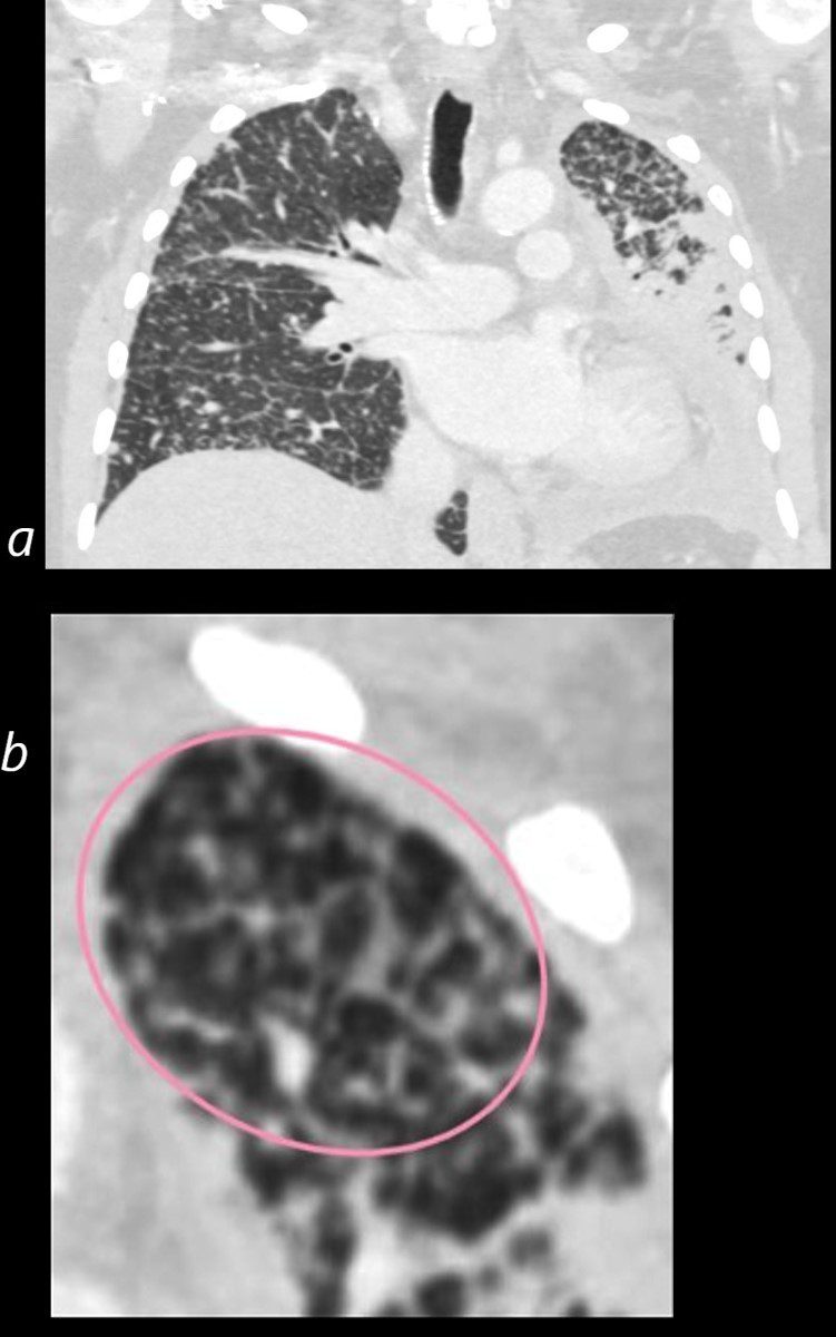
50 year old female with primary adenocarcinoma with the primary lesion presenting as pneumonic consolidation of the left lower lobe, and diffuse reticulonodular changes bilaterally
Image b is a magnified view of the left upper lobe and shows nodular thickening of the interlobular septa representing lymphatic spread along the lymphovascular bundles (pink oval)
The right lung shows interlobular septal thickening centrilobular nodules, and nodular thickening of the minor fissure
These findings are consistent with the diagnosis of lymphangitis carcinomatosa
Ashley Davidoff MD TheCommonVein.net 158Lu 131023c01L
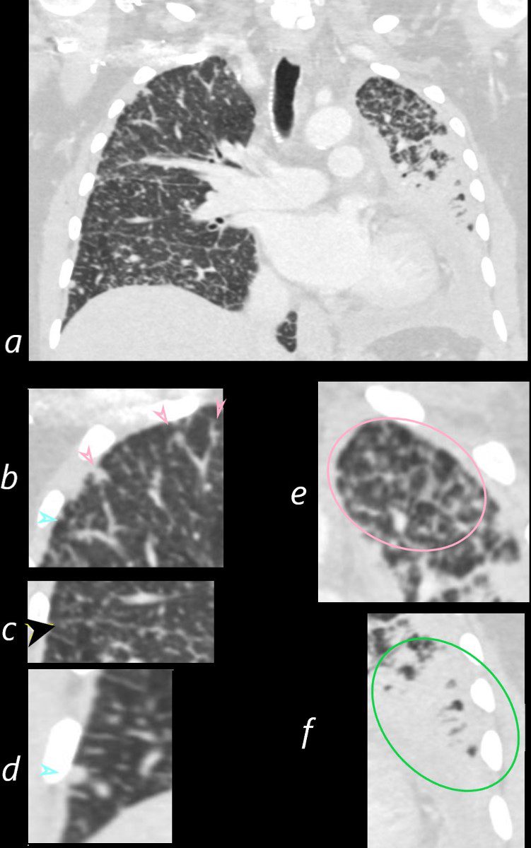
50 year old female with primary adenocarcinoma of the left lung presenting with pneumonic consolidation, and diffuse reticulonodular changes bilaterally
Images from the CT in a coronal plane (a, and magnified b-e)
Image b shows intralobular nodules (teal arrowheads) and nodular changes along the interlobular septa (pink arrowheads)
Image c shows nodular changes along the minor fissure (black arrowheads)
Image d shows a large nodule – likely centrilobular (teal arrowhead).
Image e shows diffuse nodular thickening of the interlobular septa representing lymphatic spread along the lymphovascular bundles (pink oval)
Image f shows the primary carcinoma presenting as a pneumonic consolidation (green oval). These findings are consistent with the diagnosis of lymphangitis carcinomatosa
Ashley Davidoff MD TheCommonVein.net 158Lu 131023cL

50 year old female with primary adenocarcinoma of the left lung presenting with pneumonic consolidation of the left lung, and diffuse reticulonodular changes bilaterally
CT in a coronal plane shows intralobular nodules, nodular changes along the fissures, interlobular septa, and diffuse nodular thickening of the interlobular septa. These findings represent lymphatic spread along the lymphovascular bundles and are consistent with the diagnosis of lymphangitis carcinomatosa
Ashley Davidoff MD TheCommonVein.net 158Lu 131024

50 year old female with primary adenocarcinoma of the left lung presenting with pneumonic consolidation of the left lung, and diffuse reticulonodular changes bilaterally
CT in a coronal plane shows intralobular nodules, interlobular septa, thickening of the peribronchial tissue, and thickening of the interlobular septa. These findings represent lymphatic spread along the lymphovascular bundles and are consistent with the diagnosis of lymphangitis carcinomatosa The primary tumor in the right lower lobe presents as a consolidation.
Ashley Davidoff MD TheCommonVein.net 158Lu 131025
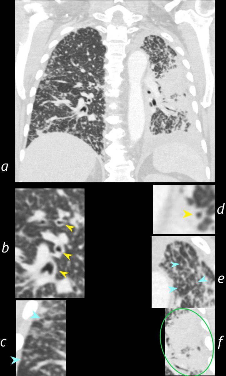
50-year-old female with primary adenocarcinoma of the left lung presenting with pneumonic consolidation, and diffuse reticulonodular changes bilaterally
Images from the CT in a coronal plane (a, and magnified b-f)
Image b shows peribronchial thickening (yellow arrowheads) likely reflecting lymphovascular spread..
Image c shows centrilobular nodular changes (teal arrowheads)
Image d shows peribronchial thickening (yellow arrowhead) likely reflecting lymphovascular spread..
Image e shows centrilobular nodules (teal arrowheads) and diffuse nodular thickening of the interlobular septa representing lymphatic spread along the lymphovascular bundles
Image f shows the primary carcinoma presenting as a pneumonic consolidation.(green oval)
Ashley Davidoff MD TheCommonVein.net 158Lu 131025cL

50-year-old female with primary adenocarcinoma of the left lung presenting with pneumonic consolidation, and diffuse reticulonodular changes in the right lung
The right lung is characterized by innumerable large centrilobular nodules (teal arrowheads) and a few foci of intralobular peripheral consolidations. Thickening of the peribronchial tissues likely reflects carcinomatosis of the peribronchial tissues. These findings are consistent with the diagnosis of lymphangitis carcinomatosa
The left lung shows diffuse consolidation of the posterior aspect of the left lower lobe
Ashley Davidoff MD TheCommonVein.net 158Lu 131026
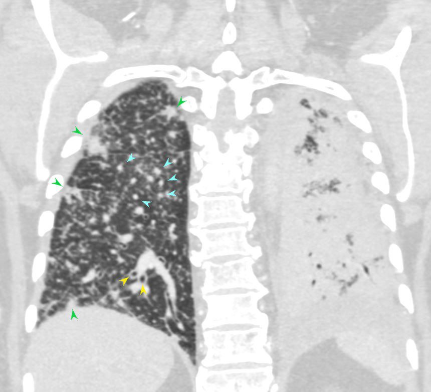
50-year-old female with primary adenocarcinoma of the left lung presenting with pneumonic consolidation, and diffuse reticulonodular changes on the right side.
The right lung is characterized by innumerable large centrilobular nodular changes (teal arrowheads) and a few foci of intralobular consolidation (green arrowheads) Thickening of the peribronchial tissues likely reflects carcinomatosis of the peribronchial tissues (yellow arrowheads) These findings are consistent with the diagnosis of lymphangitis carcinomatosa
The left lung shows diffuse consolidation of the posterior aspect of the left lower lobe
Ashley Davidoff MD TheCommonVein.net 158Lu 131026L
Bilateral Lymphangitic Spread Lung Apices
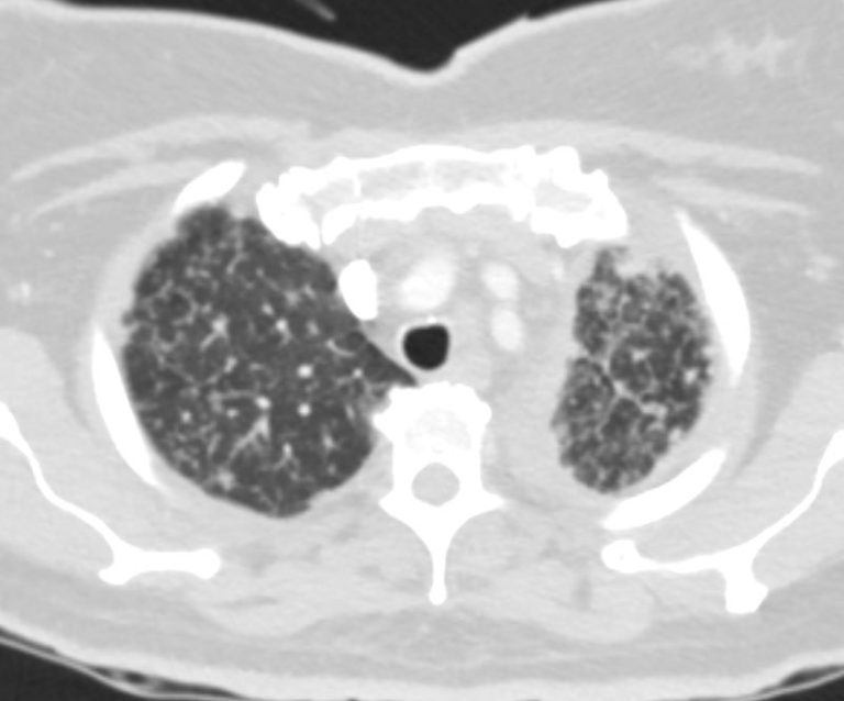
50-year-old female with primary adenocarcinoma of the left lung presenting with pneumonic consolidation, and diffuse reticulonodular changes bilaterally.
CT in the axial plane of the lung apices shows bilateral coarsened interlobular septa and prominent centrilobular nodules. These findings are consistent with the diagnosis of lymphangitis carcinomatosa. Consolidative changes are noted abutting the mediastinum on the left
Ashley Davidoff MD TheCommonVein.net 158Lu 131027
Bilateral Lymphangitic Spread Upper Lung Fields
50-year-old female with primary adenocarcinoma of the left lung presenting with pneumonic consolidation, and diffuse reticulonodular changes bilaterally.
CT in the axial plane of the upper lung fields shows bilateral coarsened interlobular septa prominent centrilobular nodules, peribronchial thickening and regions of consolidation in the left upper lobe These findings are consistent with the diagnosis of lymphangitis carcinomatosa.
Ashley Davidoff MD TheCommonVein.net 158Lu 131028
Bilateral Lymphangitic Spread
Mid Lung Fields
Fissural and Subsegmental Airway Involvement

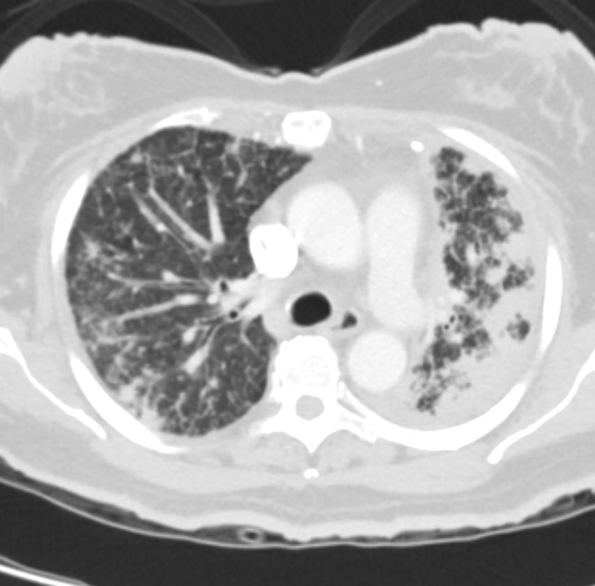
50-year-old female with primary adenocarcinoma of the left lung presenting with pneumonic consolidation of the left lower lobe, and diffuse reticulonodular changes bilaterally
The axial CT through the mid chest shows nodular changes along the major fissure and thickening of the peribronchial tissues, which likely reflect carcinomatosis of the peribronchial tissues. These findings are consistent with the diagnosis of lymphangitis carcinomatosa.
The left lung shows consolidation in the posterior aspect of the left lower lobe
Ashley Davidoff MD TheCommonVein.net 158Lu 131029
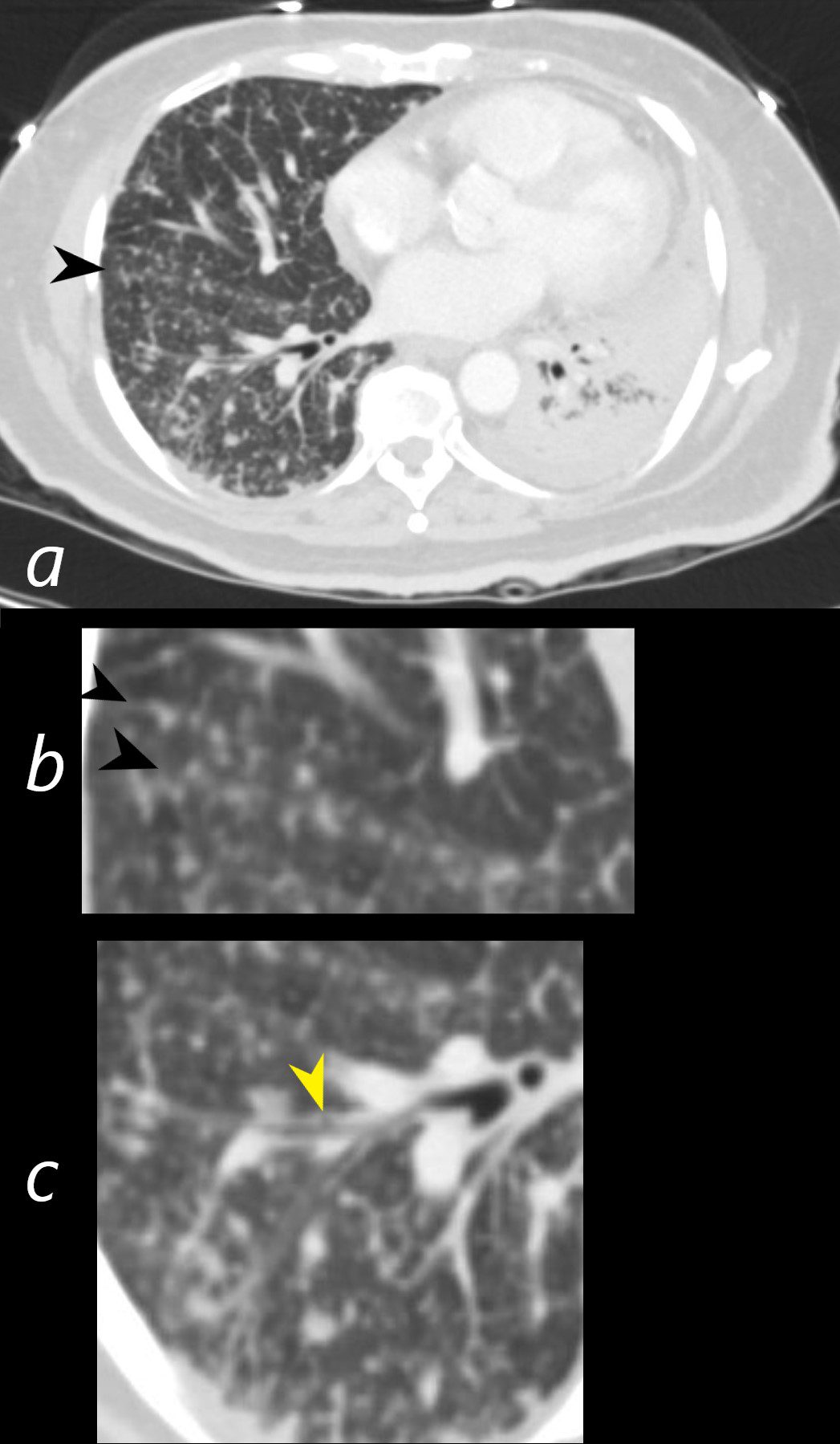
50-year-old female with primary adenocarcinoma of the left lung presenting with pneumonic consolidation of the left lower lobe, and diffuse reticulonodular changes bilaterally
The axial CT through the mid chest shows nodular changes along the major fissure (a and b, black arrowheads) and thickening of the peribronchial tissues (c yellow arrowhead) which likely reflects carcinomatosis of the peribronchial tissues. These findings are consistent with the diagnosis of lymphangitis carcinomatosa.
The left lung shows consolidation in the posterior aspect of the left lower lobe
Ashley Davidoff MD TheCommonVein.net 158Lu 131029cL
Centrilobular Nodules and Peribronchial Infiltration
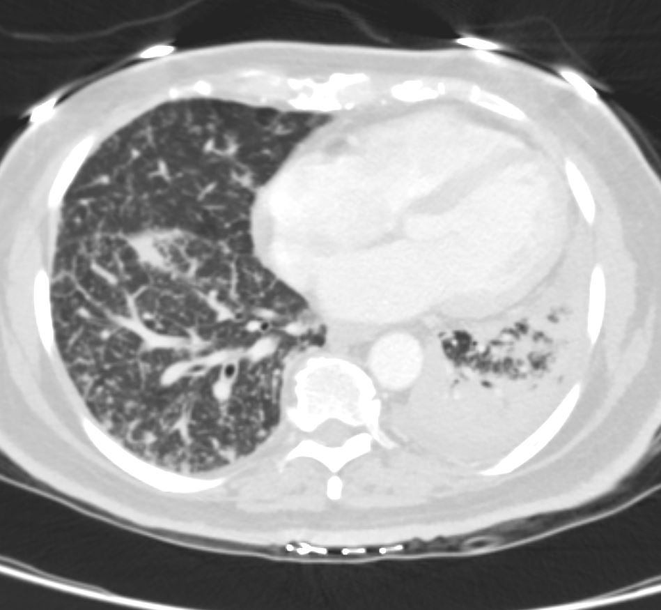
50-year-old female with primary adenocarcinoma of the left lung presenting with pneumonic consolidation of the left lower lobe, and diffuse reticulonodular changes bilaterally
The axial CT through the mid to lower chest shows thickening of the peribronchial tissues which likely reflects carcinomatosis of the peribronchial tissues. In addition, there are enlarged centrilobular nodules. Both these findings likely reflect manifestation of lymphovascular spread consistent with lymphangitis carcinomatosa
The left lung shows consolidation in the posterior aspect of the left lower lobe
Ashley Davidoff MD TheCommonVein.net 158Lu 131030
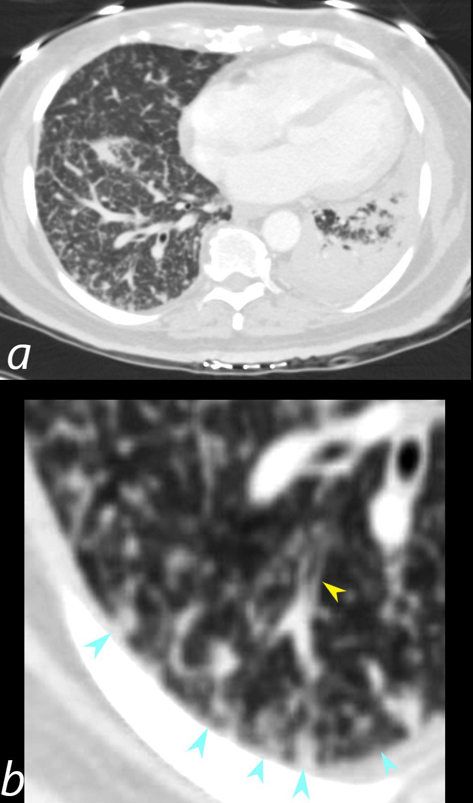
50-year-old female with primary adenocarcinoma of the left lung presenting with pneumonic consolidation of the left lower lobe, and diffuse reticulonodular changes bilaterally
The axial CT through the mid to lower chest shows thickening of the peribronchial tissues (b yellow arrowhead) which likely reflects carcinomatosis of the peribronchial tissues. In addition, there are enlarged centrilobular nodules (b, teal arrowheads). Both these findings likely reflect manifestation of lymphovascular spread consistent with lymphangitis carcinomatosa
The left lung shows consolidation in the posterior aspect of the left lower lobe
Ashley Davidoff MD TheCommonVein.net 158Lu 131030cL