
- is an imaging and physiologic term to
- retained air in a part or parts of the lung
- more easily identified during expiration
- caused by
- obstruction
- often small airway disease
- chronic bronchitis
- asthma
- Hypersensitivity pneumonitis
- sarcoidosis
- bronchiolitis
- cystic fibrosis/bronchiectasis
- ILD
- obesity
- often small airway disease
- abnormality in lung compliance
- sometimes seen in normal people
- 50% of CT scans
- obstruction
-

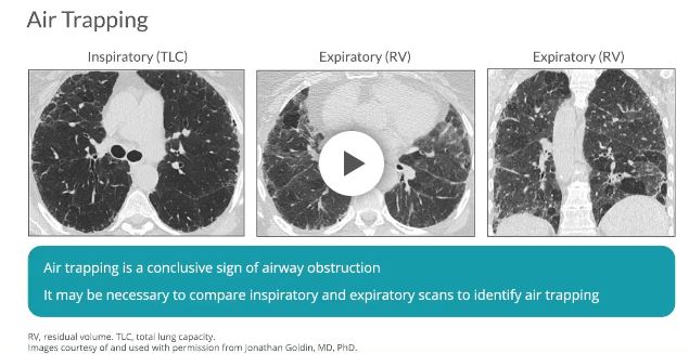
Air Trapping
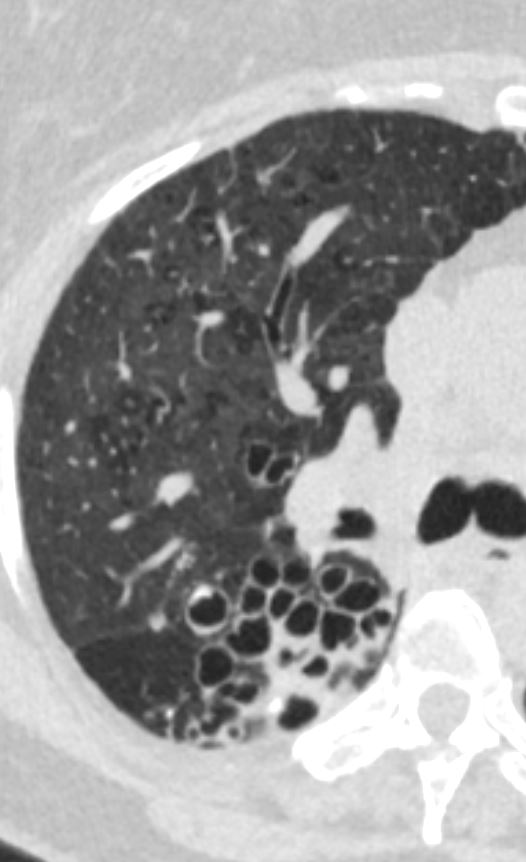
Air trapping is a conclusive sign of airway obstruction 6 and appears as areas of decreased attenuation resulting from the presence of gas retained in the lung parenchyma.7 It may be necessary to compare between inspiratory and end-expiratory HRCT scans to determine the extent of air trapping, especially if the degree of air trapping is minimal.7 Patients with normal inspiratory scans frequently have signs of air trapping on expiratory scans, suggesting that expiratory scans should be performed as part of a standard evaluation.6
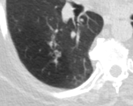
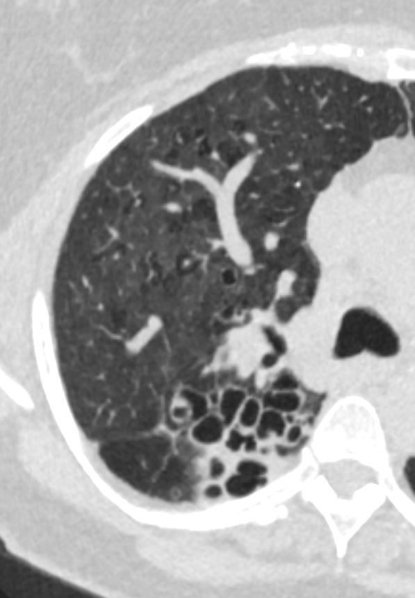
Mosaic Attenuation Caused by Obstruction of Small Airways
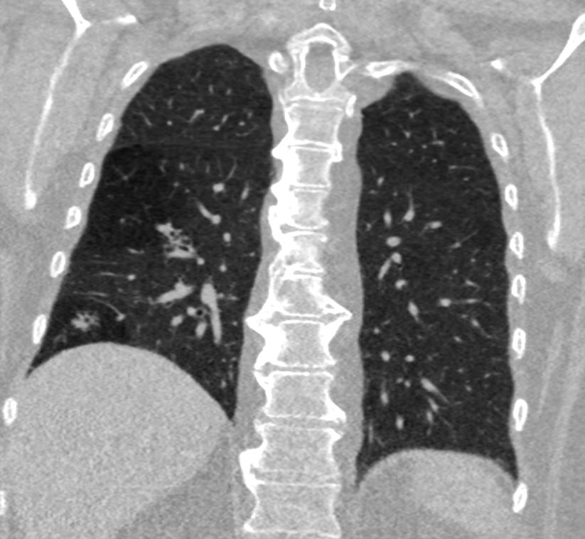
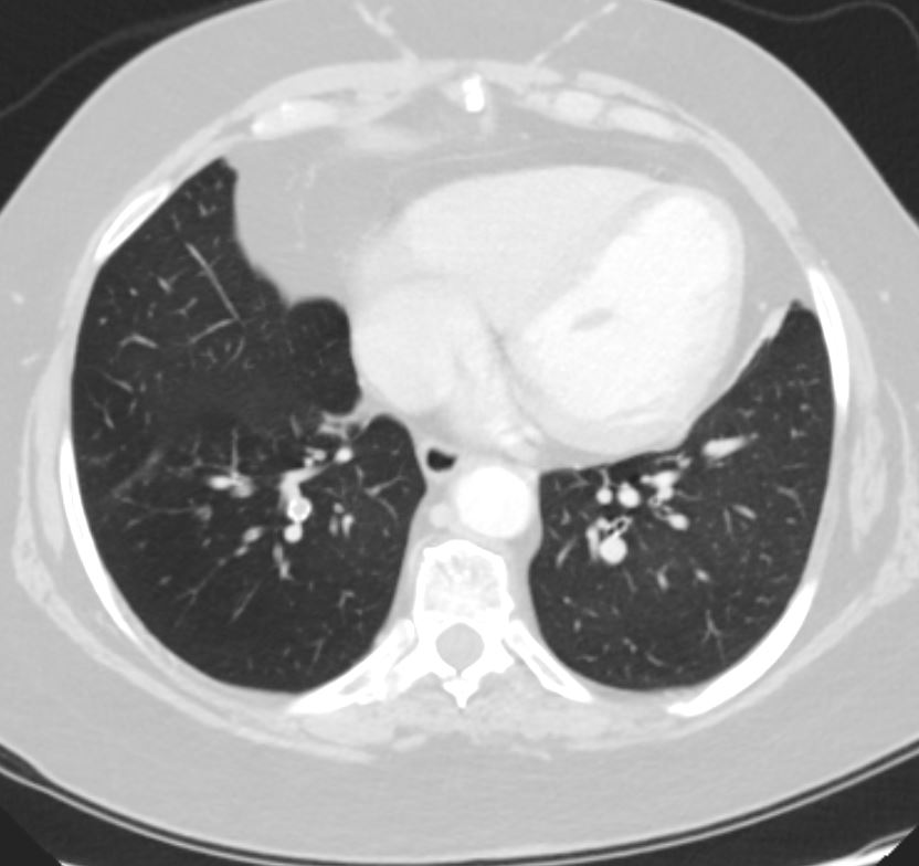
Medium Sized Airways and Smaller Airways are Filled with Mucus in a patient with COPD – Note Centrilobular Impaction of Mucus
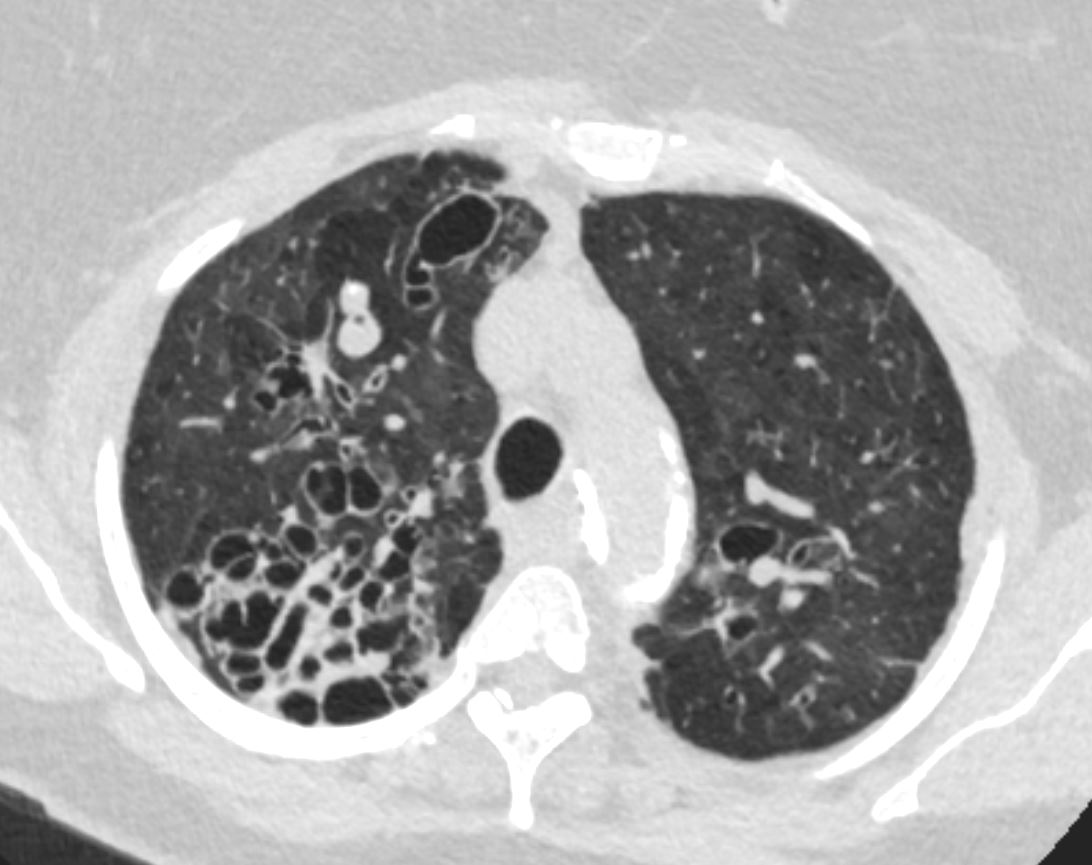
Medium Sized Airways and Smaller Airways are Filled with Mucus – Note Centrilobular Impaction of Mucus
-
- Mosaic attenuation is an
- imaging pattern
- variable lung attenuation
- results in a heterogeneous appearance of the parenchyma.
- sometimes it is caused by air trapping
- sometimes by perfusion abnormalities
- sometimes normal
- imaging pattern
- Mosaic attenuation is an
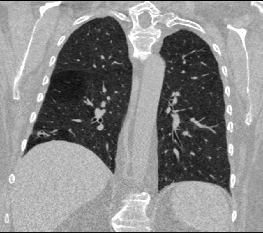
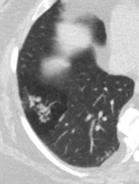
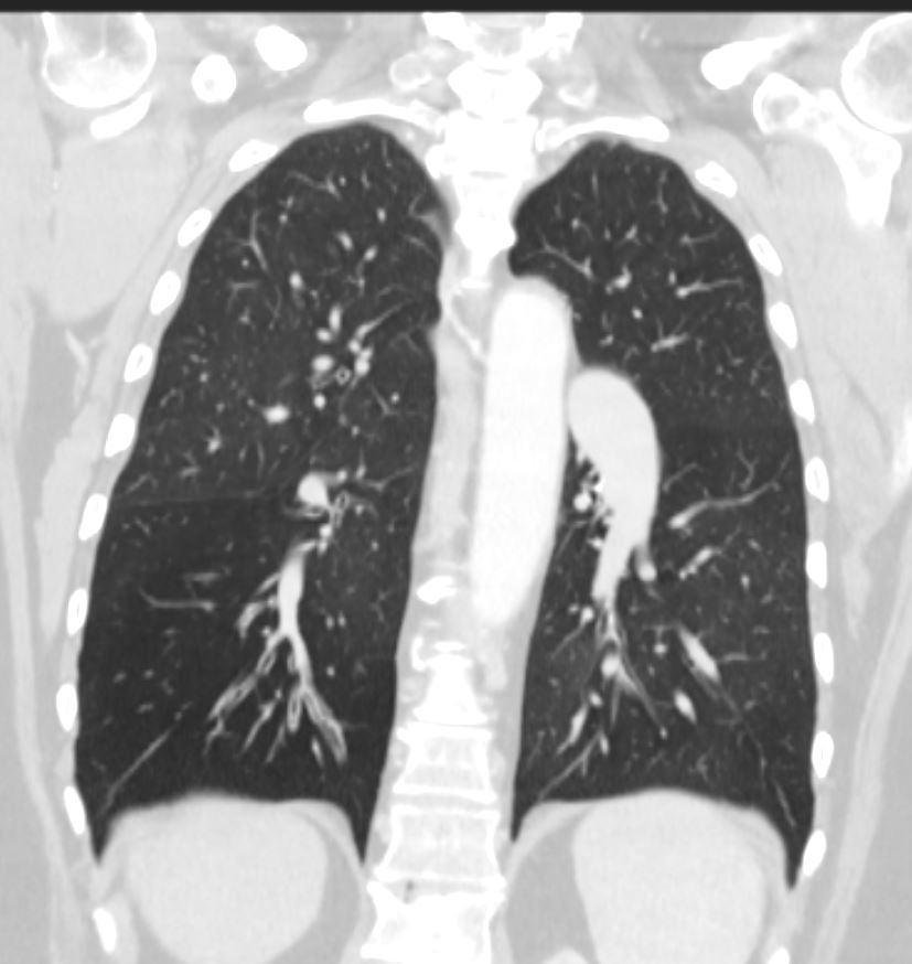
Lobar Air Trapping Due to Mucus Impaction
Note Realative Lucency of the RLL compared to the LLL
Links and References
Fleischner Society
air trapping
Pathophysiology.—Air trapping is retention of air in the lung distal to an obstruction (usually partial).
CT scans.—Air trapping is seen on end-expiration CT scans as parenchymal areas with less than normal increase in attenuation and lack of volume reduction. Comparison between inspiratory and expiratory CT scans can be helpful when air trapping is subtle or diffuse (,11,,12) (,Fig 4). Differentiation from areas of decreased attenuation resulting from hypoperfusion as a consequence of an occlusive vascular disorder (eg, chronic thromboembolism) may be problematic (,13), but other findings of airways versus vascular disease are usually present. (See also mosaic attenuation pattern.)