
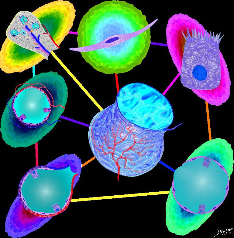
Alveolus
Parts and Bonds
Ashley Davidoff MD
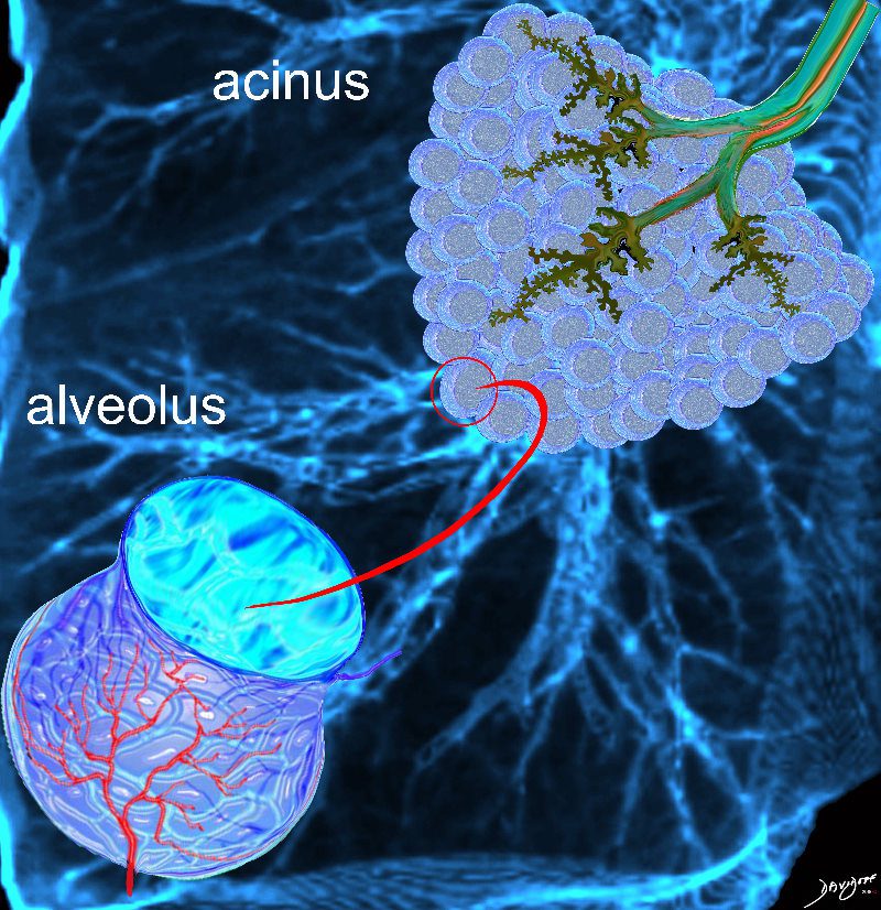
- Alveoli
- 300 million alveoli 20,000 acini
- Alveoli
- major component of the lung
- make up approximately
- 50% of lung volume.
- Found outer 1/3
- situated dominantly in the periphery
- tubular transport systems are located centrally by the hilum .
- During inspiration the
- radius of the alveolus
- doubles
- from about 0.05 mm
- to 0.1 mm
- make up approximately
- major component of the lung
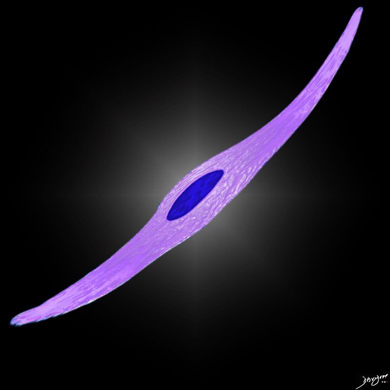
Component of the Acinus
-
-
-
-

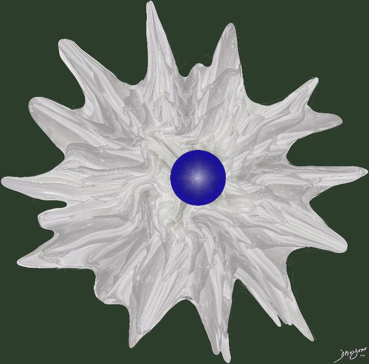
Alveolus as a Part of the Acinus
Ashley Davidoff MD TheCommonvein.net lungs-0056Functional Unit For Gas Exchange
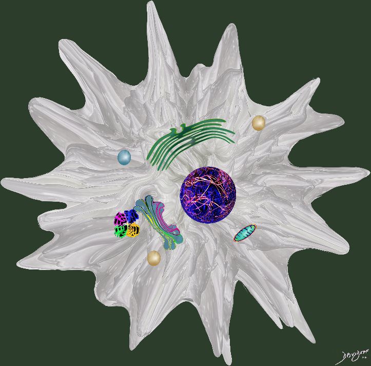
-
-
-
Ashley Davidoff TheCommonVein.net 42438b03
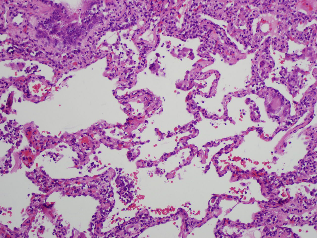
Normal Histology

Lower magnification of the lung with H and E stain shows cup-shaped alveolar spaces outlined by delicate thin alveolar capillary membrane.
key words
lung, pulmonary, normal alveolus, alveoli, histology, interstitium, interstitial
Courtesy Armando Fraire MD. 32819
Cells

The diagram shows an alveolus, lined by a single layer of squamous cells,
Ashley Davidoff MD TheCommonVein.net lungs-0705-lo res
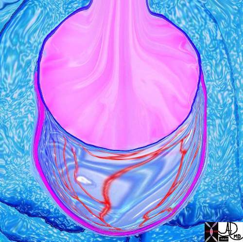
Capillary Network
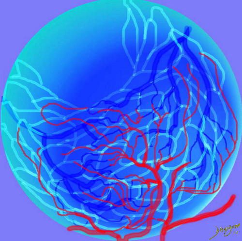
This drawing demonstrates the open mouth view of the alveolus, which is surrounded by its capillary network. The lining single layer of squamous cells (pneumocytes) can be seen peaking through the vessels.
Ashley Davidoff MD. TheCommonVein.net 32166
Capillary
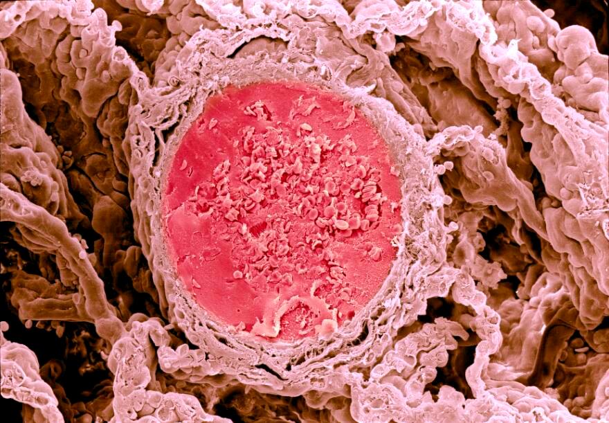
Credit: David Gregory & Debbie Marshall
Licence: Attribution 4.0 International (CC BY 4.00 Wellcome London
The Wall
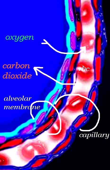
The diagram shows an alveolus, lined by a single layer of squamous cells, surrounded by a capillary with red cells which is also lined by a single layer of squamous endothelial cells . The images show exchanges of oxygen and carbon dioxide through the alveolar membrane .
Ashley Davidoff MD TheCommonVein.net lungs-0028b-low res
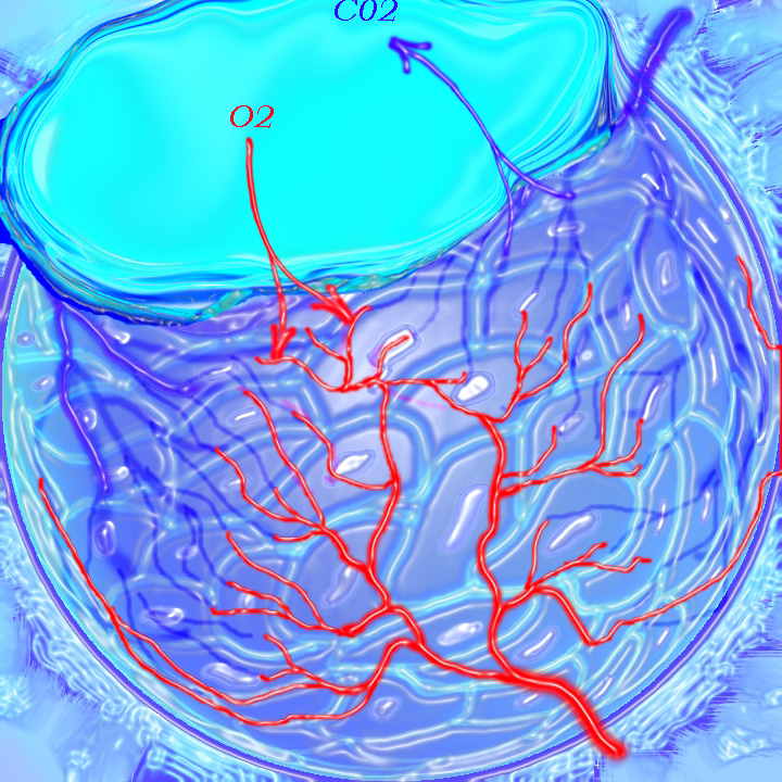
O2 Saturations
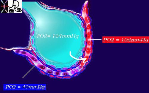
This diagram again shows the alveolus in teal, the arteriolar component of the capillary with red cells in blue and venular component replenished by oxygen in red. As noted above, the PO2 of the arterial blood is 40mmHg while the inspired air is 104mmHg. A pressure gradient thus exists and diffusion from the high to the low pressure occurs with a net movement of oxygen into the blood to equilibrate the pressure. Venous blood is now rich in oxygen with a PO2 of 104mmHg.
Ashley Davidoff MD TheCommonVein.net 42445b08b
PCO2 Saturations
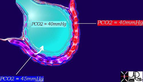
This diagram shows the PCO2 of the arterial blood at 45mmHg with red cell again showing telling signs of blue deoxygenation while the inspired air has a PCO2 of 40mmHg. There is therefore diffusion from the high to the low pressure and a net movement of carbon dioxide into the alveolus to equilibrate the pressure of 40mmHg. The venous blood is thus relatively depleted of CO2 with a PCO2 of 40mmHg. |
Ashley Davidoff MD TheCommonVein.net 42445b11
Surfactant
The alveolus is lined by a complex detergent type solution called surfactant (pink) which reduces the surface tension in the alveolus, making it easier for the alveolus to expand during inspiration and preventing alveolar collapse on expiration.
Ashley Davidoff TheCommonVein.net 42530b05b09b01a12
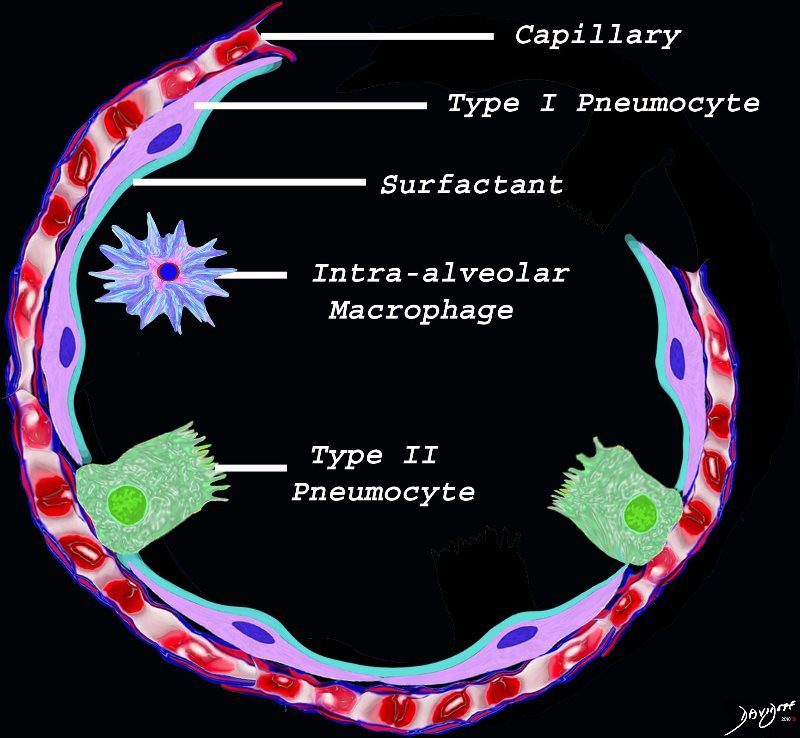
The diagram shows the lining of the normal alveolus composed of type 1 pneumocyte squamous in nature and the cuboidal cell (type pneumocyte) which rest on a lamina propria, and basement membrane (not shown) shared with the inner endothelial layer of the capillary. Intra-alveolar macrophage lies within the alveolar lumen
Ashley Davidoff
TheCommonVein.net
Pulmonary system provides the most intimate interface with the external environment . epresents the largest body surface area exposed to the environment
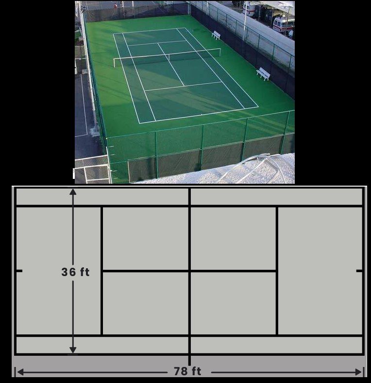
SizeThe surface area of the alveoli is equal to about 1/2 a tennis court. A tennis court is 78 by 36 feet equal to an area of 2,808 sq feet. Estimates for the surface area for the alveoli are between 800-1100 sq feet.
The skin in comparison of an adult is approximately 22 square feet
Each day, the lungs are exposed to 7,000 L of air and all it contains.
- At the level of the alveoli where gas exchange occurs, the biological barrier presents as an extremely attenuated interface composed of the
- surfactant
- 1 cell layer thick membranes
- alveolar lining and its lamina base
- endothelial lining and its fused basal lamina.
-
Cellular Makeup of the Normal Alveolus
The diagram shows the lining of the normal alveolus composed of type 1 pneumocyte squamous in nature and the cuboidal cell (type pneumocyte) which rest on a lamina propria, and basement membrane (not shown) shared with the inner endothelial layer of the capillary. Intra-alveolar macrophage lies within the alveolar lumen
Ashley Davidoff
TheCommonVein.net
- Surfactant
- first line of defense against immunological, biological and non-biological threats
- thickness
- less than 0.1 μm
- major components are
- phospholipids – 90%
- proteins
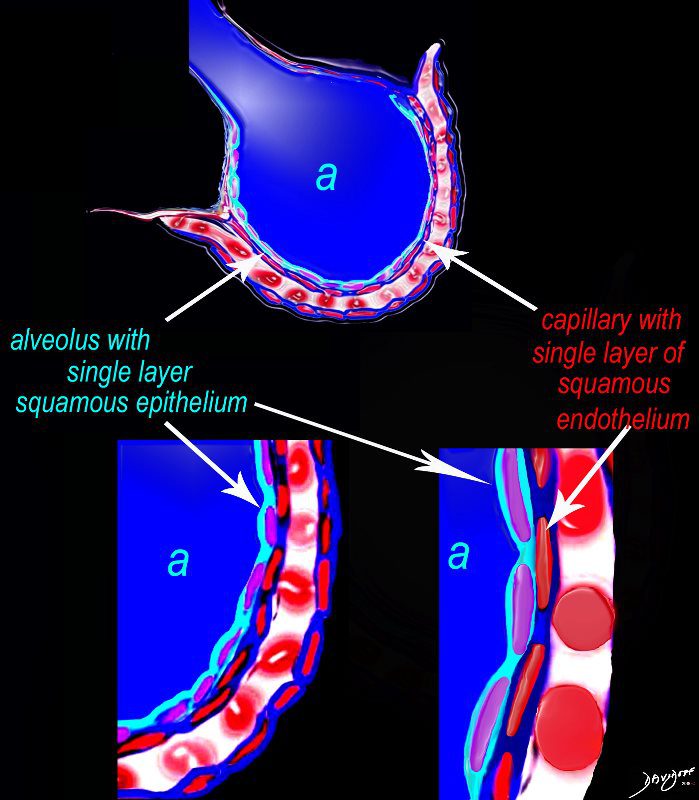
The diagram shows an alveolus (a) above, lined by a single layer of squamous cells, surrounded by a capillary with red cells which is also lined by a single layer of squamous endothelial cells . The images below show progressive magnification of the alveolar wall demonstrating the two thin layer of the alveolar membrane .
Courtesy Ashley Davidoff 2019
lungs-0028-low res
Type 1 Pneumocyte
Type II Pneumocyte

Macrophage
Ashley Davidoff MD
Alveolar Septum
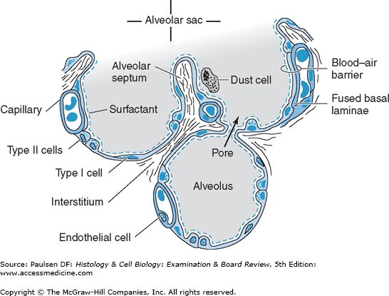
From Paulsen DF Histology and Cell Biology Copyright McGraw Hill
Diseases
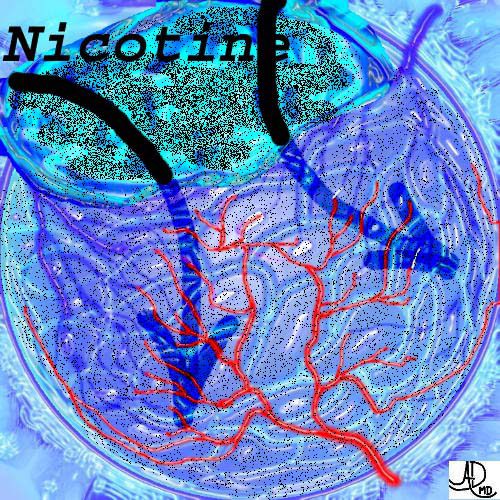
Smoking and the Alveolus
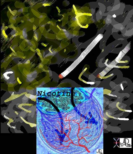
These diseases are all about cigarettes and the garbage that they deposit in our lungs.
Courtesy Ashley Davidoff MD. 32646d The CommonVein.net
An alveolus subjected day and night for 20 years to black smog from a human chimney. It had no choice but to react.
Courtesy Ashley Davidoff MD. 32166f
The CommonVein.net
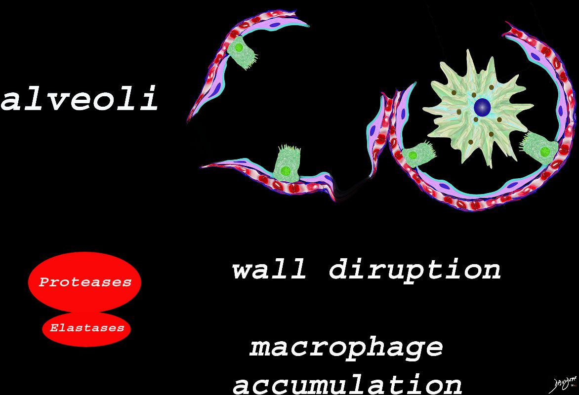
The effect of the proteases and and elastases cause destruction of the alveoli and loss of elasticity, and therefore overall function. The destruction leads to bullous disease
The accumulation of smokers macrophage, and in the case of Langerhans cell histiocytosis leads to space occupation of the alveoli also reducing function
Ashley Davidoff TheCommonVein.net lungs-00687-lo res
Hyaline Membrane Disease

A hyaline membrane evolves covering the damaged surface of the alveolus. This impedes gas exchange
Ashley Davidoff TheCommonVein.net
Chronic Hypersensitivity Pneumonitis
Courtesy Wikiw and
web lungs 435