
- Caused by
- complete obstruction
- neoplasm,
- mucus plugging
- foreign bodies
- complete obstruction
- Result
- air
- no new air can enter lung distal to the obstruction
- trapped air that is absorbed into the capillaries, l
- pleura
- cannot separate
- vacuum and
- traction of mediastinal structures and diaphragm
- mediastinal shift and elevated diaphragm
- air
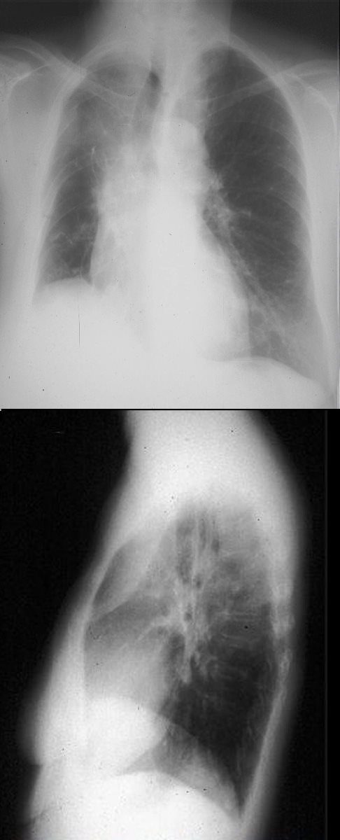
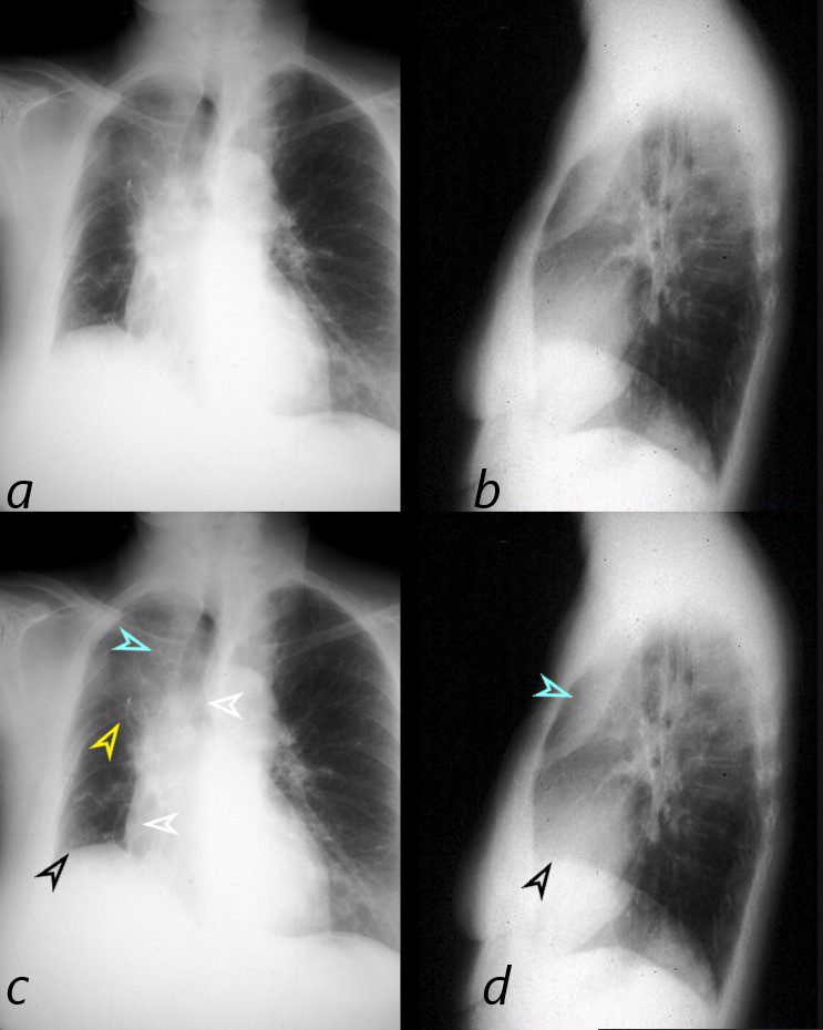
Frontal and lateral views of the chest show signs of volume loss characterized by elevation of the right hemidiaphragm (black arrowhead), rightward tracheal and mediastinal shift and elevation of the minor fissure contributing to the reverse S sign of Golden. There is a vague infiltrate in the right upper lobe correlating with an anterior pie shaped density on the lateral examination, consistent with collapse of the anterior segment of the RUL. This combination of images is consistent with a malignant mass in the hilum causing obstruction of the right mainstem bronchus.
Ashley Davidoff MD TheCommonVein.net 32292c01
-
-
-
-

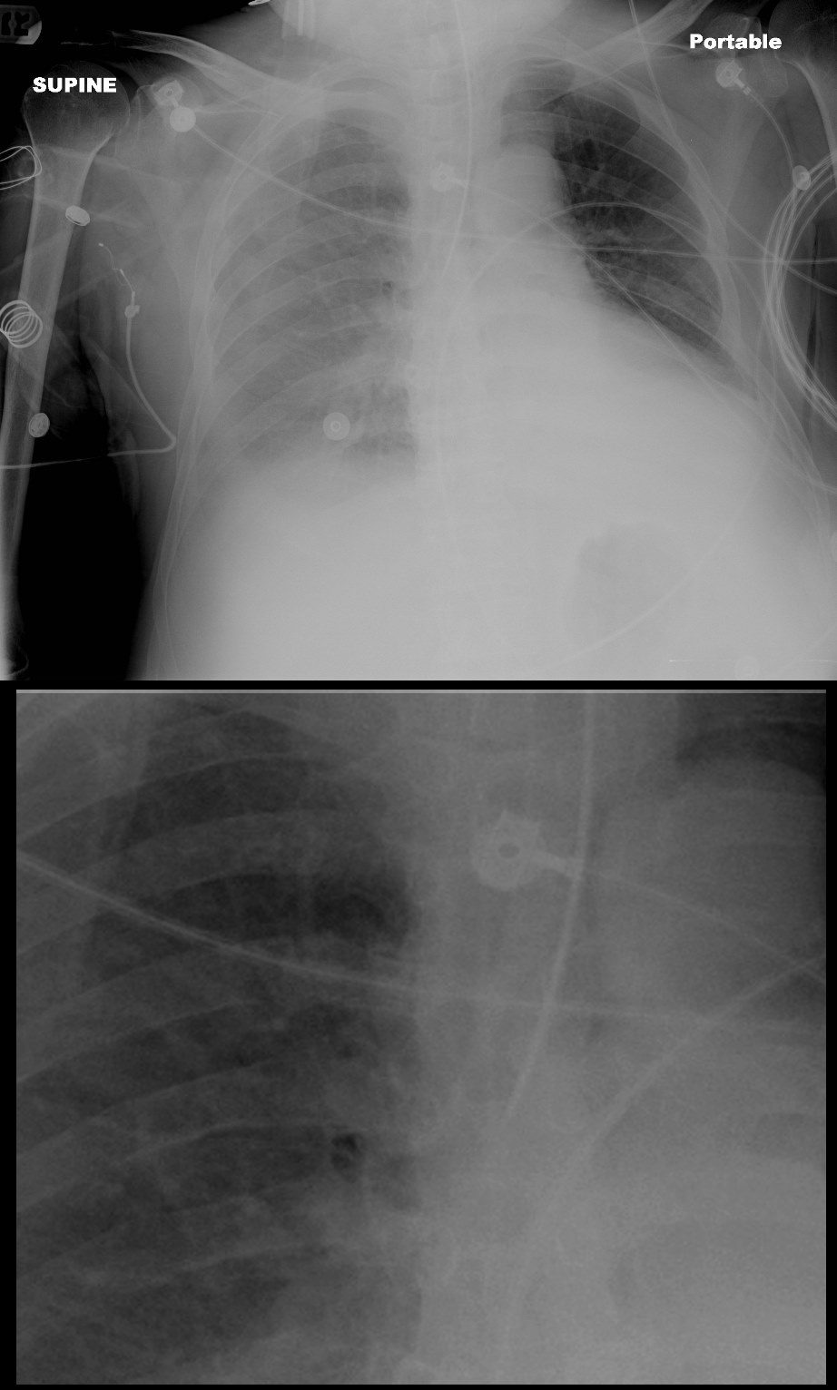
Reversed S sign of Golden
The scout film performed prior to a CT scan from a 76-year-old man with chest pain and shortness of breath. The appearance suggests atelectasis of the right upper lobe with the normal position of the minor fissure (yellow) altered so that the upper portion (light green above the yellow line) is shifted upward caused by volume loss of an atelectatic right upper lobe (pink). The lower portion of the fissure (light green below the yellow line) is bulging rightward and outward caused by an implied mass (dark green). The “reversed S sign of Golden” is demonstrated in this case and infers a central mass causing obstruction and resulting in the shape described by the light green line of the minor fissure.
Courtesy: Ashley Davidoff, M.D.
-
-
-

Post
Obstructive Atelectaisis of the left Lower Lobe
Ashley Davidoff MD TheCommonVein.net
42077
Ashley Davidoff MD TheCommonVein.net
70231cL
57-year old male presents with a cough. CXR shows silhouetting of the left hemidiaphragm and leftward mediastinal shift. CT scan shows an airless consolidation with leftward shift consistent with atelectasis.
Ashley Davidoff MD TheCommonVein.net
Bilateral Lower Lobar Atelectasis with
Occlusion of the Right Main Stem Bronchus
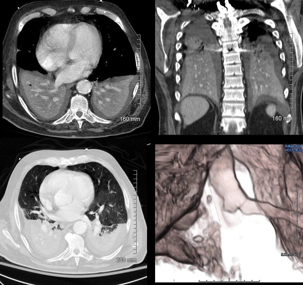
74 year old male alcoholic with bilateral basilar lobar atelectasis caused by bilateral aspiration
CT scan shows airless lower lobes with small bilateral effusions. 3D reconstruction shows total obstruction of the right mainstem bronchus, and patent proximal mainstem bronchus
Ashley Davidoff MD TheCommonVein.net
Whole Lung Atelectasis Due to Obstruction
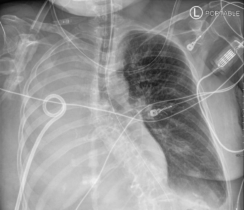
59F shows total white out caused by collapse of right lung with an
occluded right main step bronchus associated with a
large right sided effusion. The occlusion is likely due to proximal cancer. A pigtail drain has been placed to drain the effusion
Ashley Davidoff MD TheCommonVein.net 104 Lu
59F shows total collapse of left lung with an
occluded right main step bronchus(top right image)associated with a
right sided effusion. The occlusion is likely due to proximal cancer
Ashley Davidoff MD TheCommonVein.net 104 Lu
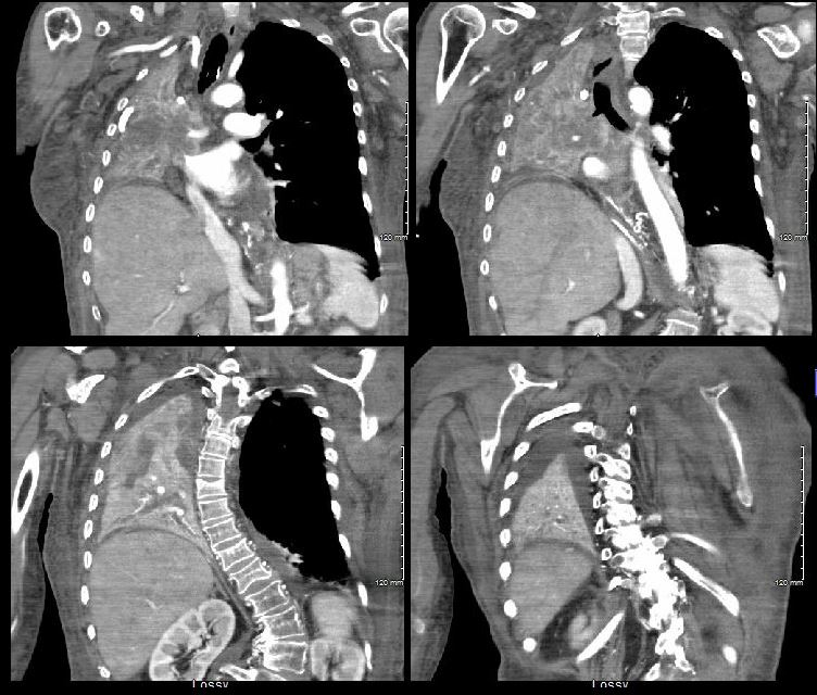
Occluded Right Main Stem Bronchus and Right Upper Lobe Collapse by Cancer with Pathology Correlation
This combination of images shows the manifestations of a malignant mass in the hilum causing compression of the right mainstem bronchus. The PA CXR shows signs of volume loss (atelectasis characterized by elevation of the right hemidiaphragm (black arrowhead), rightward tracheal and mediastinal shift (white arrowheads) and elevation of the minor fissure contributing to the reverse S sign of Golden. There is a vague infiltrate in the right upper lobe correlating with an anterior pie shaped density of the lateral (blue arrowheads), consistent with collapse of the anterior segment of the RUL
32292cL01
Ashley Davidoff MD TheCommonVein.net
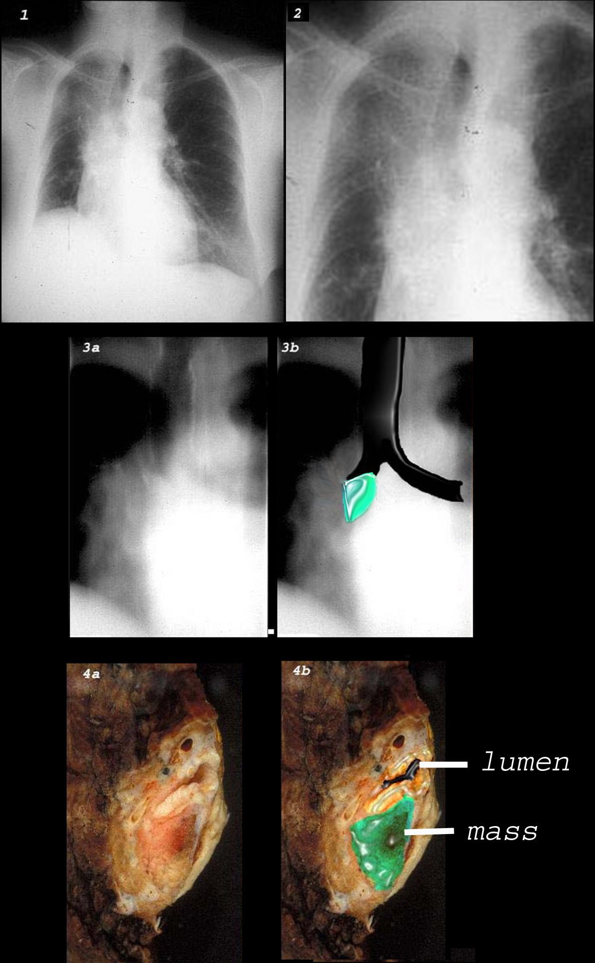
This combination of images shows the manifestations of a malignant mass in the hilum causing compression of the right mainstem bronchus. There is elevation of the right hilum on the CXR, associated with collapse of the anterior segment of the RUL seen as a vague density in the P-A . The tomogram (3a) shows an abrupt cut off of the right mainstem bronchus while the overlay in 3b shows the occlusion of the right mainstem bronchus, the implied tumor overlaid in green. Images 4a and 4b are the correlative gross pathology images showing the tumor in green pushing and occluding the right mainstem
Ashley Davidoff MD TheCommonVein.net
Right Upper Lobe Collapse
Squamous Cell Causing
Obstruction but Airways Filled with
Tumor or Infection or Mucus

CXR shows right upper lobe (RUL) atelectasis. Final diagnosis was a central RUL proximal squamous cell carcinoma with extensive filling of the distal bronchi-ectatic segmental and subsegmental airways
Ashley Davidoff TheCommonVein.net

CXR shows right upper lobe (RUL) atelectasis. Final diagnosis was a central RUL proximal squamous cell carcinoma with extensive filling of the distal bronchi-ectatic segmental and subsegmental airways
Ashley Davidoff TheCommonVein.net

Ashley Davidoff TheCommonVein.net

Ashley Davidoff TheCommonVein.net

Ashley Davidoff TheCommonVein.net
Subsegmental Obstruction of the
Apical Segment of the Right Upper Lobe
Carcinoid Tumor Causing Obstruction
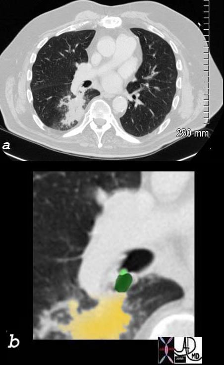
65 year old female presents with a cough. CT shows a mass (green) in proximal portion of the right lower lobe bronchus with post obstructive atelectasis in the superior segment of the right lower lobe (yellow) pathology revealed carcinoid tumor
Ashley Davidoff MD TheCommonVein.net
75679c02
Active TB Causing Obstruction
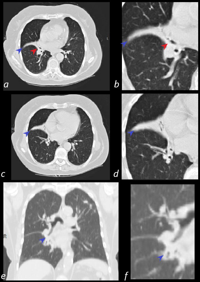
80-year-old Russian woman who initially presented with a cavitating LUL nodule that was biopsied and thought to represent sarcoidosis, nut subsequently shown to be TB. Axial CT’s shows thickening around the bronchovascular bundles of the middle lobe (red arrowhead – a, b) with post obstructive atelectasis of the lateral segment of the RML (blue arrowhead , a-f)
Ashley Davidoff MD Ashley Davidoff MD TheCommonVein.net
31645cL
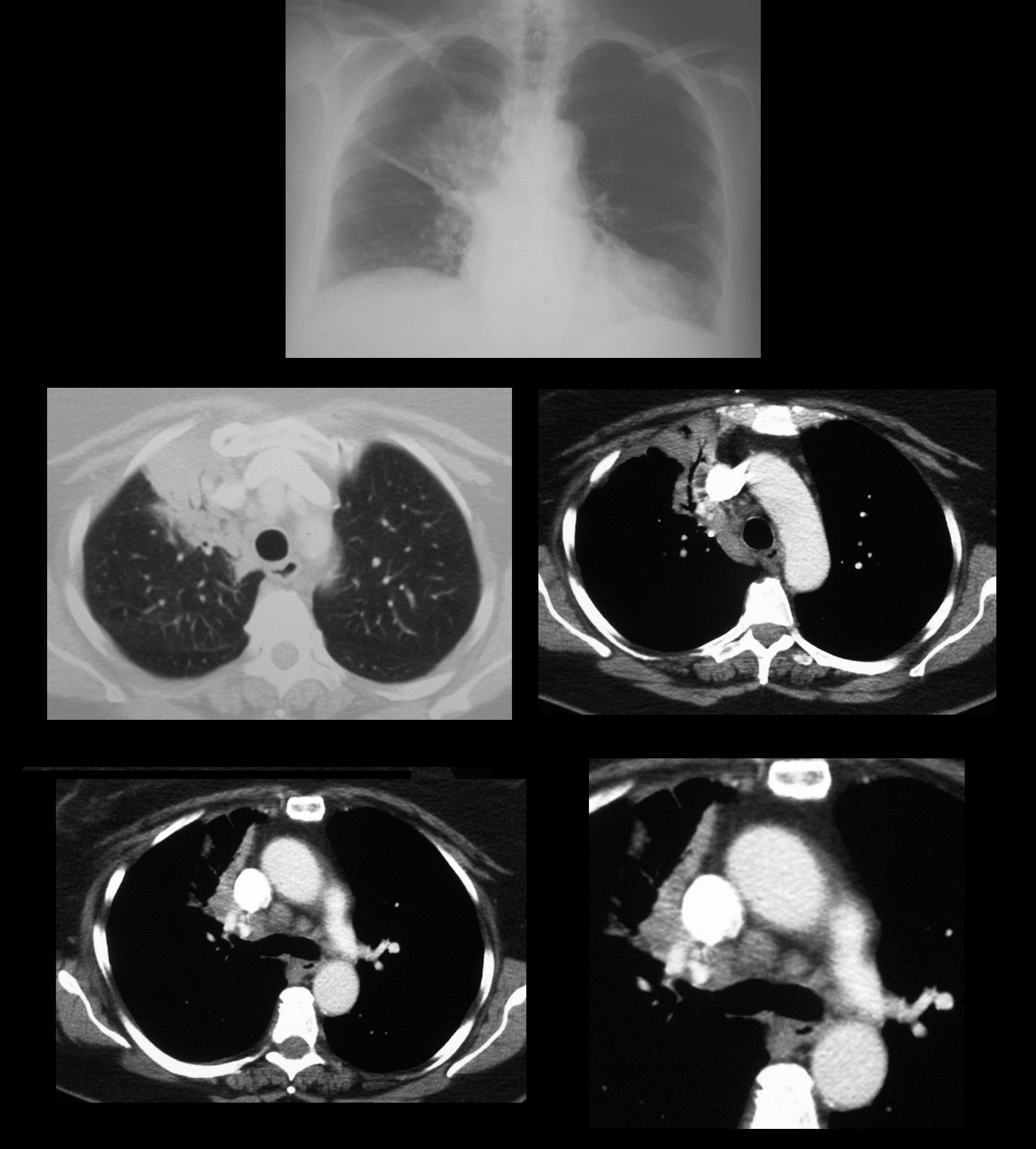
59year old male presents with cough. Chest Xray reveals signs of segmental right upper lobe atelectasis with elevation of the minor fissure and right hemidiaphragm confirms the presence of atelectasis and an air bronchogram. A proximal obstructing lesion was suspected. Slightly enlarged lymph nodes are noted in the azygous region (last row)
Ashley Davidoff MD TheCommonVein.net
Links and References
-
TCV