
- is
- inflammation and/or fibrosis involving (
- airways smaller than 2 mm in diameter, and/or
- the alveolar ducts
- inflammation and/or fibrosis involving (
- pathology
- cellular –
- inflammatory cells
- constrictive
- fibrosis
- submucosa and or
- adventitia
- fibrosis
- cellular –
- imaging
- CXR
- ill-defined opacities
- CT scan
- centrilobular nodules with or without
- air trapping
- CXR
- Types
- cellular
- primary bronchiolar disease
- acute bronchiolitis
- usually viral infections,
- infants and children
- usually viral infections,
- respiratory bronchiolitis,
- smoking adults
- follicular bronchiolitis,
- connective tissue disease,
- immunodeficiency disorders, or
- chronic infections.
- diffuse panbronchiolitis,
- Asian population (particularly in the Japanese).
- diffuse aspiration bronchiolitis,
- mineral dust airway disease
- inorganic dusts, – occupation-related
- silica,
- silicates,
- asbestos,
- iron oxide,
- aluminum oxide,
- talc,
- mica, and
- coal. The disorder is primarily
- inorganic dusts, – occupation-related
- acute bronchiolitis
- constrictive or obliterative bronchiolitis
-
- bronchiolitis obliterans syndrome
- bone marrow transplant
- lung transplant
- collagen diseases
- inhalational diseases
- autoimmunity
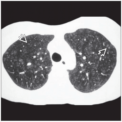
- CT air trapping mosaic attenuation
- bronchiolitis obliterans syndrome
-
- primary bronchiolar disease
- cellular
- related to ILD
- smoking related
- respiratory bronchiolitis-associated interstitial lung disease (RB-ILD),
- desquamative interstitial pneumonia (DIP),
- hypersensitivity pneumonitis (HP),
- cryptogenic or secondary organizing pneumonia (OP),
- sarcoidosis, and
- idiopathic pulmonary fibrosis (IPF).
- smoking related
- Types
- primary bronchiolar disease
- acute bronchiolitis
- usually viral infections,
- infants and children
-

Histopathology of acute bronchiolitis characterized by dense peribronchiolar infiltrate of acute and chronic inflammatory cells associated with an intraluminal exudate rich in neutrophils
Ryu, J Pulmonary Medicine Bronchiolitis Pulmonology Advisor
- usually viral infections,
- respiratory bronchiolitis,
- smoking adults
-
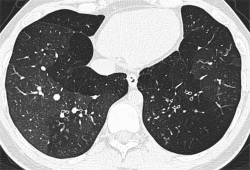
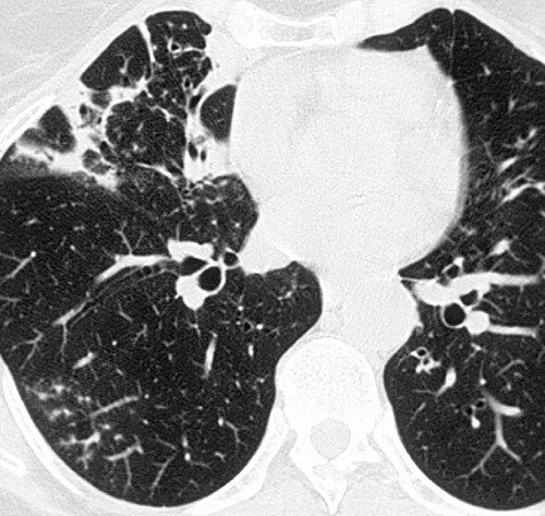
Mosaic attenuation pattern. CT scan through lower lobes shows the indirect sign of constrictive obliterative bronchiolitis, in this case the sequel to a severe viral lower respiratory tract infection.
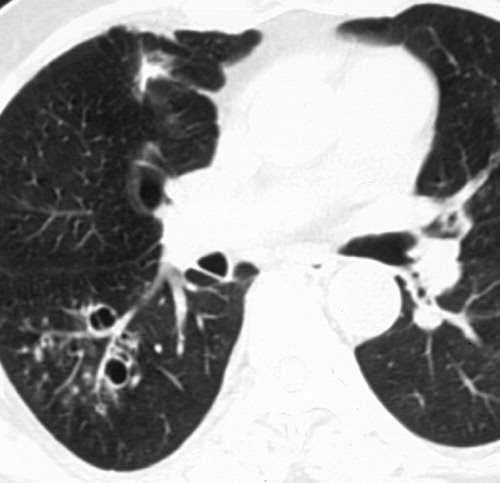
https://pubs.rsna.org/doi/full/10.1148/radiol.13120908Tree-in-bud pattern. CT scan through lower lobes shows the direct sign of exudative bronchiolitis in a 42-year-old man with a sino-bronchial sepsis syndrome.
https://pubs.rsna.org/doi/full/10.1148/radiol.13120908
-
- smoking adults
- constrictive or obliterative bronchiolitis
- bronchiolitis obliterans syndrome
- bone marrow transplant
- lung transplant
-
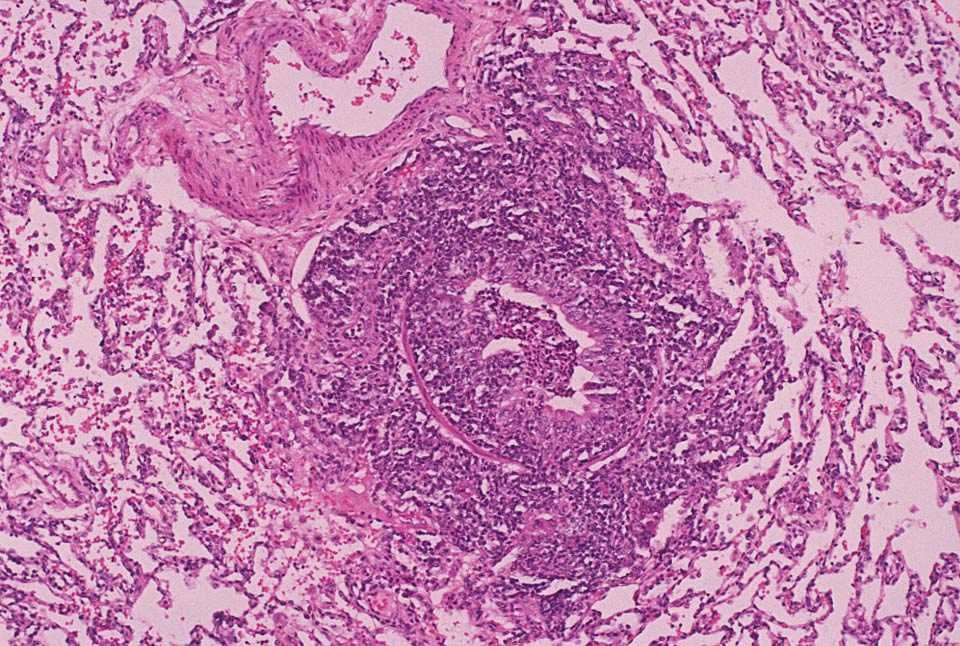
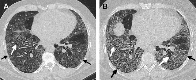
Obliterative bronchiolitis after bone marrow transplantation in a 47-year-old man with myeloma. (a) Expiratory high-resolution CT scan shows diffuse centrilobular nodules connected to branching linear opacities bilaterally. Note the air trapping in the right lower lobe. (b) Photomicrograph (original magnification, ×200; hematoxylin-eosin stain) of a specimen from open lung biopsy shows the bronchiolar walls surrounded by concentric chronic inflammatory infiltrates (arrows).
Rossi, SE et al Tree-in-Bud Pattern at Thin-Section CT of the Lungs: Radiologic-Pathologic Overview RadioGraphics Vol. 25, No. 3 2005 - collagen diseases
-
Sjögren syndrome in a 54-year-old woman. Thin-section CT scan shows peripheral tree-in-bud patterns in the right lower lobe. Note the bronchial dilatation, bronchial wall thickening, and consolidation.
Rossi, SE et al Tree-in-Bud Pattern at Thin-Section CT of the Lungs: Radiologic-Pathologic Overview RadioGraphics Vol. 25, No. 3 2005
-
- inhalational diseases
-
Inhalation bronchiolitis in a 56-year-old man after accidental exposure to sulfur dioxide. High-resolution CT scan shows bronchiectasis in combination with the tree-in-bud pattern in the right lower lobe.
Rossi, SE et al Tree-in-Bud Pattern at Thin-Section CT of the Lungs: Radiologic-Pathologic Overview RadioGraphics Vol. 25, No. 3 2005
-
- CT air trapping mosaic attenuation
-
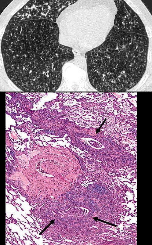
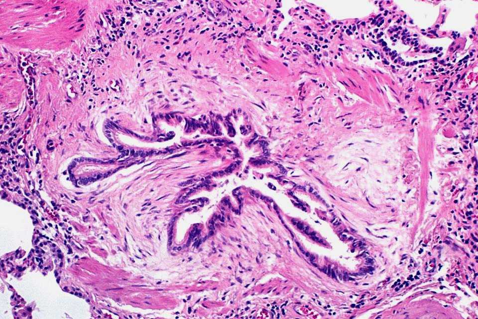
Histopathology of constrictive bronchiolitis characterized by fibroblast proliferation and stromal edema with associated collagen deposition in the submucosa, resulting in airway luminal constriction
Ryu, J Pulmonary Medicine Bronchiolitis Pulmonology Advisor
- bronchiolitis obliterans syndrome
- follicular bronchiolitis,
- connective tissue disease,
- immunodeficiency disorders, or
- chronic infections.
- diffuse panbronchiolitis,
- Asian population (particularly in the Japanese).
-
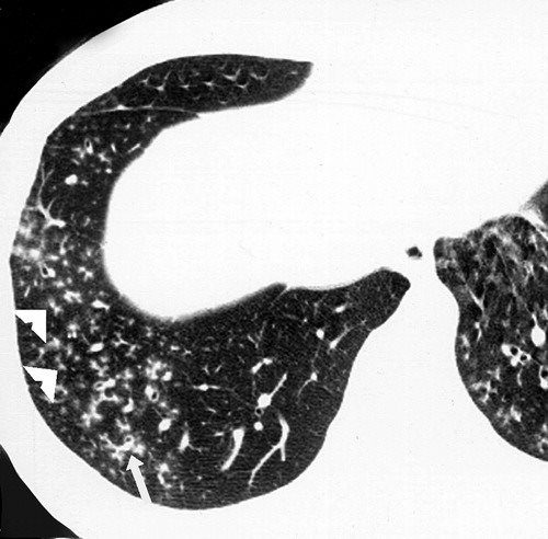
Diffuse panbronchiolitis in a 44-year-old Japanese man. High-resolution CT scan shows diffuse small centrilobular nodules and branching linear opacities (arrow), which resemble the objects used in the game of jacks. Note the bronchiolar dilatation and mucoid impaction (arrowheads).
Rossi, SE et al Tree-in-Bud Pattern at Thin-Section CT of the Lungs: Radiologic-Pathologic Overview RadioGraphics Vol. 25, No. 3 2005
- diffuse aspiration bronchiolitis,
- mineral dust airway disease
- inorganic dusts, – occupation-related
- silica,
- silicates,
- asbestos,
- iron oxide,
- aluminum oxide,
- talc,
- mica, and
- coal. The disorder is primarily
- inorganic dusts, – occupation-related
- acute bronchiolitis
- primary bronchiolar disease
- related to ILD

Hellemons M etal Eur Respir Rev 2020; 29: 190181. – September 30, 2020
Kligerman S et al Clinical-Radiologic-Pathologic Correlation of Smoking-Related Diffuse Parenchymal Lung Disease 2016
- hypersensitivity pneumonitis (HP)
-
Head cheese or brawn is a cold cut terrine or meat jelly, often made with flesh from the head of a calf or pig (less commonly a sheep or cow), typically set in aspic, that originated in Europe. Usually eaten cold, at room temperature, or in a sandwich, the dish is, despite the name, not a dairy cheese. The parts of the head used in the dish vary, though commonly do not include the brain, eyes or ears of the animal. The tongue, and sometimes the feet and heart of the animal may be included; the dish is also made using trimmings from more commonly eaten cuts of pork and veal, with the addition of gelatin as a binding agent. Head cheese may also be made without using the flesh from the head of an animal.
Courtesy Rainer Zenz source Wiki -
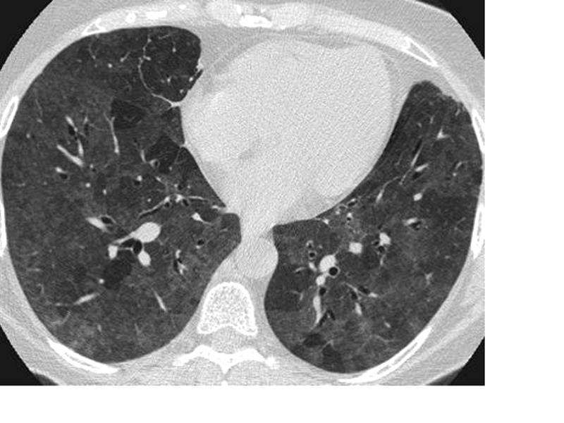
Hypersensitivity Pneumonitis
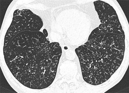
High-resolution CT: increase in density in areas of ground glass and air trapping in lower lobes in patients with hypersensitivity pneumonitis
Courtesy Mluisamtz11 -
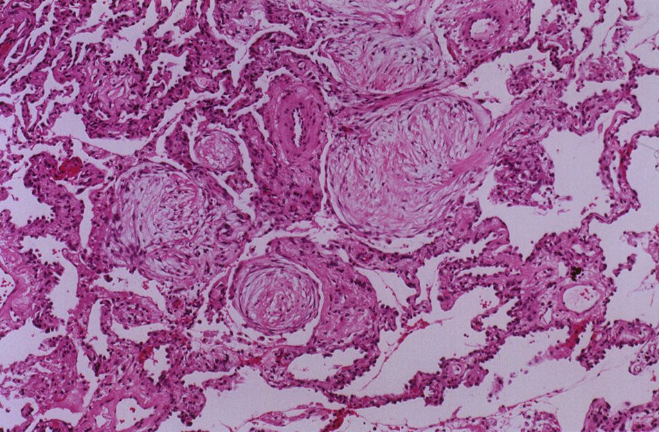
- cryptogenic or secondary organizing pneumonia (OP),
-
Histopathology of organizing pneumonia (or BOOP), characterized by intraluminal plugs of proliferating fibroblasts that fill distal airways and peribonchiolar air spaces
Ryu, J Pulmonary Medicine Bronchiolitis Pulmonology Advisor - sarcoidosis, and
- idiopathic pulmonary fibrosis (IPF).

Links and References
Ryu, J Pulmonary Medicine Bronchiolitis Pulmonology Advisor
Winningham P, J. et al Bronchiolitis: A Practical Approach for the General Radiologist RadioGraphicsVol. 37, No. 3 2017
Links and References
TCV
Small Airway Disease Introduction
Fleischner Society
bronchiolitis
Pathology.—Bronchiolitis is bronchiolar inflammation of various causes (,33).
CT scans.—This direct sign of bronchiolar inflammation (eg, infectious cause) is most often seen as the tree-in-bud pattern, centrilobular nodules, and bronchiolar wall thickening on CT scans. (See also small-airways disease, tree-in-bud pattern.)