
CT Left Lower Lobe Bronchopneumonia
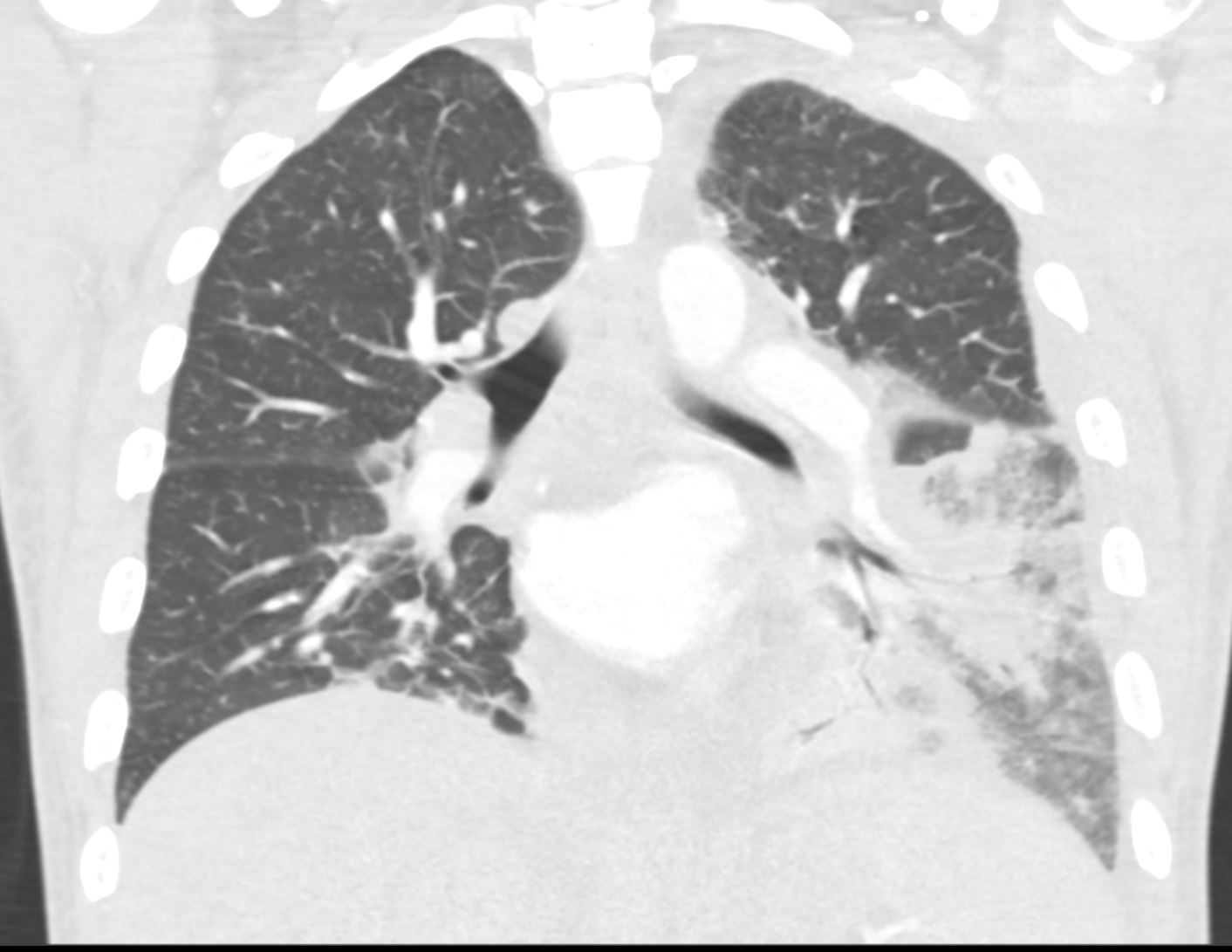
35-year-old woman, presents with dyspnea and fever. Coronal CT shows a bronchocentric pneumonic infiltrate in the superior segment of the left lower lobe and extending into the medial basal segment, and silhouetting the medial aspect of the diaphragm. There is an adjacent region of ground glass infiltrate. These findings are consistent with a bronchopneumonia in the left lower lobe.
Courtesy Ashley Davidoff MD TheCommonVein.net 289Lu 136560
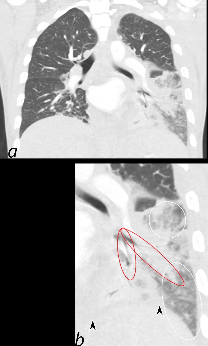
35-year-old woman, presents with dyspnea and fever. Coronal CT shows a bronchocentric pneumonic infiltrate in the superior segment of the left lower lobe (red rings) and extending into the medial basal segment, resulting in silhouetting of the medial aspect of the left hemidiaphragm (between the black arrowheads). There is an adjacent region of ground glass infiltrate. (b, white rings) These findings are consistent with a bronchopneumonia in the left lower lobe.
Courtesy Ashley Davidoff MD TheCommonVein.net 289Lu 136560cL
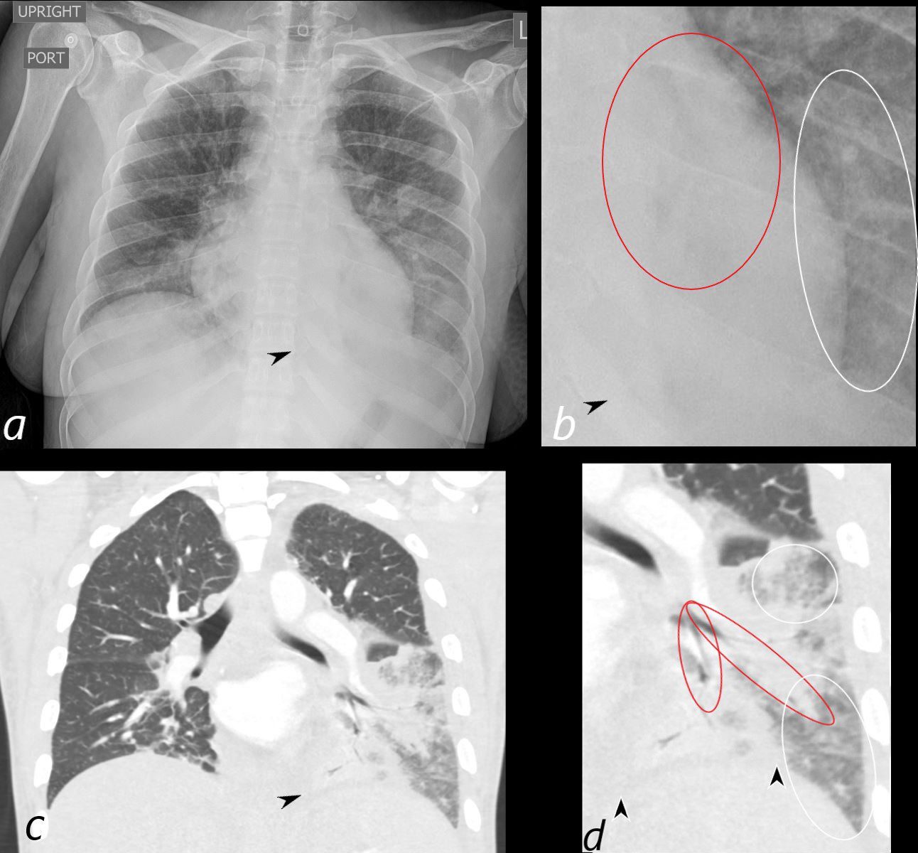
35-year-old woman, presents with dyspnea and fever. Frontal CXR (a, and magnified in b) shows a retrocardiac infiltrate, with suggestion of air bronchograms, ground glass changes (b, white ring) and silhouetting of the diaphragm medially (a, b, black arrowheads)
Coronal CT shows a bronchocentric pneumonic infiltrate in the superior segment of the left lower lobe and extending into the medial basal segment, (red rings), consistent with a bronchopneumonia. There is an adjacent region of ground glass infiltrate (d, white rings)
Courtesy Ashley Davidoff MD TheCommonVein.net 289Lu 136562cL
COP vs NSIP
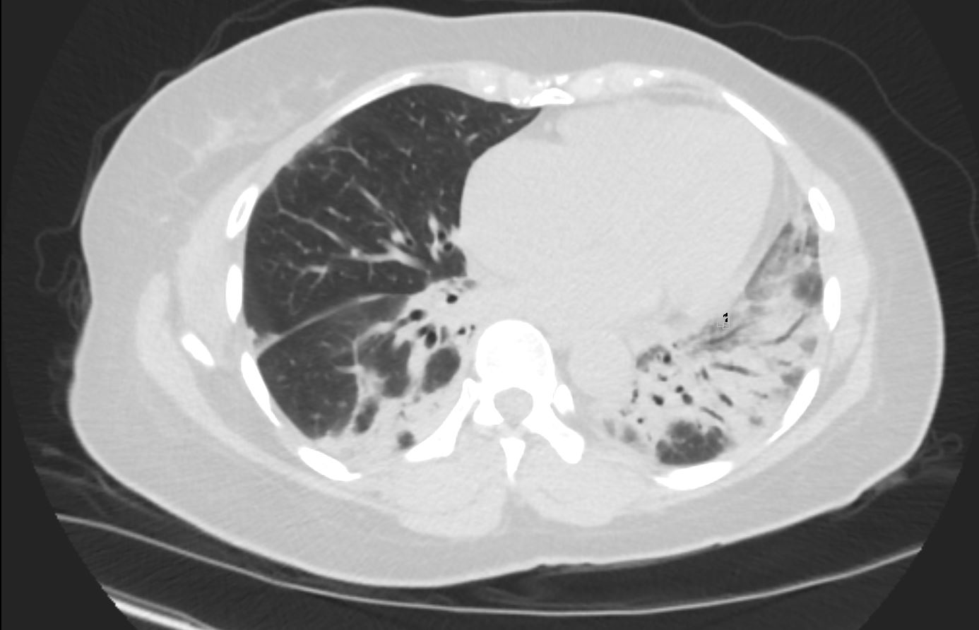
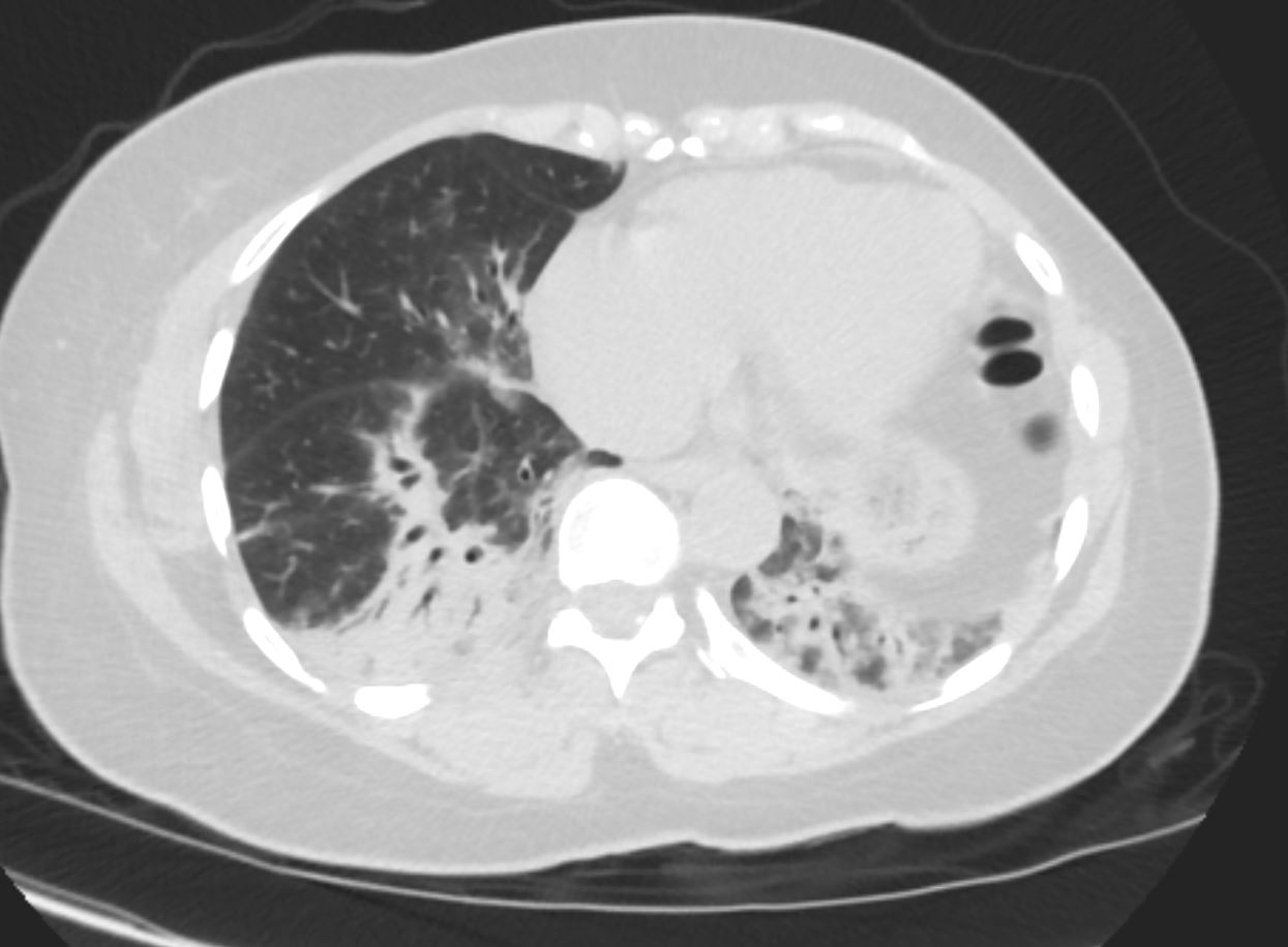
56 year old female presents with CT findings of basilar bronchovascular infiltrates, almost symmetrical, associated with mediastinal and axillary adenopathy.
Pathological report was complex but suggested a diagnosis of cryptogenic organizing pneumonia
56 year old female presents with CT findings of basilar bronchovascular infiltrates, almost symmetrical, associated with mediastinal and axillary adenopathy.
Pathological report was complex but suggested a diagnosis of cryptogenic organizing pneumonia
Ashley Davidoff MD
56 year old female presents with CT findings of basilar bronchovascular infiltrates, almost symmetrical, associated with mediastinal and axillary adenopathy.
Pathological report was complex but suggested a diagnosis of cryptogenic organizing pneumonia
Ashley Davidoff MD
1 Year Later
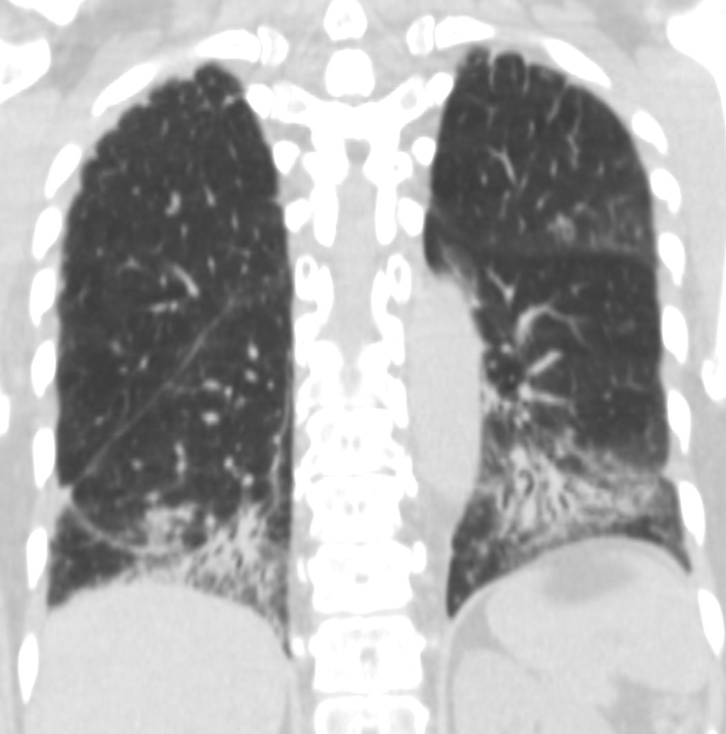
57 year old female presents 1 year later with similar CT findings of basilar bronchovascular infiltrates, almost symmetrical, associated with mediastinal and axillary adenopathy.
Pathological report was complex but suggested a diagnosis of cryptogenic organizing pneumonia
Ashley Davidoff MD
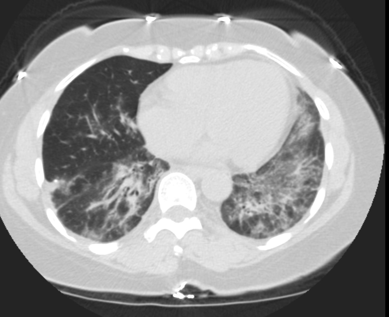
57 year old female presents 1 year later with similar CT findings of basilar bronchovascular infiltrates, almost symmetrical, associated with mediastinal and axillary adenopathy.
Pathological report was complex but suggested a diagnosis of cryptogenic organizing pneumonia
Ashley Davidoff MD