Ashley DAvidoff
TheCommonVein.net

Ashley Davidoff
The CommonVein.net
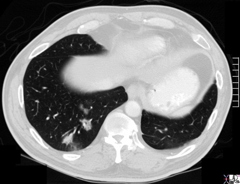
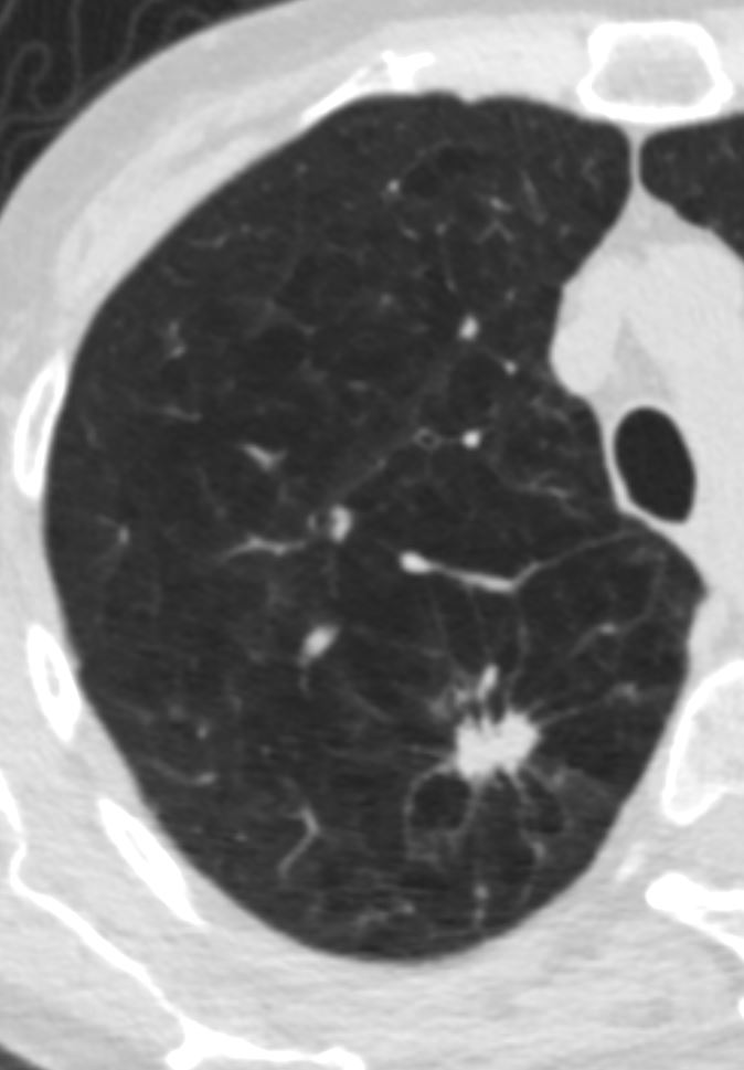
Spiculated and Cavitating Nodule
Ashley Davidoff
TheCommonVein.net
Ashley Davidoff
TheCommonVein.net
28979c
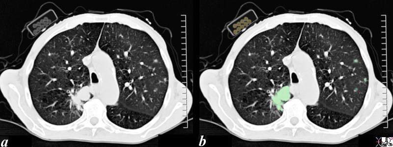
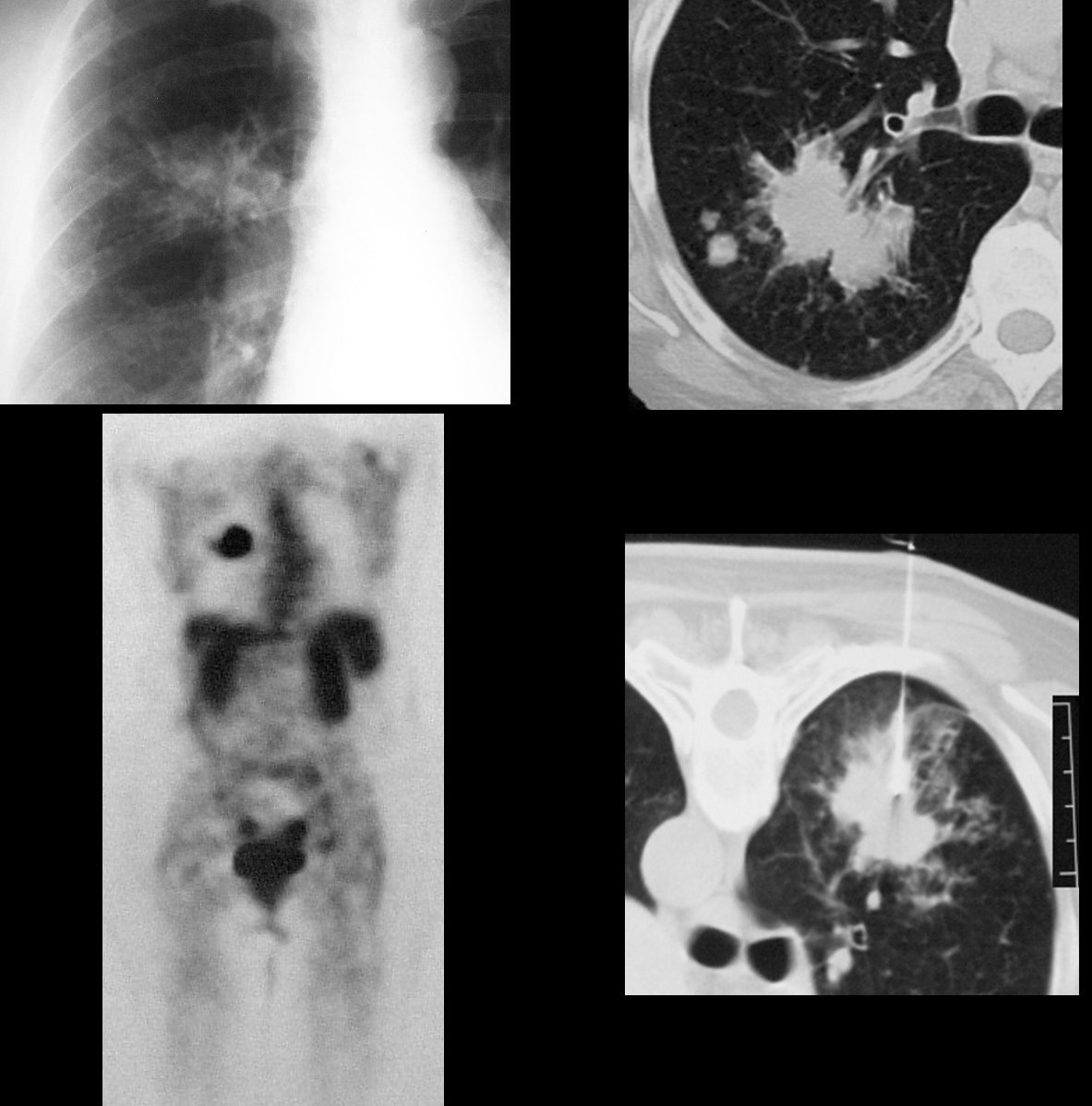
The CT scan through the chest of an 80 year old male shows a large lung mass in the posterior aspect of the right upper lobe (overlaid in green) and3 small nodules in the left upper lobe (overlaid in green). The patient is obviously a smoker and the incriminating pack of cigarettes is identified in his right shirt pocket containing 9 cigarettes. The lung cancer was shown to be an adenocarcinoma The pathology of the nodules may either represent metastatic disease or multicentric foci of bronchioloalveolar cell carcinoma. Associated finding of a thinned anterior junction line suggests hyperinflation and emphysema, and the thickened bronchial walls noted in the right lung suggest chronic bronchitis. Saber shaped trachea is also reminiscent of emphysema. The patient is emaciated, a finding that relates both to his chronic lung disease and his cancer.
Ashley Davidoff MD 87831c01b.8s
TheCommonVein.net