
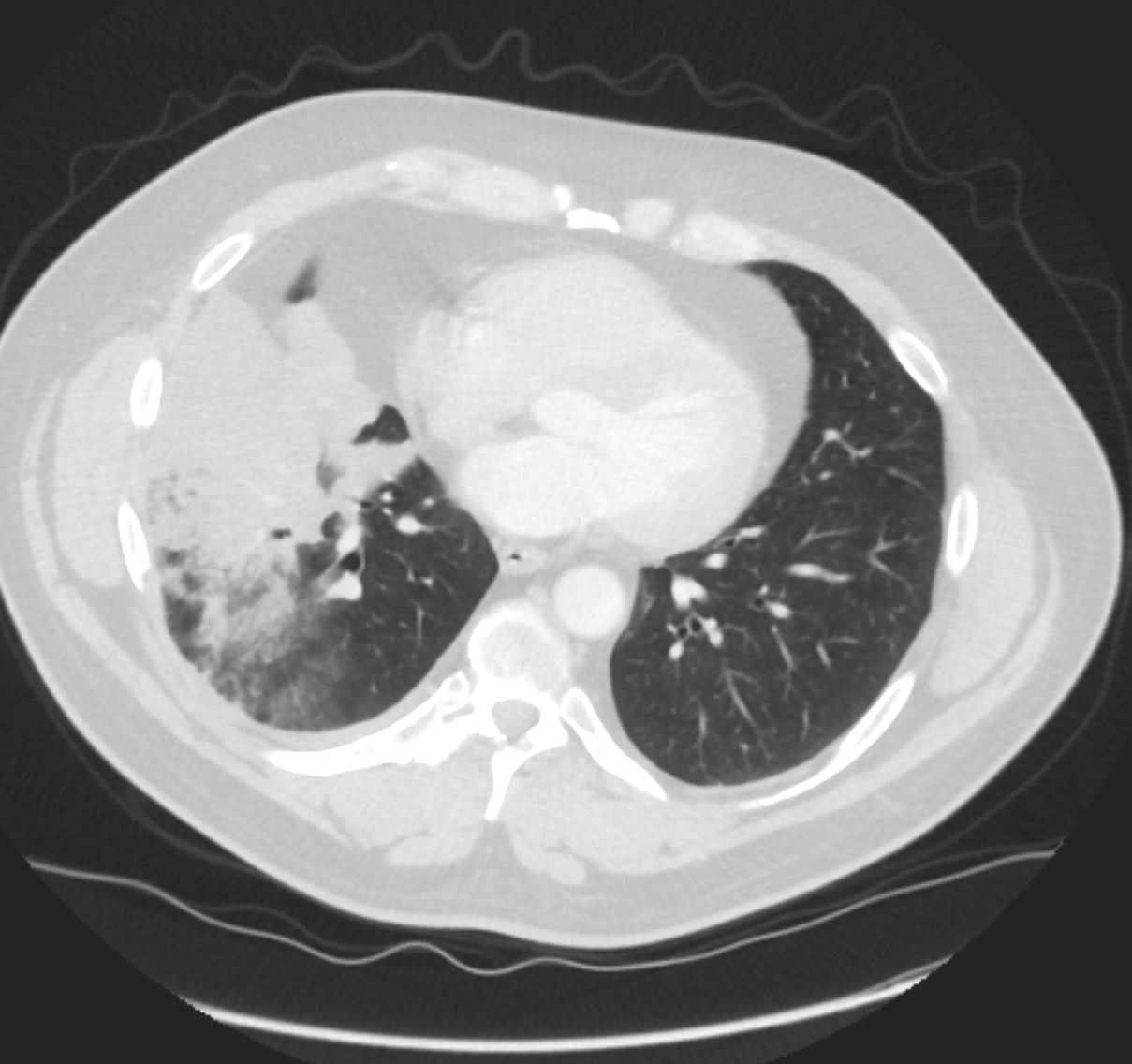
43 year old male with ground glass consolidation on CXR
43 year old male with pneumonic consolidation of malignant origin
Ashley Davidoff MD
TheCommonVein.com
31791c
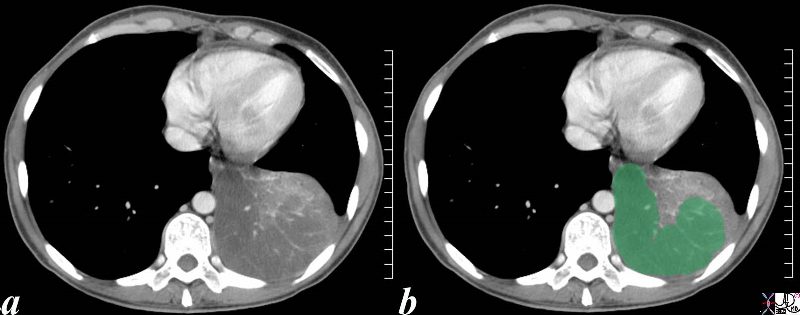
This is an unusual appearance of an adenocarcinoma showing a large lobar consolidation without significant distortion of the vessels. The vessels in the infiltrated section (green overlay) are spread apart. Infiltrative adenocarcinoma was identified on biopsy.
Courtesy Ashley Davidoff MD
TheCommonVein.net
46589c02.8s
70 year old male presents with a cough
There is patchy opacity in the right upper lung
which may represent post obstructive atelectasis from the right hilar mass or multifocal airspace disease.
Ashley Davidoff MD
TheCommonVeiin.net
70M lung ca 001

There is patchy opacity in the right upper lung
which may represent post obstructive atelectasis from the right hilar mass or multifocal airspace disease. The minor fissure is elevated and there is a suggestion of SVC encasement
Ashley Davidoff MD
TheCommonVeiin.net
70M lung ca 006
There is an infiltrate in right upper lung
which may represent post obstructive atelectasis from the right hilar mass or multifocal airspace disease. There is narrowing of the right mainstem bronchus and the SVC. These findings are consistent with a primary carcinoma
Ashley Davidoff MD
TheCommonVeiin.net
70M lung ca 002
There is patchy opacity in the right upper lung
which may represent post obstructive atelectasis from the right hilar mass or multifocal airspace disease. There is encasement of the right upper lobe pulmonary artery suggesting a malignant process.
Ashley Davidoff MD
TheCommonVeiin.net
70M lung ca 005

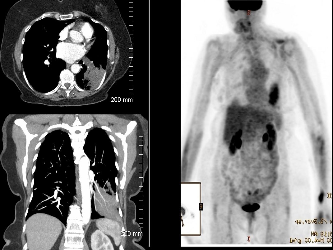
There is patchy opacity in the right upper lung. Right upper lobe lung mass is moderately hypermetabolic
with re-demonstration of mediastinal invasion and at least 3 cm of
contact with the chest wall, very concerning for primary lung
malignancy. There is new complete post-obstructive of the right upper lobe which has progressed
Hypermetabolic ipsilateral right supraclavicular, right upper and
lower paratracheal, subcarinal and right middle lobe segmental lymph nodes are most consistent with nodal metastases. No evidence of metastatic adenopathy on the contralateral left side.
Ashley Davidoff MD
TheCommonVeiin.net
70M lung ca 016 PET
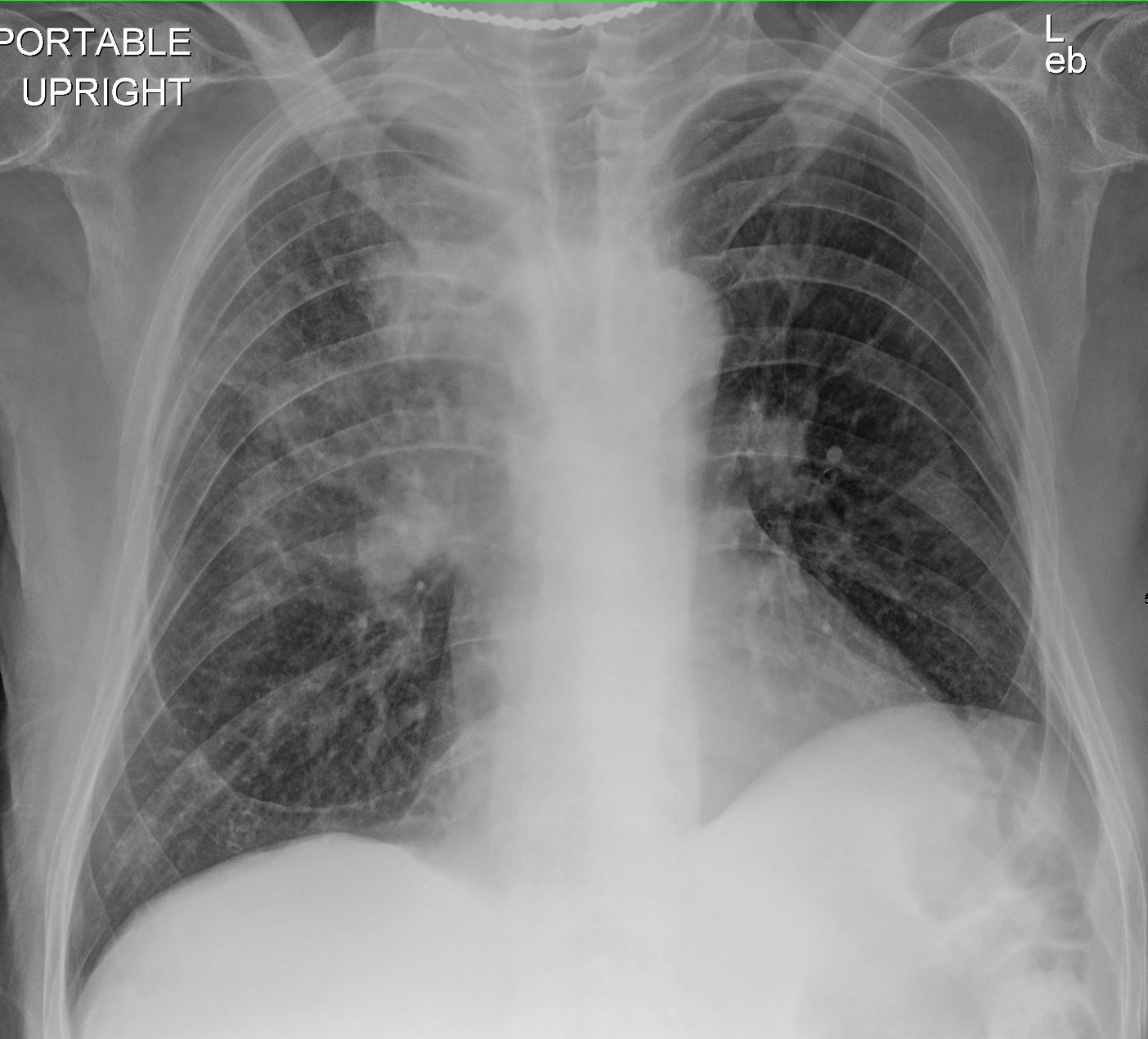
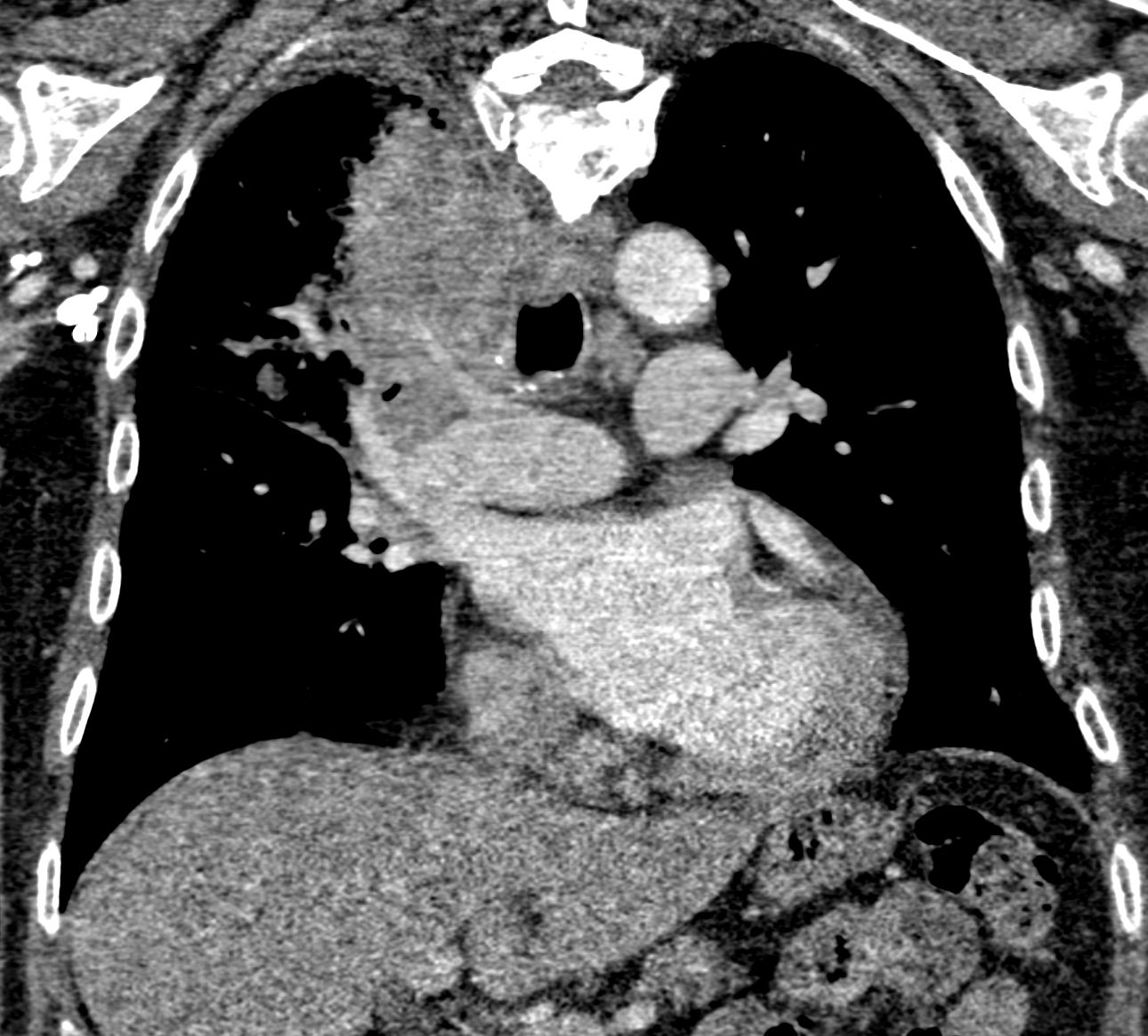
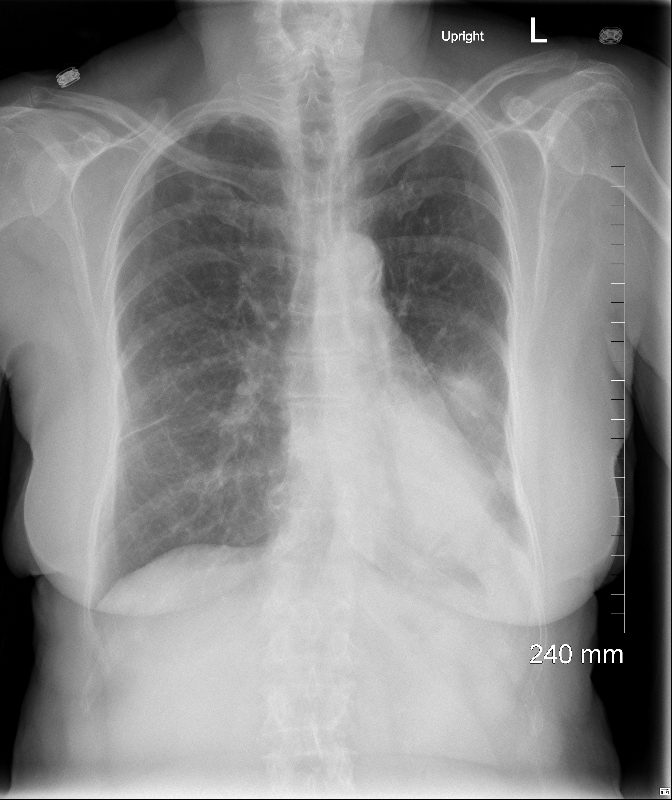
56 year old male with Adenocarcinoma presenting with a Consolidation
CXR showing right lower and middle lobe infiltrate
Ashley Davidoff MD TheCommonVein.net
56 pneumonic mucinous adenoca 001
CXR showing right lower and middle lobe infiltrate
Ashley Davidoff MD TheCommonVein.net
56 pneumonic mucinous adenoca 002
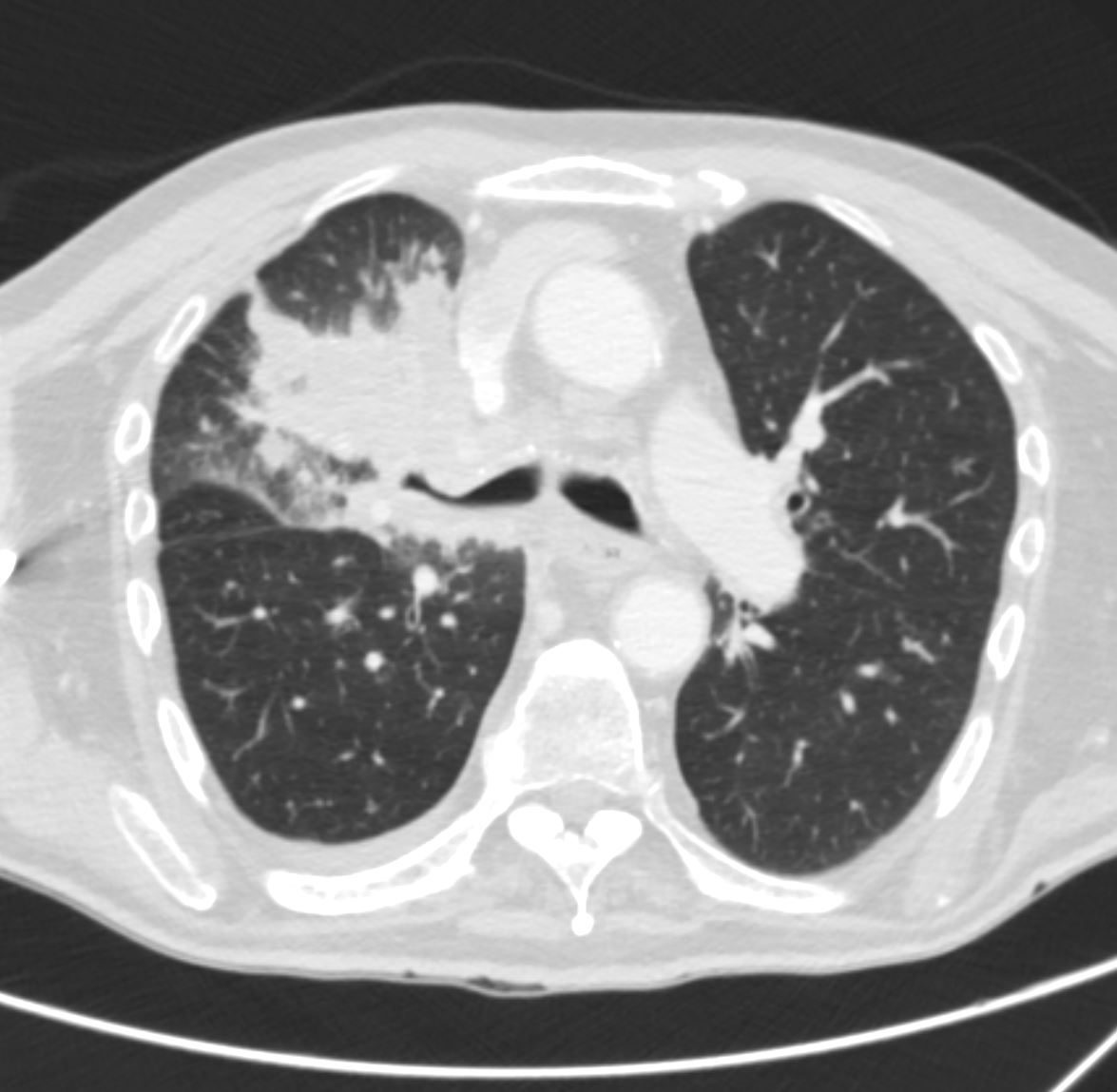
CT shows a right lower and middle lobe infiltrate
Ashley Davidoff MD TheCommonVein.net
56 pneumonic mucinous adenoca 005
CT shows a right lower and middle lobe infiltrate
Ashley Davidoff MD TheCommonVein.net
56 pneumonic mucinous adenoca
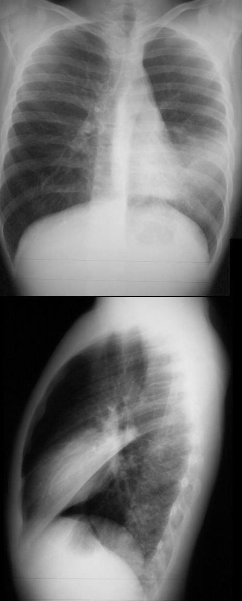
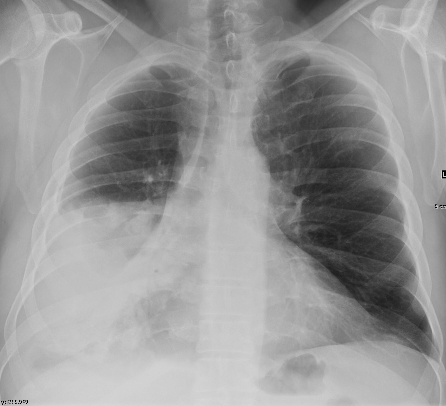
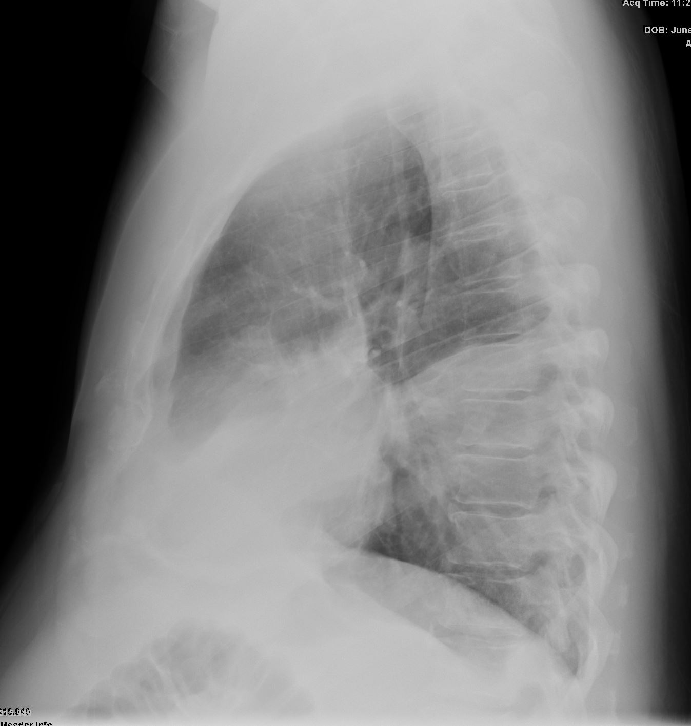
75 year old female presents with a left lower lobe infiltrate on CXR.
Ashley Davidoff MD TheCommonVein.net
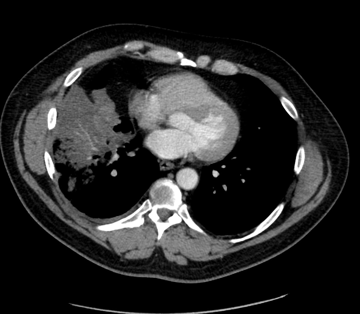
75 year old female presents with a left lower lobe infiltrate on CT scan and PET CT positivity
Ashley Davidoff MD TheCommonVein.net
Links and References