
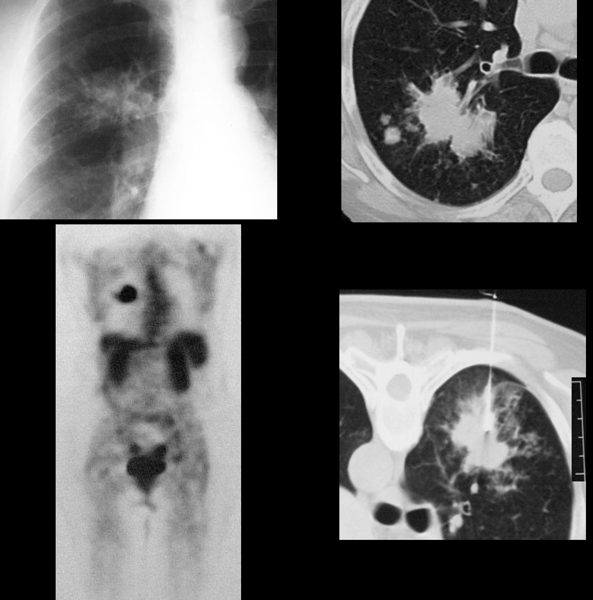
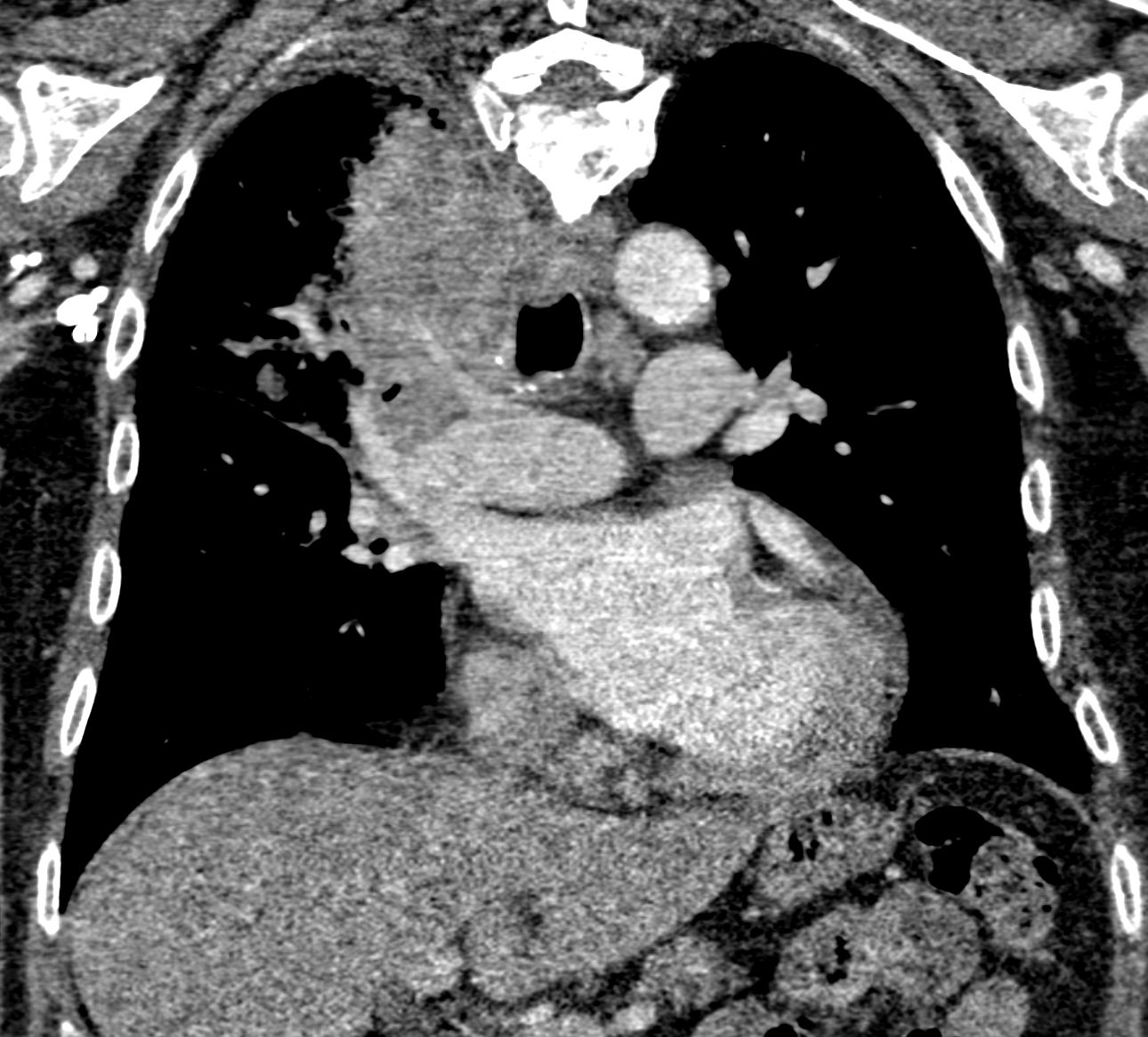
The plain film and CT features of this mass in a 66 year female smoker are concerning for the following reasons; size greater than 3cms, shape with spiculated borders and satellite nodules seen in b. The PET scan was positive and biopsy confirmed the diagnosis. Ashley Davidoff
TheCommonVein.net
28979c

Ashley Davidoff MD
TheCommonVein.net
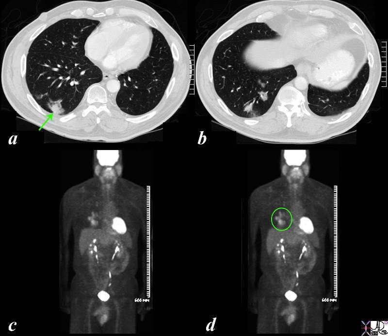
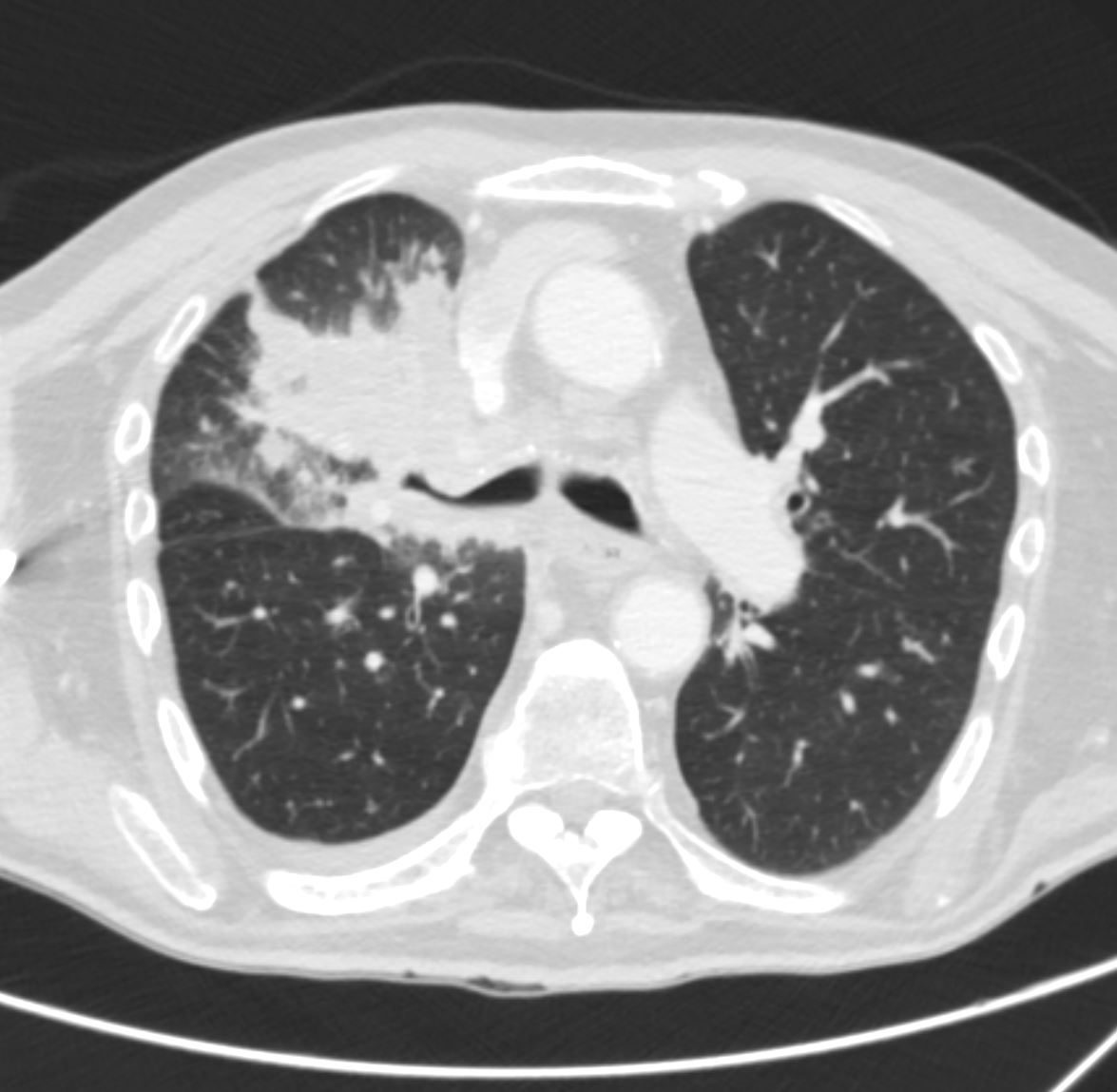
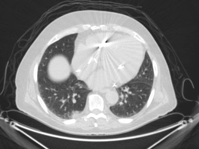
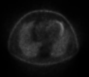
The CT is from a 51 year old male who is a smoker who shows a ground glass opacity (GGO) mixed with a solid mass (arrow in a) and multicentric BAC in a lower cut (b) characterised by three areas in the right lower lobe of partial alveolar opacification. Ground glass appearance is the result of partial opacification of the alveoli. Two of the GGOs measure close to 2cms each and the third more anterior measures about 8mms. In this instance a PET scan was positive in the area (green ring in d) It is likely that the solid component in image a represents the transformation of BAC – (really a carcinoma in situ) into adenocarcinoma.
Courtesy Ashley Davidoff MD
TheCommonVein.net
87769c02b.8s
Ashley Davidoff MD
TheCommonVein.net
32269b see 680249
keywords
lungs pulmonary mass RUL neoplasm malignant primary lymphatics lymphangitis imaging plain film CXR CTscan PETscan
70 year old male presents with a cough
There is patchy opacity in the right upper lung
which may represent post obstructive atelectasis from the right hilar mass or multifocal airspace disease.
Ashley Davidoff MD
TheCommonVeiin.net
70M lung ca 001

There is patchy opacity in the right upper lung
which may represent post obstructive atelectasis from the right hilar mass or multifocal airspace disease. The minor fissure is elevated and there is a suggestion of SVC encasement
Ashley Davidoff MD
TheCommonVeiin.net
70M lung ca 006
There is an infiltrate in right upper lung
which may represent post obstructive atelectasis from the right hilar mass or multifocal airspace disease. There is narrowing of the right mainstem bronchus and the SVC. These findings are consistent with a primary carcinoma
Ashley Davidoff MD
TheCommonVeiin.net
70M lung ca 002
There is patchy opacity in the right upper lung
which may represent post obstructive atelectasis from the right hilar mass or multifocal airspace disease. There is encasement of the right upper lobe pulmonary artery suggesting a malignant process.
Ashley Davidoff MD
TheCommonVeiin.net
70M lung ca 005

There is patchy opacity in the right upper lung. Right upper lobe lung mass is moderately hypermetabolic
with re-demonstration of mediastinal invasion and at least 3 cm of
contact with the chest wall, very concerning for primary lung
malignancy. There is new complete post-obstructive of the right upper lobe which has progressed
Hypermetabolic ipsilateral right supraclavicular, right upper and
lower paratracheal, subcarinal and right middle lobe segmental lymph nodes are most consistent with nodal metastases. No evidence of metastatic adenopathy on the contralateral left side.
Ashley Davidoff MD
TheCommonVeiin.net
70M lung ca 016 PET
False Negatives
Ashley Davidoff THECOMMONVEIN.net 77M adenocarcinoma neg PET
Ashley Davidoff THECOMMONVEIN.net 77M adenocarcinoma neg PET