
Introduction
The result is space occupying disease and depending on the location and degree of structural change symptomatology clinical and radiological findings will differ. Partial obstruction may cause focal emphysema while a total obstruction may lead to segmental or subsegmental atelectasis. Secondary infection may result in bronchitic, pneumonic change and even abscess formation
Venous structures can also be compressed the most dramatic of which is the involvement of the SVC and resulting SVC syndrome.
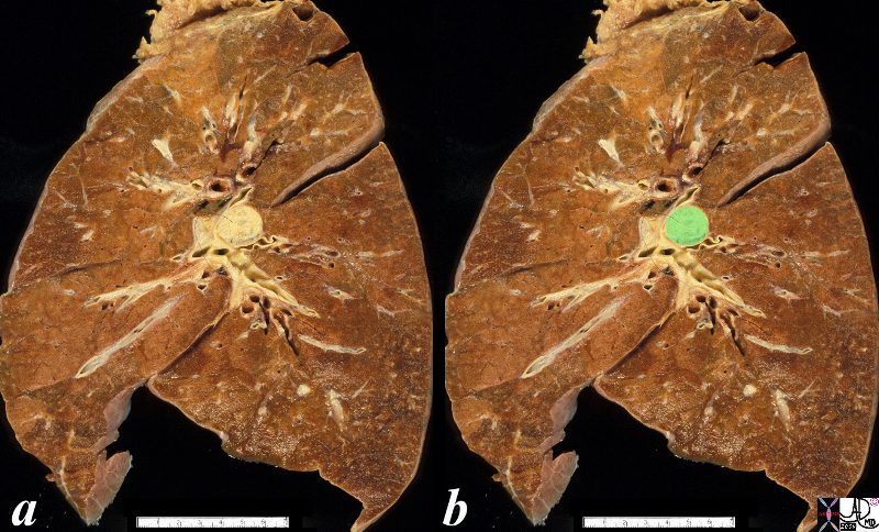
The anatomical specimen shows a 2cms central lesion near the hilum of the left lung causing extrinsic impression on the central bronchovascular bundle. The relatively small size and central location suggests a squamous cell carcinoma though a non small cell carcinoma is also possible
Ashley Davidoff
TheCommonVein.net
32323c02.8s
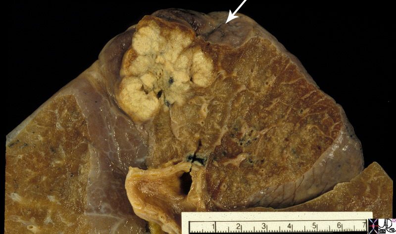
The 3.5cms carcinoma is peripherally situated and is sub-pleural in location. It reveals fibroblastic (aka desmoplastic) effect as witnessed by the puckering of the pleura (arrow). It is remote from central bronchovascular structures and therefore remains relatively asymptomatic for an extended duration unless it causes pleuritic pain. The location and desmoplastic puckering of the pleura are characteristic of an adenocarcinoma.
Ashley Davidoff
TheCommonVein.net
32201b.81s
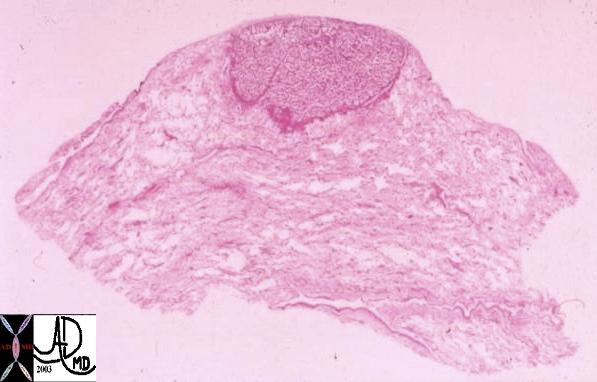
This whole mount of a part of the periphery of the lung reveals a peripherally located nodule without evidence puckering of overlying pleura. Microscopically this proved to be a papillary carcinoma.
Courtesy Armando Fraire MD. 32812

An axial and a sagittal view of a centrally placed carcinoma that lies on the major fissure of the right lung code lung mass fissure likely carcinoma central probability of it being a squamous cell carcinoma or a small carcinoma is high because of its central location.
keywords lung mass nodule major fissure cancer carcinoma central CTscan
Ashley Davidoff
TheCommonVein.net
75675c01.8s

The CT scan of the centrally positioned small cell carcinoma has structural implication as a result of its central location close to large arteries, veins, airways and lymphatics. In this case the centrally placed tumor (dark green in image b) is pushing on the right mainstem bronchus (shown with white arrow) and the lymphatics with peribronchial thickening (image d light green) and extension into the interlobular septa (bright green in d) Subcarinal nodal involvement and left hilar involvement (light green in b)together with small right effusion (a,b) are also noted suggesting advanced disease.
Ashley Davidoff
TheCommonVein.net
87711c01.8s
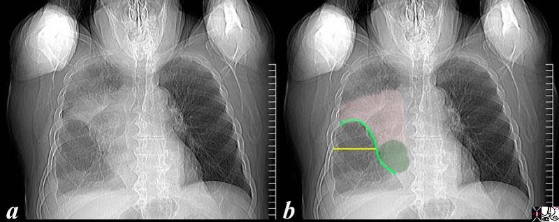
The scout film performed prior to a CT scan from a 76 year old man with chest pain and shortness of breath. The appearance suggests atelectasis of the right upper lobe with the normal position of the minor fissure (yellow) altered so that the upper portion (light green above the yellow line) is shifted upward caused by volume loss of atelectatic right upper lobe. (pink). The lower portion of the fissure (light green below the yellow line) is bulging rightward and outward caused by an implied mass (dark green). The “reversed S sign of Golden” is demonstrated in this case and infers a central mass causing obstruction and resulting in the shape described by the light green line of the minor fissure.
Ashley Davidoff
TheCommonVein.net
87740c02.8s

What is the Approach to this Patient?
The CTscan is from a middle aged male with lung carcinoma in the left upper lobe (b in green) characterized by spiculated border (c) extending to the pleura, and an ipsilateral A-P window node (overlaid in green in d) and associated with profound emaciation. code lung cancer malignant mass spiculated A-P window node aortopulmonary node emaciation CTscan 3D volume rendering
Ashley Davidoff
TheCommonVein.net
78088c038.8s