
40 or more pulmonary cell types have been identified making the lung by far the most heterogeneous of organs
Cells in the Conducting Airway
Cells of the Bronchioles
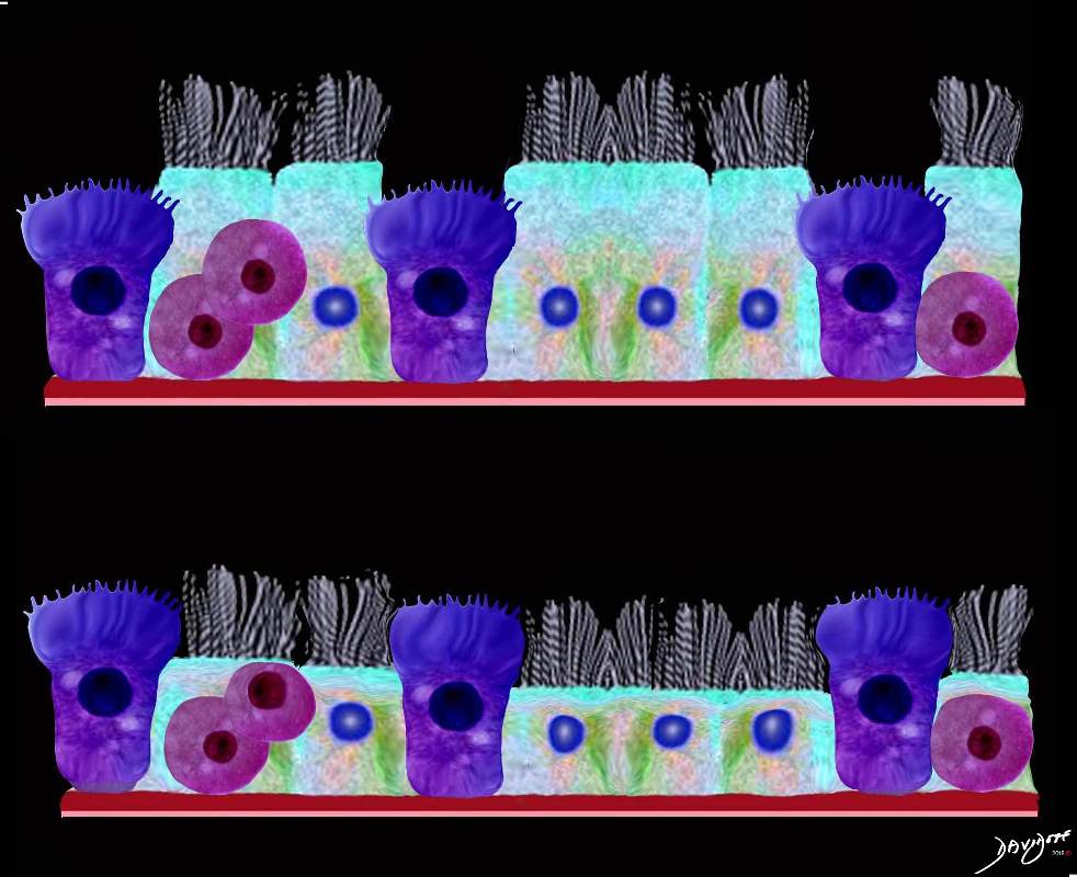
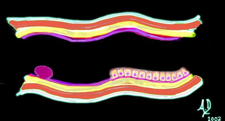
This upper diagram shows the ciliated columnar epithelium present throughout the 20- 25 generations of branching, until the airways start to transition their function from a transport system to a gas exchange system at the respiratory bronchiole level The ciliated columnar epithelium becomes a ciliated cuboidal epithelium. There are no goblet cells in the bronchioles In addition to the ciliated cells there are 2 other types of cells including the club cell (purple) and the neuroendocrine cell. The club cells Purple with dome shaped superior aspects – formerly Clara Cell) have many functions. The neuroendocrine cell (NE) (dark pink and round ) can be seen as a single cell (NE) and sometimes seen in a cluster, known as a neuroendocrine body (NEB) . The cells rest on a basement membrane, with prominent muscle layer (maroon) as well as elastic tissue (pink). There is no cartilage
Ashley Davidoff MD TheCommonVein.net
lungs-0741n
The Club Cell
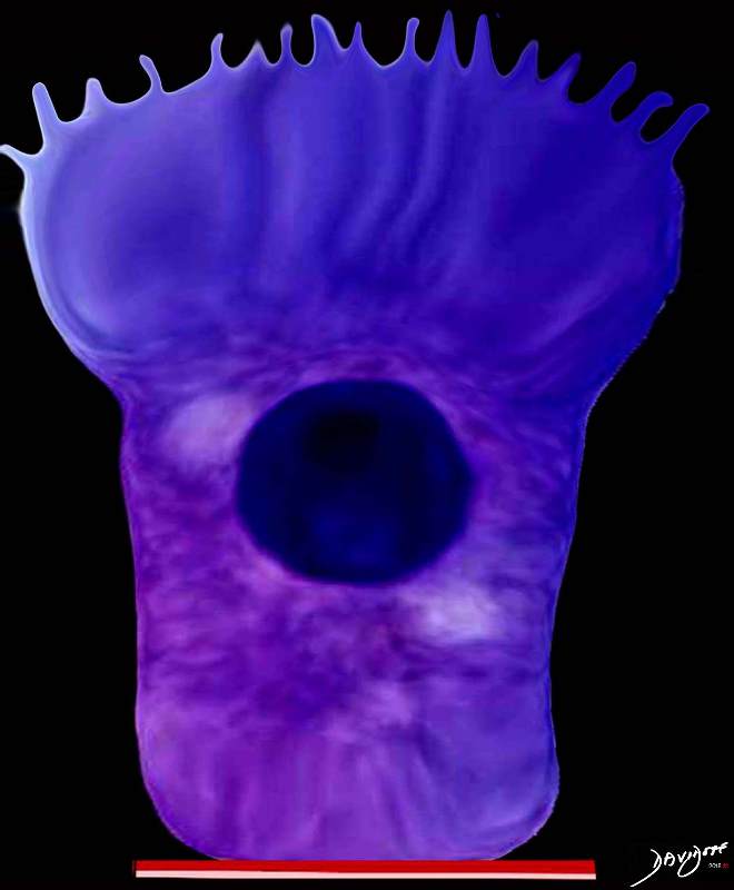
The club cell aka bronchiolar exocrine cells formerly known as the Clara cell is a low columnar cell with short microvilli and are most abundant in the bronchioles
Ashley Davidoff MD TheCommonVein.net lungs-0743
Cells in the Alveoli
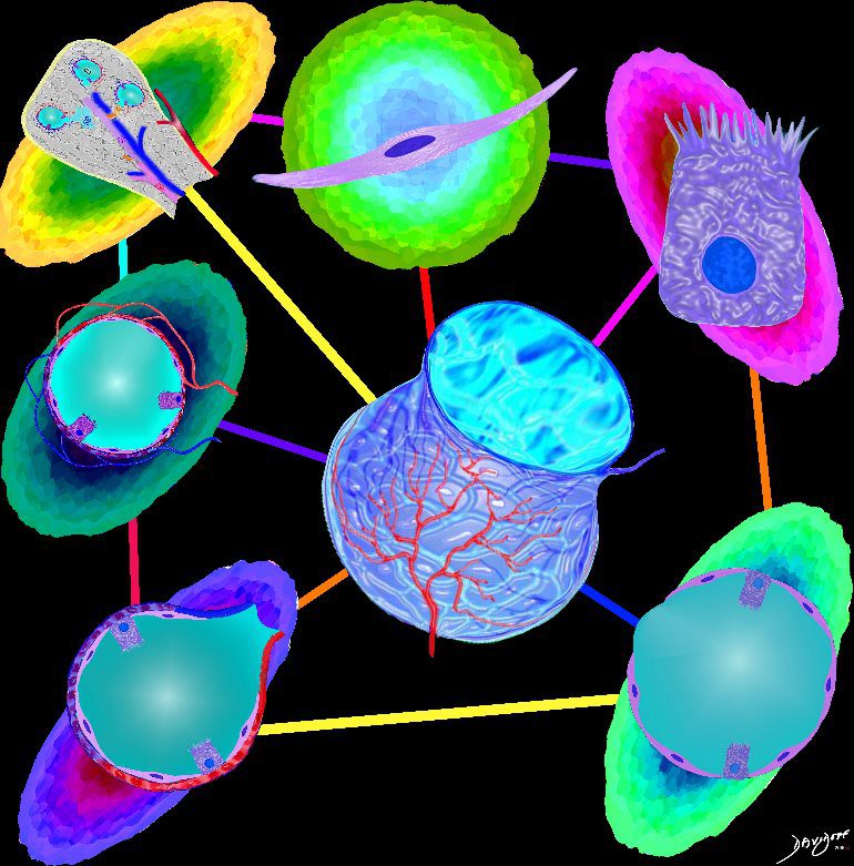
Parts and Bonds
Ashley Davidoff MD
Cellular Environment
In order to protect this thin surface from the chemical and biological environment, the immune mechanisms at the interface have to be quite sophisticated There are about 40 different cell types have been described in the lining tissues, bronchial tree and respiratory tissues
Ashley Davidoff MD
The Buck Ends Here
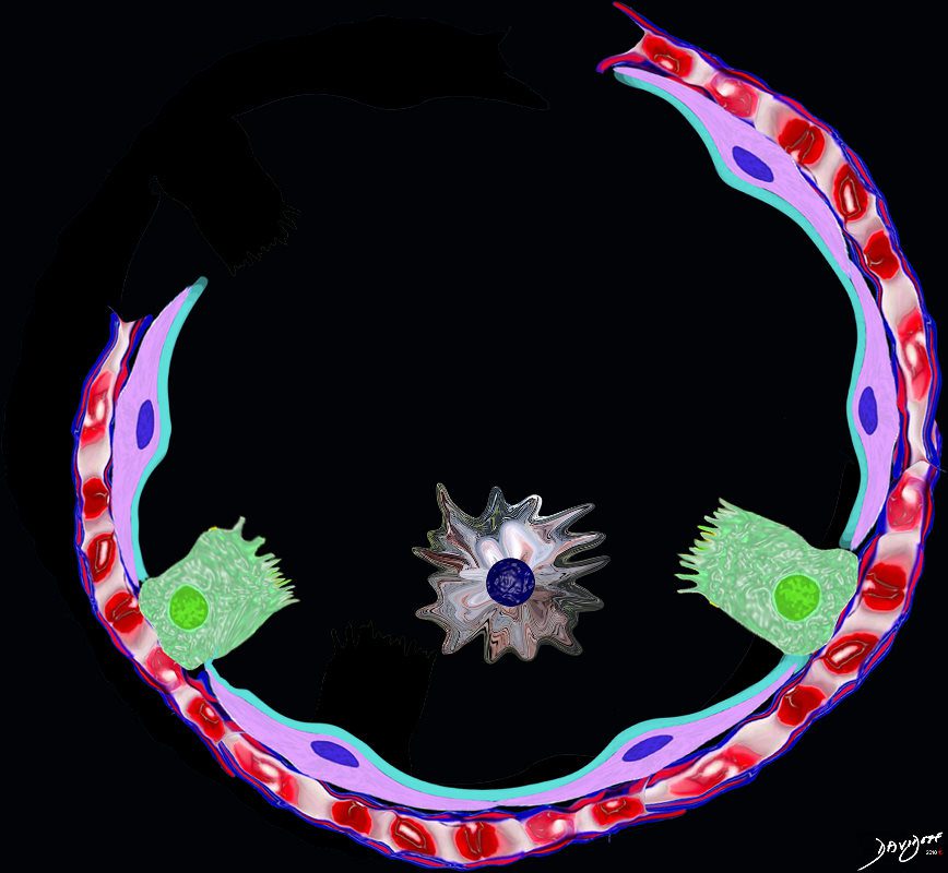
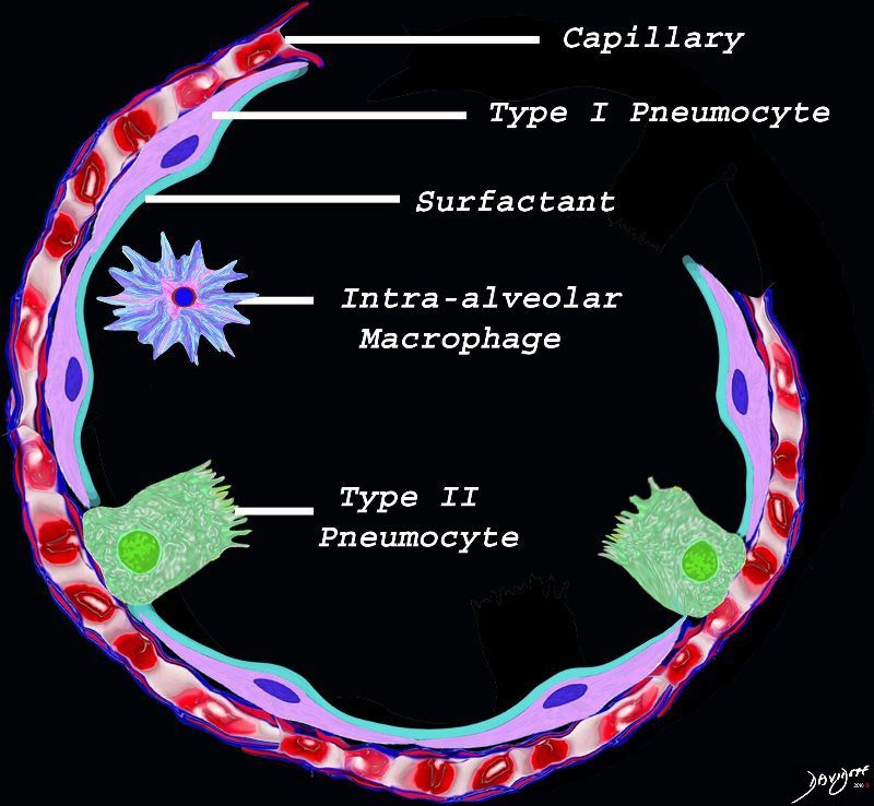
The alveolus is lined by a simple epithelium – one cell layer thick. There are two types of lining cells; Type 1 pneumocytes are squamous cells that cover 90% of the surface of the inner lining of the lung , and type II cuboidal pneumocytes that are in fact much more numerous than Type I. They are involved in the production of surfactant . In the lumen there are resident macrophages which play a crucial role in the immune system. The mucosa is grounded by a basement membrane and a lamina propria, and connected to the lamina propria and basement membrane of the surrounding capillary. The alveolus is lined by a thin layer of surfactant. (teal blue)
Ashley Davidoff
TheCommonVein.net
The diagram shows the lining of the normal alveolus composed of type 1 pneumocyte squamous in nature and the cuboidal cell (type pneumocyte) which rest on a lamina propria, and basement membrane (not shown) shared with the inner endothelial layer of the capillary. Intra-alveolar macrophage lies within the alveolar lumen
Ashley Davidoff
TheCommonVein.net
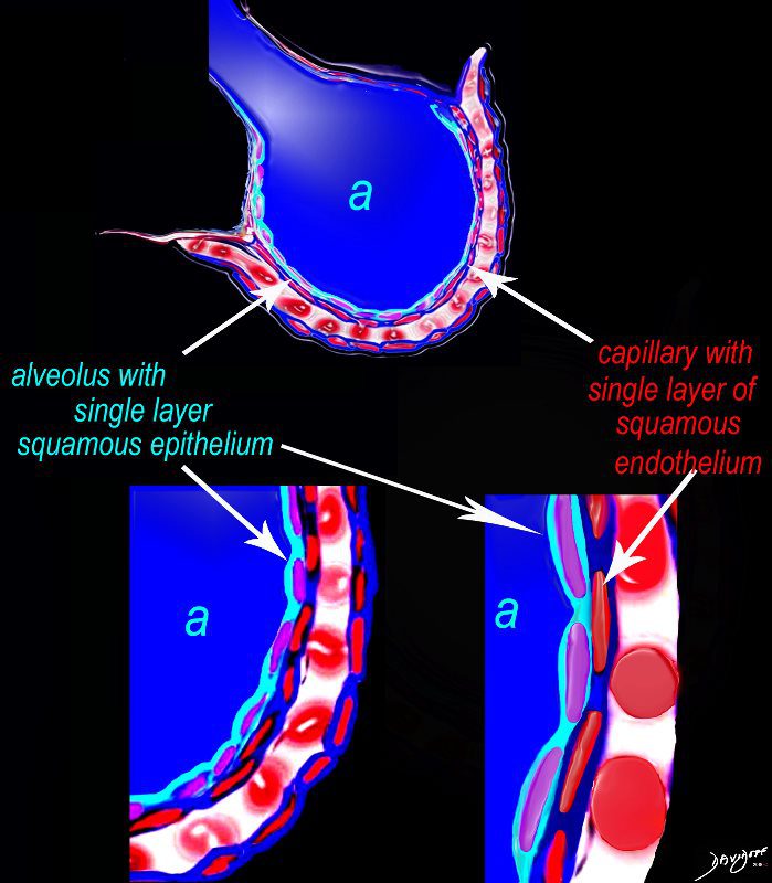
The diagram shows an alveolus (a) above, lined by a single layer of squamous cells, surrounded by a capillary with red cells which is also lined by a single layer of squamous endothelial cells . The images below show progressive magnification of the alveolar wall demonstrating the two thin layer of the alveolar membrane .
Courtesy Ashley Davidoff 2019
lungs-0028-low res
- At the level of the terminal respiratory units, the alveoli,
- relatively simple cellular composition
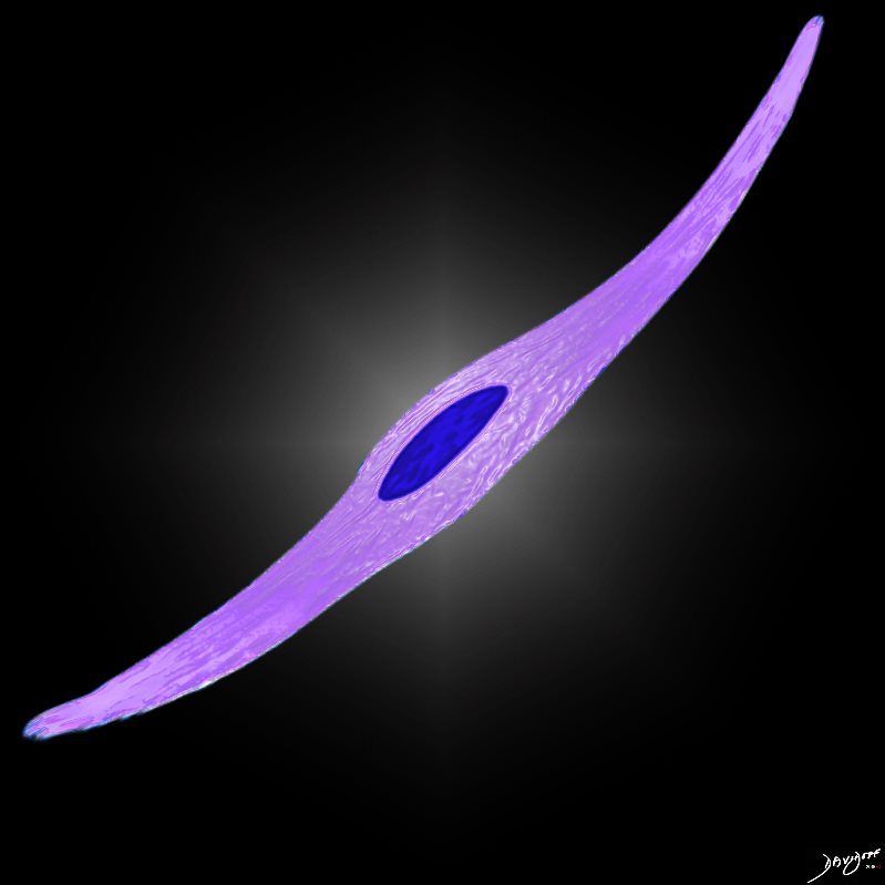
- Type I Alveolar Cell
- major lining cells which present an extremely large surface area to inhaled air and therefore any toxins ar
- covers some 90% of the gas exchange area
- very susceptible to injury
-

Simple squamous epithelium with pale staining cytoplasm
– flattened for gas exchange, forms a part of the Blood-Gas Barrier
Ashley Davidoff
TheCommonVein.net
- Type II Alveolar cell
- Cuboidal.
- very active metabolically
- are the source of the majority of the components of the surfactant , (together with non-ciliated bronchiolar (Clara) cells)
-
Simple cuboidal cell with reddish foamy and sometimes vacuolated cytoplasm
It produces the phospholipid – part of the surfactant that reduces surface tension and allows the alveoli to remain open
Ashley Davidoff TheCommonVein.net lungs-0062-low-resAlveolar Macrophages
First line of defense against infections of the lung.
Reside in alveolar walls, lymphatic channels and lymph node.
Originate in the bone marrow and are part of the Mononuclear Phagocytic System.
Ashley Davidoff
TheCommonVein.net
- Type I Alveolar Cell
-
Smoking and the Macrophage
-
Smokers Macrophage
Light brown granules in the macrophage is characteristic of the smokers macrophage
Ashley Davidoff
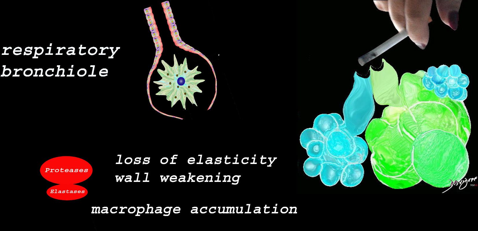
TheCommonVein.netMembranous airways (respiratory bronchiole, alveolar ducts, alveolar sacs)
At the level of the membranous airways the effect is predominantly related to the loss of elasticity, and aberrant accumulation of smoking related macrophages.
The weakening and destruction results in emphysema and the abnormal accumulation of smoking related macrophages relates to DIP
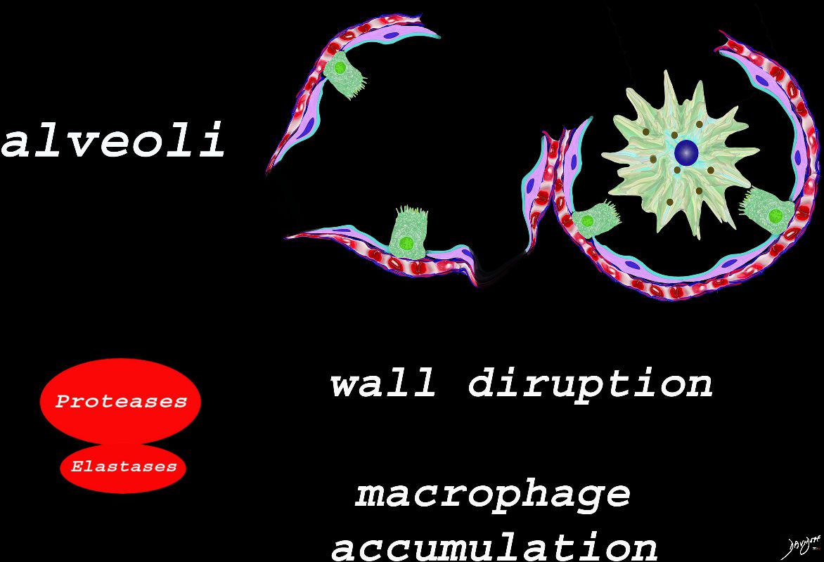
Ashley Davidoff TheCommonVein.netSmoking and the Alveolus –
The effect of the proteases and and elastases cause destruction of the alveoli and loss of elasticity, and therefore overall function. The destruction leads to bullous disease
The accumulation of smokers macrophage, and in the case of Langerhans cell histiocytosis leads to space occupation of the alveoli also reducing function
Ashley Davidoff TheCommonVein.net lungs-00687-lo resCancer
-
Mucosal Lesion
32347d01 key words mucosa submucosa muscularis adventitia serosa mucosal mass polyp neoplasm carcinoma acute angles with the lumen histopathology imaging diagnosis Ashley Davidoff TheCommonVein.netHistopathological Classification
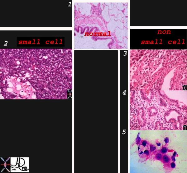
Classification system of carcinoma of the lung based on histological subtype. This collage of histopatholgy images of lung carcinoma shows part of a normal bronchiole and a few alveoli (1), followed by a small cell carcinoma (left lower 2) and non small cell carcinoma grouped together on the right side with squamous cell (3), adenocarcinoma with glandular formation (4), and undifferentiated large cell derived from pleural aspirate (5). 1-4
Courtesy Dr Armando Fraire MD, and image 6 courtesy of tumorboard.com 32501cL02Squamous Cell Carcinoma of Lung.
Cell Block of aspirate biopsy. Note glassy cytoplasm, nucleoli, and intercytoplasmic spaces with bridges.
Courtesy tumorboard.com 54405-
Adenocarcinoma of the Lung. Tumor nest in a lymphatic vessel.
Courtesy tumorboard.com
54411 Keywords
lung pulmonary lymphatics neoplasm malignant malignancy primary lung adenocarcinoma histopathology
Adenocarcinoma with Lepidic Growth – Histopathology
Histological sections of a group of alveoli filled with nests of malignant cells originating from and spreading along the alveolar epithelium without destroying the alveolar walls.(dark pink overlay) The cells seem to be almost hanging from the alveolar epithelium like washing from a washing line. Note groups of tall, columnar, mucin producing cells spreading along preexisting alveolar walls, which is typical of bronchioalveolar carcinoma.
Courtesy Dr Armando Fraire MD TheCommonVein.net
32828c08.8sDiffuse mucinous bronchioloalveolar carcinoma in a 78-year-old man. (a) High-resolution CT scan shows a bilateral crazy-paving pattern and centrilobular nodules. (b) Photomicrograph (original magnification, 400; hematoxylin-eosin stain) of a specimen from open lung biopsy shows replacement of the alveolar epithelium
by epithelial neoplastic cells with abundant intracytoplasmic mucin (arrows).
Rossi, S.E et al “Crazy-Paving” Pattern at Thin-Section CT of
the Lungs: RadiologicPathologic Overview Radiographics Volume 23 – Number 6, 2003 -
-
Links and References
Scott, J.E The Pulmonary Surfactant: Impact of Tobacco Smoke and Related Compounds on Surfactant and Lung DevelopmentTob Induc Dis. 2004; 2(1): 3–25.
- relatively simple cellular composition


 1
1