
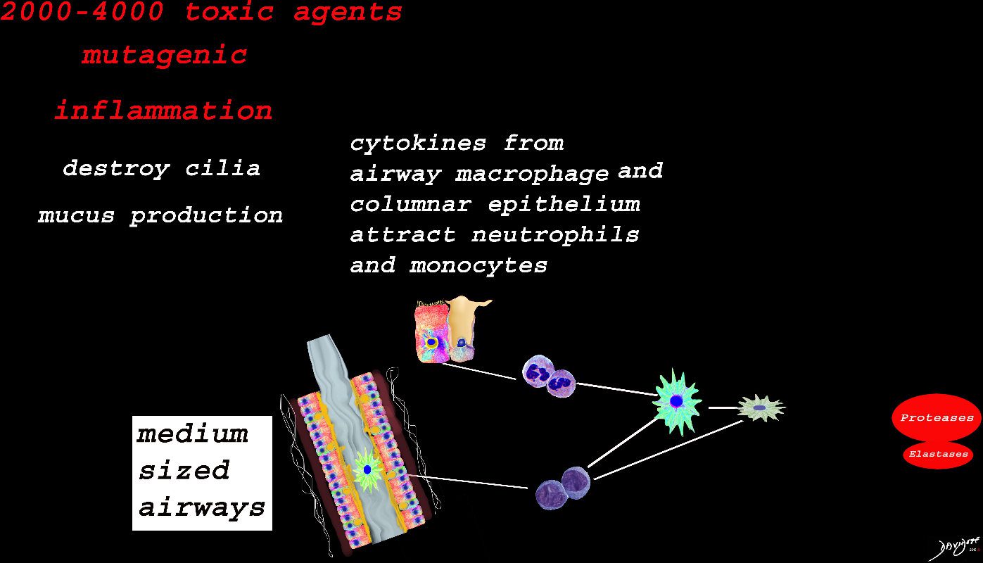
Inhaled smoke contains between 2000-4000 toxic substances. The cancer producing properties are well known. The smoke causes inflammation of the medium sized airways resulting in activation of the resident macrophage in the lumen of the airway, hyperemia of the mucosa, activation of the neutrophils, monocytes, nonresident macrophages, and fibroblasts. Cytokines are released which also induce the secretion of proteases and elastases among many others. Structurally there is destruction of the cilia, hypersecretion of mucus, hypertrophy of the muscularis and mild fibrosis. These findings are the hallmark of chronic bronchitis
Ashley Davidoff
TheCommonVein.net
The alveolus is lined by a simple epithelium – one cell layer thick. There are two types of lining cells; Type 1 pneumocytes are squamous cells that cover 90% of the surface of the inner lining of the lung , and type II cuboidal pneumocytes that are in fact much more numerous than Type I. They are involved in the production of surfactant . In the lumen there are resident macrophages which play a crucial role in the immune system. The mucosa is grounded by a basement membrane and a lamina propria, and connected to the lamina propria and basement membrane of the surrounding capillary. The alveolus is lined by a thin layer of surfactant. (teal blue)
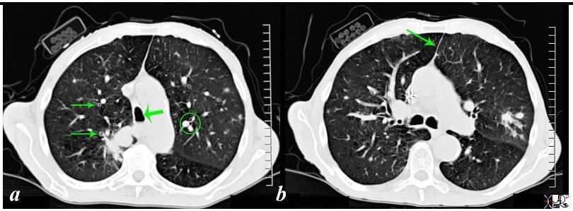
87831c02b02b.8s Among many of the associated and relevant findings in the include the thickened bronchi (arrows on the right in a) compared to the more normal appearing bronchi ringed in the left lung in a, a finding suggesting chronic bronchitis. Noted also is the unusual shape of the trachea (large arrow head in a) where the A-P dimension is significantly larger than the transverse diameter (termed saber trachea) together with a suggestion of an air fluid level in the dependent part of the trachea in a. The air fluid level suggests mucus or artifact in the trachea. The saber trachea together with the thin anterior junction line noted in b is suggestive of hyperinflated lungs that is hallmarks of emphysema. Other findings to note is the discrepancy in size of the arteries that travel with the bronchi – (ringed in a on the left), which is a sign seen in pulmonary congestion. Lastly the lack of subcutaneous tissue suggests a thin emaciated male. code lung cancer smoking anterior junction line bronchi thickened cigarettes shirt pocket mass emaciated saber trachea chronic bronchitis emphysema BAC bronchoalveolar carcinoma bronchioloalveolar carcinoma ground glass opacities GGO radiologists and detectives emaciated CT scan muscle wasting Sherlock Holmes alveolar infiltrate subsegmental
Ashley Davidoff
TheCommonVein.net
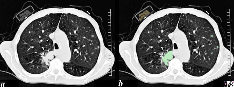
The CT scan through the chest of an 80 year old male shows a large lung mass in the posterior aspect of the right upper lobe (overlaid in green) and3 small nodules in the left upper lobe (overlaid in green). The patient is obviously a smoker and the incriminating pack of cigarettes is identified in his right shirt pocket containing 9 cigarettes. The lung cancer was shown to be an adenocarcinoma The pathology of the nodules may either represent metastatic disease or multicentric foci of bronchioloalveolar cell carcinoma. Associated finding of a thinned anterior junction line suggests hyperinflation and emphysema, and the thickened bronchial walls noted in the right lung suggest chronic bronchitis. Saber shaped trachea is also reminiscent of emphysema. The patient is emaciated, a finding that relates both to his chronic lung disease and his cancer.
Ashley Davidoff MD 87831c01b.8s
TheCommonVein.net

There is thickening of the medium sized airways and mucus in the lumen
Ashley Davidoff
TheCommonVein.net