
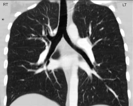
 This coronally reformatted CT scan shows the characteristic short and fat right mainstem that arises at a relatively acute angle off the trachea, such that an aspirated peanut will favor it over the left because of this angle. The left is long and thinner. Courtesy of: Ashley Davidoff, M.D.
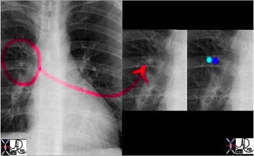 In this normal CXR a RUL segmental bronchus and artery are side by side with the lucent air filled bronchus in teal and the artery in royal blue. Note that at this stage they are the same size and they will be for many divisions until they reach the terminal bronchiole.
Courtesy of: Ashley Davidoff, M.D.
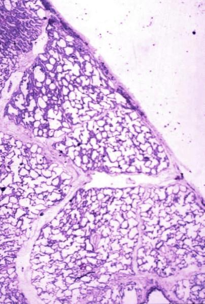
Normal lung histology
This image is a panoramic view of the lung showing secondary lobules and interlobular septa. Within the interalveolar septae, one sees small venules and lymphatics.Courtesy Armando Fraire MD. 32649b
code lung pulmonary alveoli alveolus secondary lobule interlobular septa vein lymphatic histology
interstitium interstitial
32649b
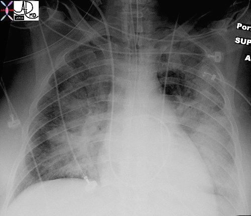
In this patient with acute congestive cardiac failure the consolidation that has hilar distribution has reminded radiologists of bat wings and is caused by alveolar edema. As a result of the fluid in the alveoli, gas exchange across the respiratory membrane is reduced and required intubation to improve the gas exchange process. Note the endotracheal tube as well as the central venous line that is used to assess the heart pressure and monitor the congestion.
Ashley Davidoff MDTheCommonVein.net 42073b01
 Heart Failure Kerley B lines Heart Failure Kerley B lines |
|
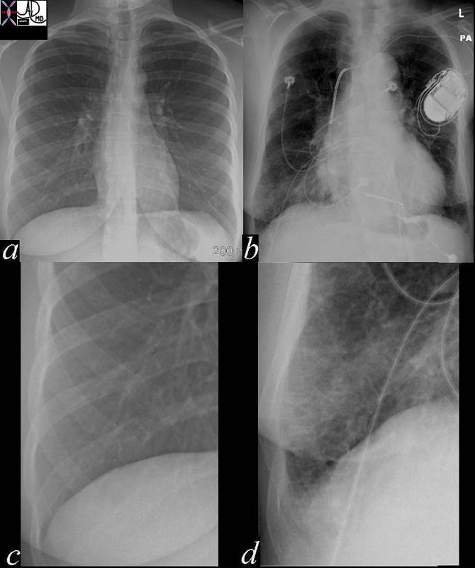
In these images. a nd c are normal and b and d represent thickened interlobular septa in a patient with congestive heart failure. These are the well known Kerley lines, often spoken about but rarely seen. They are identifiedas thin horizontal lines usually seen in the costophrenic angles, not being longer than 2cms in length and touching the pleural surface. 42545c01.800 Ashley Davidoff MD |