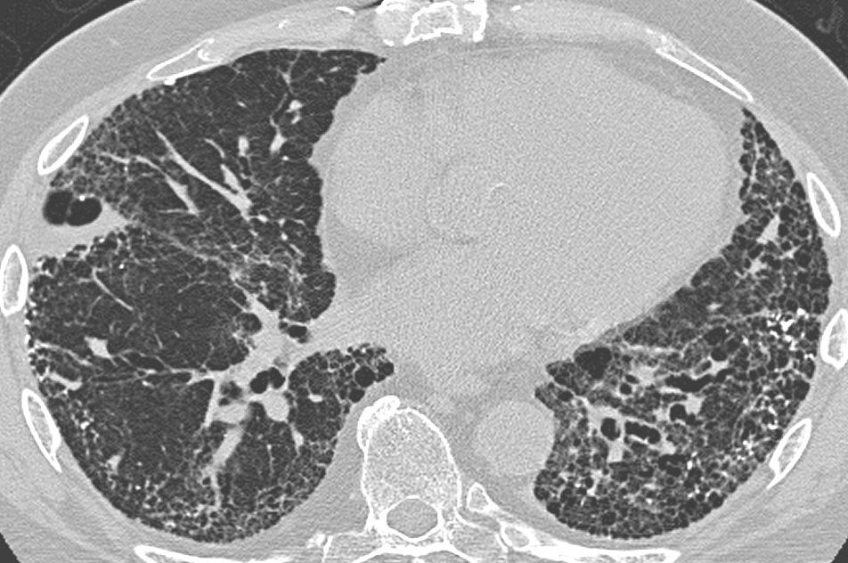
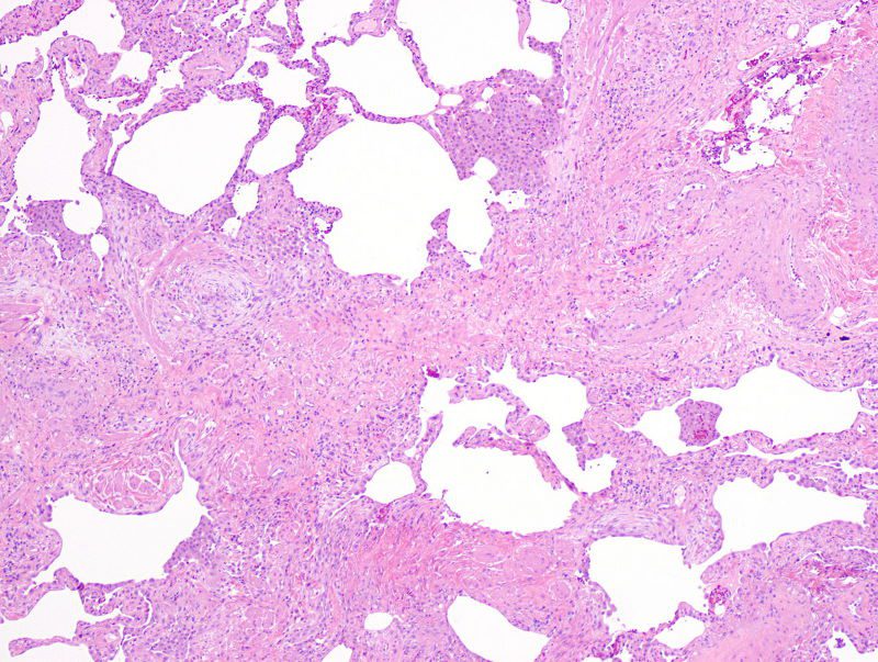
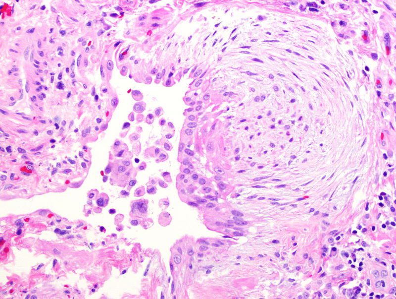
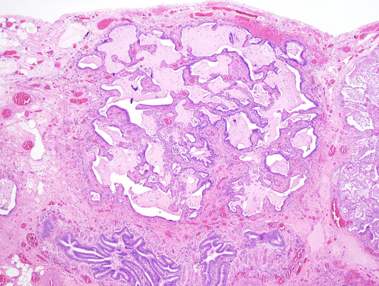
CT scan in usual interstitial pneumonia (UIP). There is interstitial thickening, architectural distortion, honeycombing and bronchiectasis. Darel Heitkamp, MDUsual interstitial pneumonia seen on CT scan. Honeycomb fibrosis is seen at the bases of both lungs. Yale RosenAppearance of usual interstitial pneumonia (UIP) in a surgical lung biopsy at low magnification. The tissue is stained with hematoxylin (purple dye) and eosin (pink dye) to make it visible. The pink areas in this picture represent lung fibrosis (collagen stains pink). Note the “patchwork” (quilt-like) pattern of the fibrosis.A fibroblast focus in a surgical lung biopsy of UIP. Hematoxylin-eosin stain, high magnification. The white space to the left is an airspace. The pale area to the right is a fibroblast focus. It is an area of active fibroblast proliferation within the interstitium of the lung.Appearance of honeycomb change in a surgical lung biopsy at low magnification. The dilated spaces seen here are filled with mucin. Hematoxylin-eosin stain, low magnification.