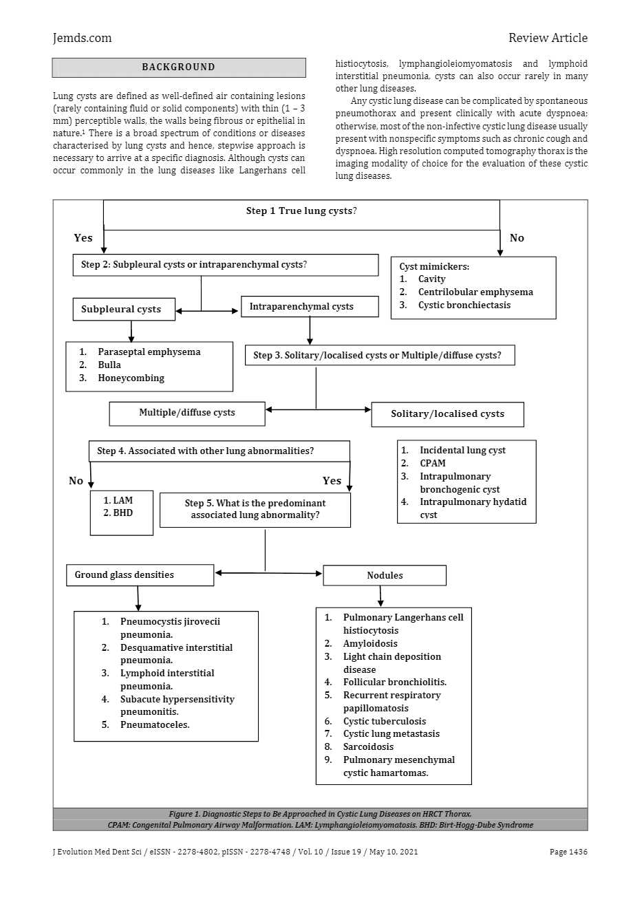
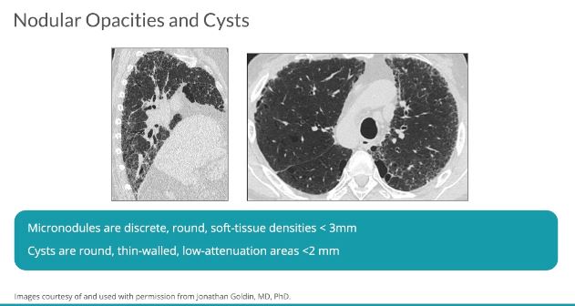
- Cyst is a
- nonspecific term for a
- thin-walled (usually <3 mm),
- well-defined and circumscribed,
- air containing lesion
- 1 cm or more in diameter.
- heterogenous group of disorders characterised by
- multiple air-filled lucencies
- surrounded by discrete walls.
- heterogenous group of disorders characterised by
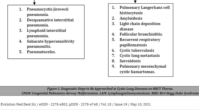
- Diseases
- Lymphangioleiomyomatosis (LAM),
- Langerhans cell histiocytosis (PLCH),
- Birt-Hogg-Dubé syndrome (BHD), and
- Lymphoid interstitial pneumonia (LIP),
- Thin walled cysts
- No central core structures
- Differential Diagnosis
-
- Lymphangioleiomyomatosis (LAM),
-

LAM
Ashley Davidoff MD TheCommonvein.net
-
- Langerhans cell histiocytosis (PLCH),
-
Pulmonary Langerhans Cell Histiocytosis
Thiin Walled Bizarre Shaped Upper Lobe Cysts
Ashley Davidoff MD TheCommonVein.net- Langerhans Cell Histiocytosis
CT scan shows multiple small cysts sometimes irregular in shape predominantly in the upper lobes. The cysts are round and air filled large and are between 5mm-8mm
- Langerhans Cell Histiocytosis
- Birt-Hogg-Dubé syndrome (BHD), and
- Lymphoid interstitial pneumonia (LIP), although the differential diagnosis can at times expand to encompass other extremely rare etiologies
- Amyloidosis
- Light Chain Deposition Disease
- Neurofibromatosis
- Lymphangioleiomyomatosis (LAM),
- Less Common
- Sjögren syndrome
- light chain deposition disease
- Ehlers Danlos syndrome type IV
- fire-eater’s lung (pneumatoceles)
- lymphomatoid granulomatosis
- neurofibromatosis
- congenital pulmonary airway (cystic adenomatoid) malformation,
- smoking related small airways injury [
- Proteus syndrom
- Look Alikes
- Emphysema
- No wall
- Central soft tissue elements
- Blebs
- Bullae
- >1 cm in diameter, bounded by a
- thin wall (<1 mm)
- background emphysema
- Bronchiectasis
- continuity with airways
- Honeycombing
- enlarged airspaces
- .3 -1.0cms (up to 2.5cms)
- thick fibrous walls
- End stage fibrotic disease
- subpleural
- stacked
- enlarged airspaces
- Pneumatoceles
- thin-walled parenchymal cyst
- Infection
- bacterial pneumonia eg children
- PCP Pneumocystis jirovecii,
- coccidioidomycosis,
- hyperimmunoglobulinemia E syndrome,
- staphylococcal pneumonia
- Chest Trauma
- Emphysema
- Look Alikes
- LAM
-
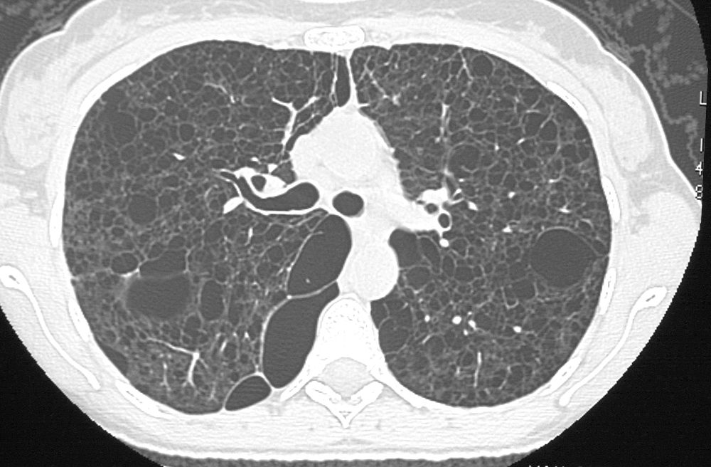
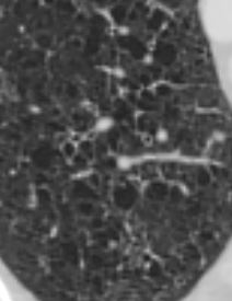
LAM and LYMPHANGIOLEIOMYOMAS in the ABDOMEN
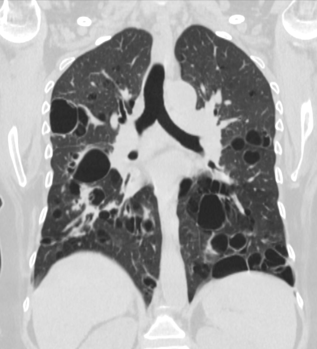
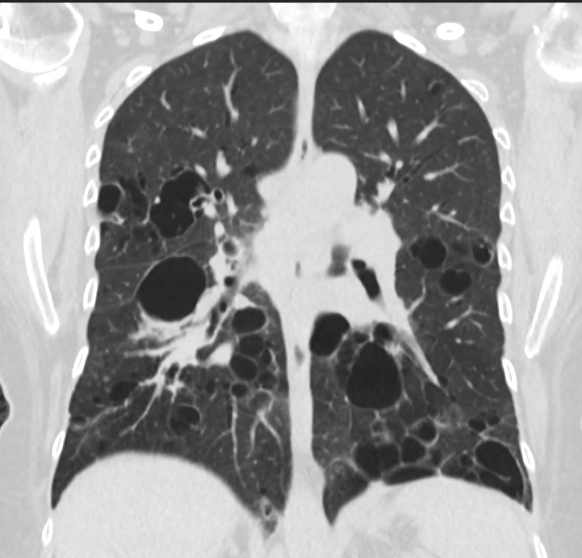
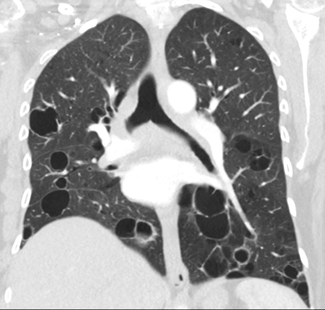
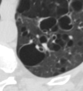
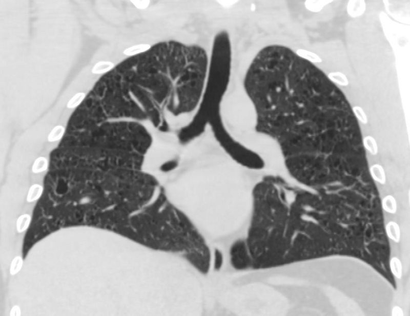
38-year-old patient with progressive dyspnea and cough
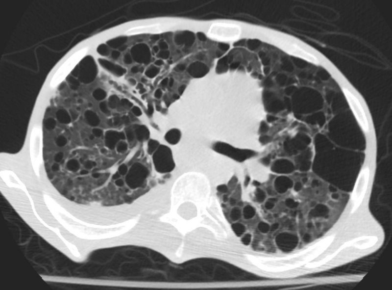
CXR (scout for CT) shows hyperinflated lungs with increased lung volumes with bilateral and extensive thin-walled cysts surrounded by very little normal lung parenchyma. The cysts are round and thin-walled except for air filled large irregular pocket in the right apex (image 27628/29) . Some of the cysts do not have walls at all and others have an irregular configuration.
In the abdomen multiple low density lymphangioleiomyomas are present that are due to lymphatic obstruction.
Ashley Davidoff MD TheCommonVein.net
-
Upper
- Infections
- PCP
-
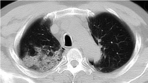
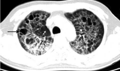
Chest CT of lung cysts dominated PCP in an AIDS patient. Numerous thin walled lung cysts in upper lobe of both lungs. The cysts have various shapes and sizes. In some cysts there are interior separations (arrow). Infiltration lesions are seen around some of the cysts
Lu, Pu-Xuan, et al Correlation between imaging features of Pneumocystis Jiroveci Pneumonitis (PCP), CD4+ T lymphocyte count, and plasma HIV viral load: A study in 50 consecutive AIDS patients Quantitative Imaging in Medicine and Surgery 2(2):124-9, June 2012Chest CT of a condominated PCP in an AIDS patient. Patchy shadows are shown in the upper lobe of right lung
Lu, Pu-Xuan, et al Correlation between imaging features of Pneumocystis Jiroveci Pneumonitis (PCP), CD4+ T lymphocyte count, and plasma HIV viral load: A study in 50 consecutive AIDS patients Quantitative Imaging in Medicine and Surgery 2(2):124-9, June 2012Complication of PCP Lung Cysts
- PTX,
- incidence 35% in patients with cysts
- frequently bilateral
- often refractory to chest tube Rx,
- frequently need surgery eg pleurodesis
- associated higher mortality rate,
- especially in patients on ventilation.
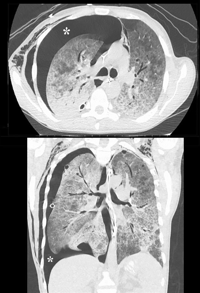
Pneumocystis carinii pneumonia. CT scans in a 32-year-old man with acquired immunodeficiency syndrome and a CD4 count of 7cells per microliter who presented with respiratory arrest. (a) Axial and (b) coronal images in lung windows demonstrate a moderate right pneumothorax (*) and widespread ground-glass and airspace opacities.
Parekh, M et al Review of the Chest CT Differential Diagnosis of Ground-Glass Opacities in the COVID Era Radiology Vol. 297, No. 3 July 2020 - PTX,
- ILD
- DIP
-
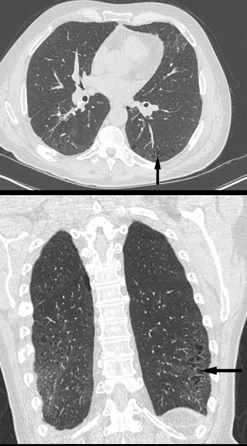
Desquamative interstitial pneumonitis. (a) Axial and (b) coronal lung window CT scans in a 64-year-old man with a history of smoking (17 pack-years) and shortness of breath. Images show small cysts in both lower lobes (arrow) with surrounding interstitial prominence.
Parekh, M et al Review of the Chest CT Differential Diagnosis of Ground-Glass Opacities in the COVID Era Radiology Vol. 297, No. 3 July 2020LIP
- Lower Lobe predominance
-
Stable Cystic Changes
47 F SLE Sjogrens LIP vs Birt-Hogg-Dube basilar thin walled cysts lymphadenopathy
Subsegmental right lower lobe infiltrate
Ashley Davidoff TheCommonVein.net47 F SLE Sjogrens LIP vs Birt-Hogg-Dube basilar thin walled cysts lymphadenopathy
Subsegmental right lower lobe infiltrate
Ashley Davidoff TheCommonVein.net47 F SLE Sjogrens LIP vs Birt-Hogg-Dube basilar thin walled cysts lymphadenopathy
Ashley Davidoff TheCommonVein.netCysts Associated with Blood Vessels in LLL
47 F SLE Sjogrens LIP vs Birt-Hogg-Dube basilar thin walled cysts lymphadenopathy
Subsegmental right lower lobe infiltrate
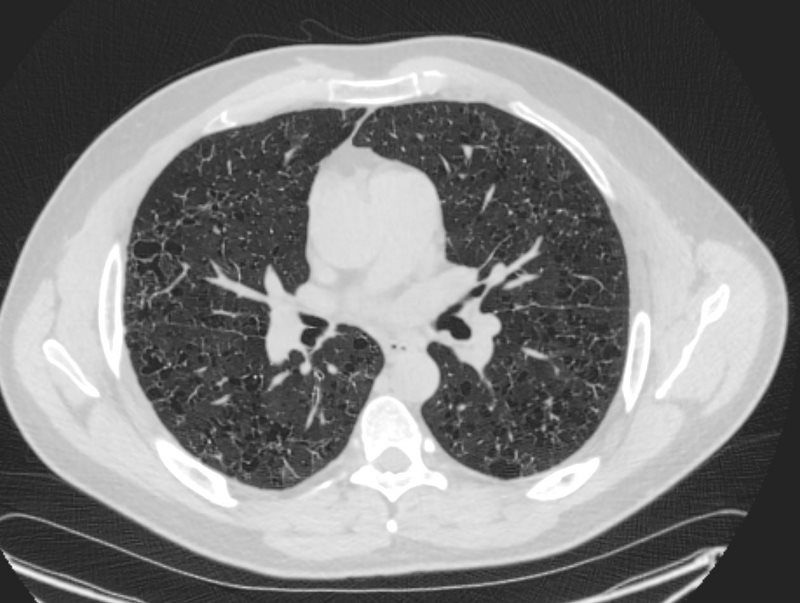
Ashley Davidoff TheCommonVein.net- LIP Lymphoid Interstitial Pneumonia
-
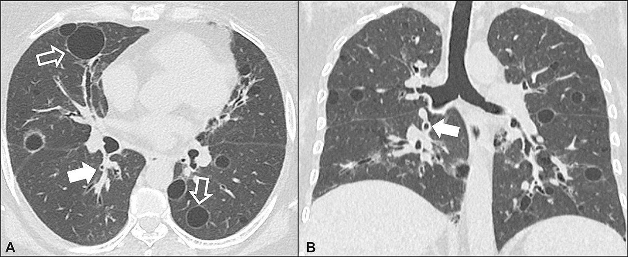
CT scan of lymphocytic interstitial pneumonia, with pulmonary cysts.
Lymphocytic interstitial pneumonia. A 62-year-old female patient with Sjögren’s syndrome. Axial high-resolution computed tomography scan of the chest (A) and coronal reformatting (B). In A, diffuse thickening of the bronchial walls (closed arrows), some ground-glass opacities and thin-walled cysts of varying sizes, with a diffuse, bilateral distribution (open arrows). In B, distribution predominantly in the lower fields.
Daniel Simões Oliveira et al
Radiologia Brasileira 51 (5): 321–327. - 017Lu 27F LIP HIV AIDS Lymphoma
028Lu LIP Emphysema
-
J Evolution Med Dent Sci / eISSN – 2278-4802, pISSN – 2278-4748 / Vol. 10 / Issue 19 / May 10, 2021 J Evolution Med Dent Sci / eISSN – 2278-4802, pISSN – 2278-4748 / Vol. 10 / Issue 19 / May 10, 2021 J Evolution Med Dent Sci / eISSN – 2278-4802, pISSN – 2278-4748 / Vol. 10 / Issue 19 / May 10, 2021 J Evolution Med Dent Sci / eISSN – 2278-4802, pISSN – 2278-4748 / Vol. 10 / Issue 19 / May 10, 2021 - LIP Lymphoid Interstitial Pneumonia