
The Lungs
Copyright 2009
Ashley Davidoff MD
Also Go to
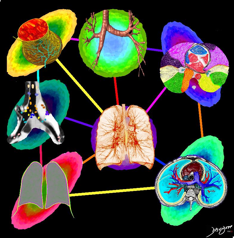
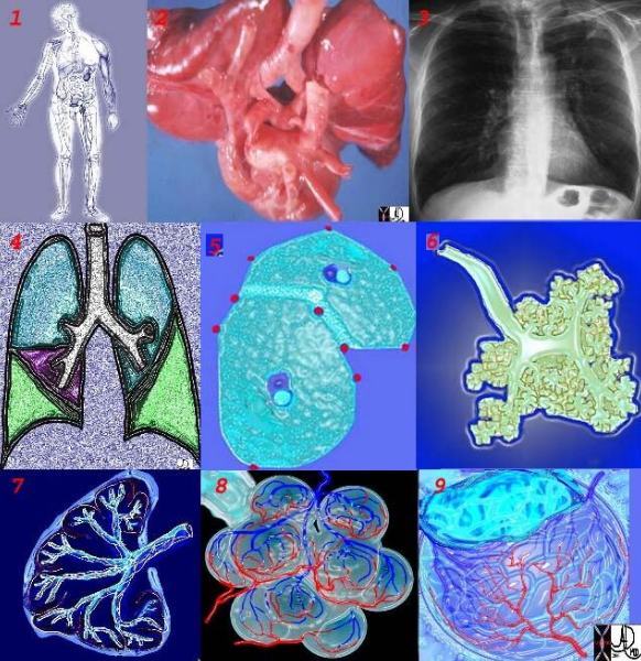
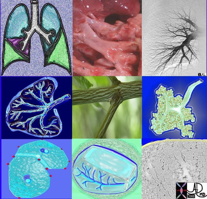
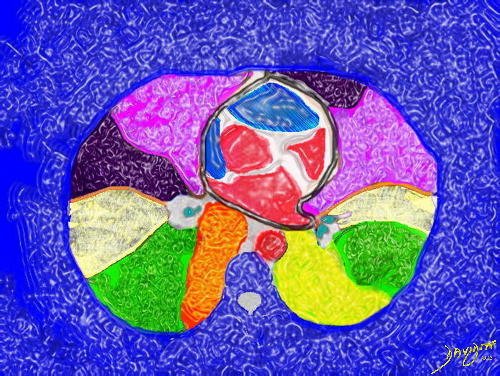
The image shows some of the major components of the lung that when bonded create a new and powerful unit – a vital organ. In the center is an example of the airways and parenchyma making up the 2 lungs. At 12 oclock the tracheo-bronchial tree with segmental and subsegmental airways. At 1 o’cloclock, is a cross section of the lungs showing some of the segments of the lung. At 5o’clock a cross section shows the arteries and veins of the lungs. At 7o’clock the drawing shows the pleura and pleural space of the lungs. At 9o’clock, a coronal reformat of the tracheobronchial tree shows the lymph node stations of the lungs. At 11 o’clock is the golden alveolus, the epicentral unit where gas exchange takes place
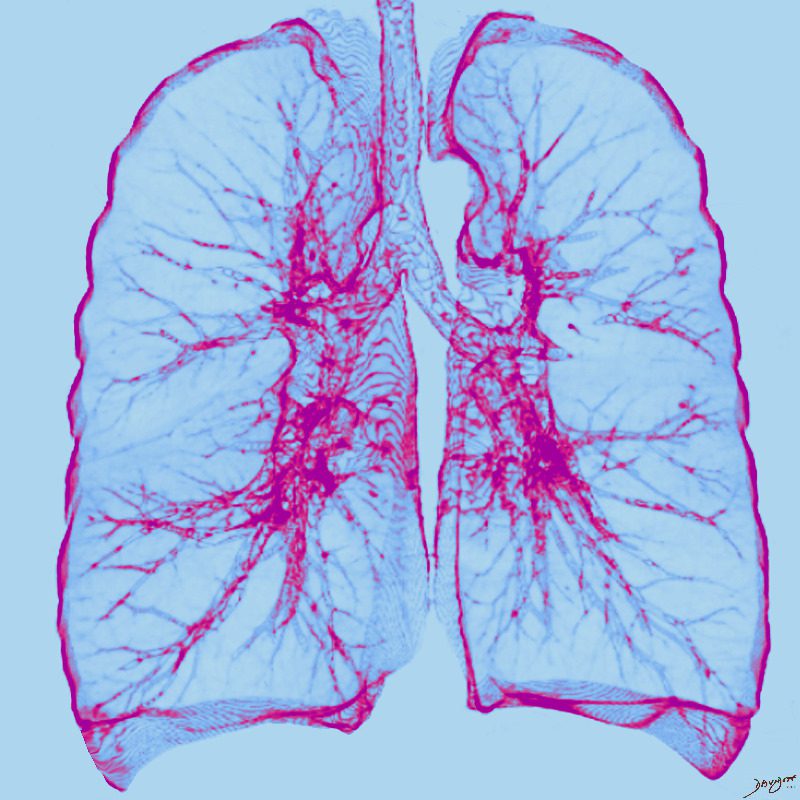
Ashley Davidoff MD TheCommonVein.net lungs-0696-lo res
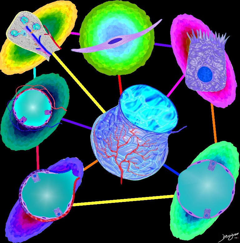 Alveolus
Parts and Bonds
Ashley Davidoff MD
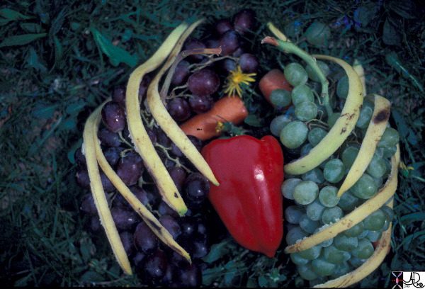
This artistic rendition of the heart and lungs uses the shape of fruit and vegetables to create an image of the chest. The lungs are made of grapes, the pulmonary arteries are made of carrots, the ribs are made of banana peel and the heat is made of a red pepper. 02032p Ashley Davidoff MD 02032p
TTagsheCommonVein.net
by Ashley Davidoff MD TheCommonVein.net
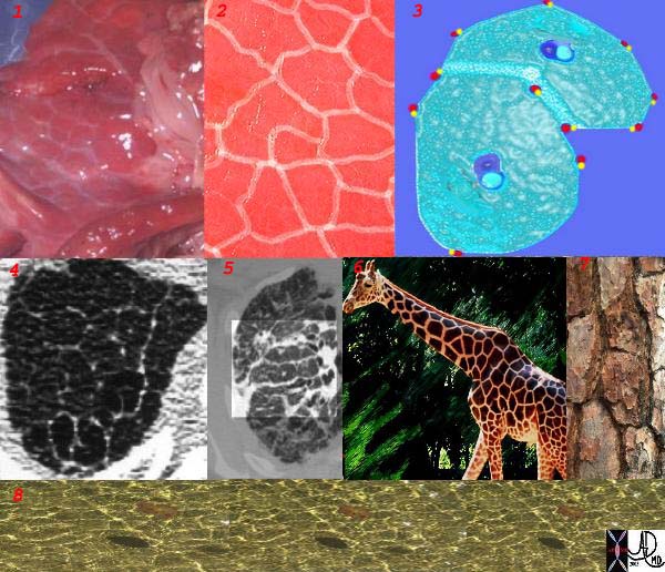
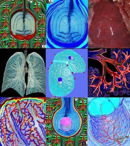
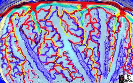
This is a series of images demonstrating the shape of the secondary lobule. The first image (1) is a post mortem specimen with congested lungs showing the interlobular septa, while the next (2), is an overlay of the septa in white showing their polygonal shape. The next drawing reveals side-by-side secondary lobules with central bronchovascular bundles and peripheral lympho-vascular bundles. Image 4 is a CT image through the apex of the lung showing thickened secondary lobules in a patient with mild emphysema, and 5 shows marked thickening of the interlobular septa in a patient with end stage sarcoidosis. 6,7,8 show the shape of the secondary lobules in the skin of a giraffe, the bark of a pine, and the ripples of the water respectively.
Ashley Davidoff MD TheCommonVein.net 31866collage
42651c
keywords lung chest
Ashley Davidoff TheCommonVein.net
82738p chest lung connective tissue pulmonary artery pulmonary vein axial interstitial tissue secondary lobule lobes segments trachea bronchi interlobular septa polygonal Ashley Davidoff MD
TheCommonVein.net
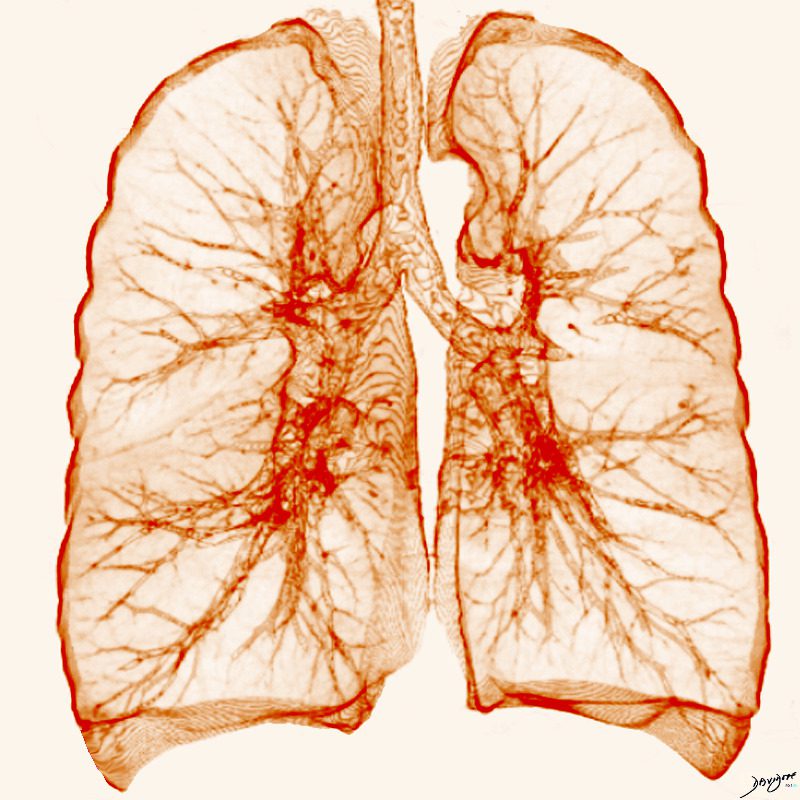
Ashley Davidoff MD TheCommonVein.net lungs-0011

Ashley Davidoff MD TheCommonVein.net lungs-0701
Ashley Davidoff MD TheCommonVein.net lungs-0702
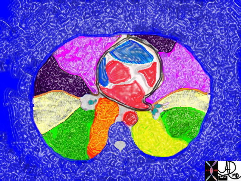
The five major layers that keep the air moving include the outer bony cage, the muscular layer represented in maroon, the pleural complex (orange yellow orange) the lung (blue) and surfactant within the alveolus. (pink)
42530b05b09b01a08
Ashley Davidoff MD
TheCommonVein.net
The chest is surrounded by a ring of muscle (maroon) made up of a various groups which work in concert. The diaphragm is the workhorse of the respiratory muscles and is shown as a thick maroon band inferiorly. 42530b05b09b14 Ashley Davidoff MD
TheCommonVein.net
Ashley Davidoff MD TheCommonVein.net lungs-0024


This cupola or dome was photographed in the church of the Villa Melzi gardens in Bellagio, Italy. If you imagine yourself in the chest cavity and you look up towards the neck, this is what you will see – the dome shaped structure of the apex of the lung and pleura.
Ashley Davidoff TheCommonVein.net 78115pb01
Ashley Davidoff TheCommonVein.net . 22071b01.800
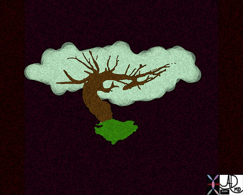
Two leaves of the coleus plant, with a pyramidal or conical shape that reminded the photographer of a set of lungs. The branching system originates from the hilum of the leaf almost at its center, but unlike the tracheobronchial tree it is not irregularly dichotomous.
Ashley Davidoff TheCommonVein.net . 42643
The chest quietly expands and contracts under basal conditions in order to serve the alveoli. At first glance it seems like a simple bellows-like process, but as one delves into the layers of detail, the complexity of the structural design unfolds as a combination of physical and chemical forces.
Ashley Davidoff MD TheCommonVein.net 42530b05b09b28
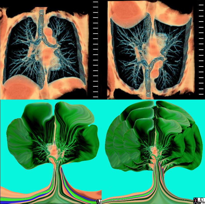
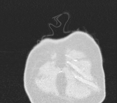
The axial CT through the level of the heart shows a few of the right and left pulmonary segments including parts of the middle lobe, lingula and of the lung bases
Ashley Davidoff MD TheCommonVein.net 32557bb03.8s

Tracheobronchial Tree
Tree, flower, tracheobronchial tree, trachea bronchi lung
Ashley Davidoff Art 32620b14.800b02p
Ashley Davidoff MD
TheCommonVein.net
Tracheobronchial Tree
lung bronchus tracheobronchial tree airway tree the common vein applied biology
Ashley Davidoff MD
TheCommonVein.net
32620c02.800

46649b04b.800 lung pulmonary artery pulmonary trunk Ashley Davidoff MD
TheCommonVein.net
46649b11.800b01 lung pulmonary Ashley Davidoff MD
TheCommonVein.net
46649c01.800 lung pulmonary artery pulmonary trunk
Ashley Davidoff MD
TheCommonVein.net

42444b18.8 lungs anatomy X-ray
Ashley Davidoff MD
TheCommonVein.net
32368 Ashley Davidoff MD
TheCommonVein.net
32647 Davidoff
Ashley Davidoff MD
TheCommonVein.net
There are two types of cells lining the alveoli: Type I alveolar cells (pneumocytes) are squamous cells. They enable gas exchange. Type 2 alveolar cells ,are cuboidal in shape and they secrete surfactant.
by Ashley Davidoff MD

Ashley Davidoff MD
TheCommonVein.net.
32697
This diagram illustrates the branching pattern of the tracheobronchial tree that extends from the bronchi to the terminal bronchioles transitioning into the alveoli via the alveolar sacs.
32645b04b04 lung D
Ashley Davidoff MD
TheCommonVein.net 32645a10.800

42474b18.800 lung trachea bronchi tracheobronchial tree
Ashley Davidoff MD
TheCommonVein.net
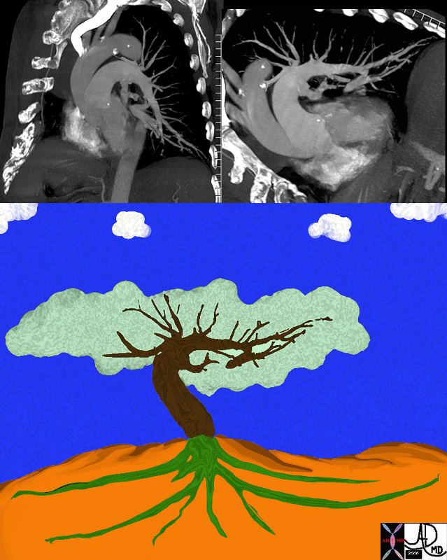
Art image derived from a parasagittal view of a reconstructed CT scan – CTA of the chest
Ashley Davidoff MD Copyright 2018
46649b04b.800
Art image derived from a parasagittal view of a reconstructed CT scan – CTA of the chest
Ashley Davidoff MD Copyright 2018
46649b11.800b01

Art image derived from a parasagittal view of a reconstructed CT scan – CTA of the chest
Ashley Davidoff MD Copyright 2018
46649c01.800
by Ashley Davidoff MD
The secondary lobules, connect, and unite, linked through the airways, blood vessels, lymphatics and nerves, to form segments in the lungs
There are ten bronchopulmonary segments in the right lung: three in the upper lobe, two in the middle lobe, and five in the lowerlobe. Some of the segments may fuse in the left lung to form usually eight to nine segments (four to five in the upper lobe and four to five in the lower lobe.
Ashley Davidoff MD
TheCommonVein.net lungs-0015catalogue-signed-small
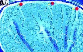
This picture shows us on the left with a white ring around us (we were the tallest) and the other couples who looked so much like us (also ringed). We called our tribe the “bronchovascular bundle” with the one part of the bundle being the progeny of the bronchus and the other the progeny of the pulmonary artery. In the distance at the periphery we could see the pairs from the other friendly tribe – the red pulmonary vein with its smaller yellow buddy the lymphatic. Behind them we could see the transparent window membrane through which we had peaked earlier. Oh my goodness!!! Look what has happened to my body!!!!!!!…… Ashley Davidoff MD. The Common Vein.net 42447b03b01
Ashley Davidoff MD TheCommonVein.net
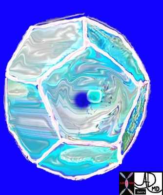
Here is a picture of the outside of the polyhedral pulmonary lobule from the side. It looked quite futuristic. Through the transparent side window we saw a couple similar to ourselves. From this vantage point the morphing did not look too different from what we had already been through – division after division – leaner and meaner. Ashley Davidoff MD. The Common Vein.net 42449b02
Courtesy Ashley Davidoff MD
lungs-0028-low res
This picture was taken just before the real drama started. The image gives a sense of what was to come. You can see here in the house of the lobule that we were all dividing into smaller parts and were getting smaller and the picture was quite colorful and rosy. I fully expected to have intimate contact with the arteriole… but it did not happen as I expected…… Ashley Davidoff MD. The Common Vein.net 42447b05b02
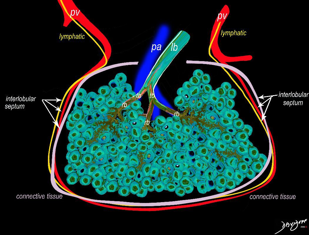
The secondary lobule is housed in a connective tissue framework in which run the lymphatic and venular tributaries . Together these 3 structures form the interlobular septum.
The lobar arteriole enters the framework, accompanied by the lobar bronchiole, and they all run together and form the interlobular septa. This structure measures between .5cms and 2cms and is visible on CT scan.
It is important in clinical radiology since many of the structures can be identified in health, and more particularly in disease, enabling the identification and characterization of many pathological processes.
Courtesy Ashley Davidoff MD The CommonVein.net
lungs-0036-low res
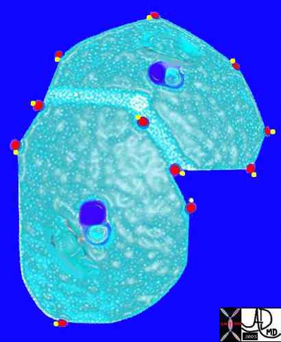
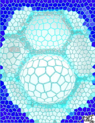
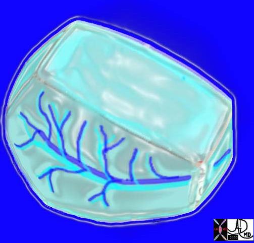
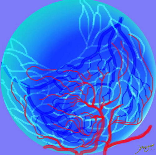
The arteries and airways pair up and travel together from the interlobular septa to the hilum. The pulmonary lobule, also called the secondary lobule is a structural unit surrounded by a membrane of connective tissue, and it is smaller than a subsegment of lung but larger than an acinus. This diagram shows two secondary lobules lying side by side. The pulmonary arteriole (royal blue) and bronchiole (pink) are shown together in the centre of the lobule (“centrilobular”), while the oxygenated pulmonary venules (red) and lymphatics (yellow) are peripheral and also form a formidable and almost inseparable pair.
42440b03
Ashley Davidoff MD
TheCommonVein.net
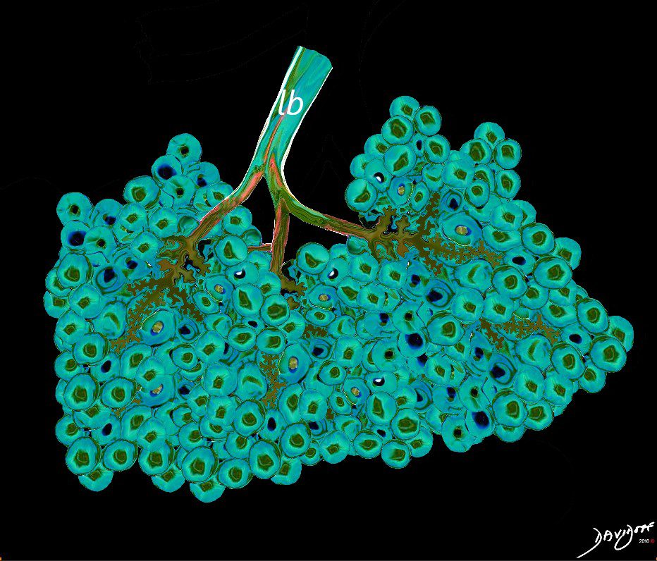
Between 10 and 30 acini combine to form a secondary lobule which is between .5- 2 cms in diameter. It is subtended by a single lobar bronchiole (lb), and is accompanied by arterioles, venules, lymphatics and connective tissue. It is important in clinical radiology since many of the structures can be identified in health, and more particularly in disease, enabling the identification and characterization of many disease processes.
Courtesy Ashley Davidoff MD
lungs-0035-low res
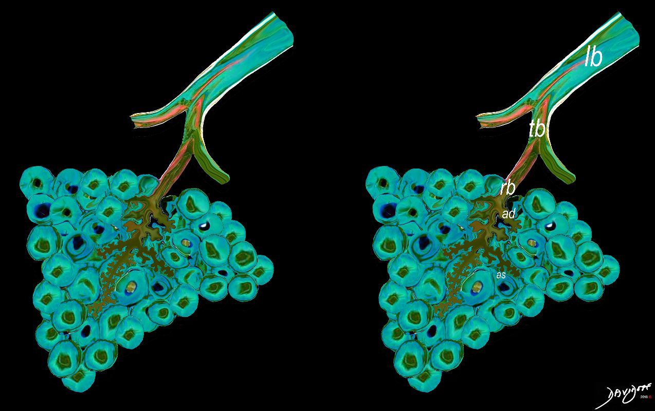
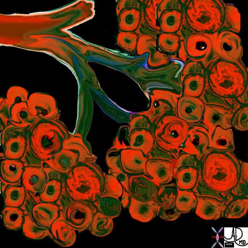
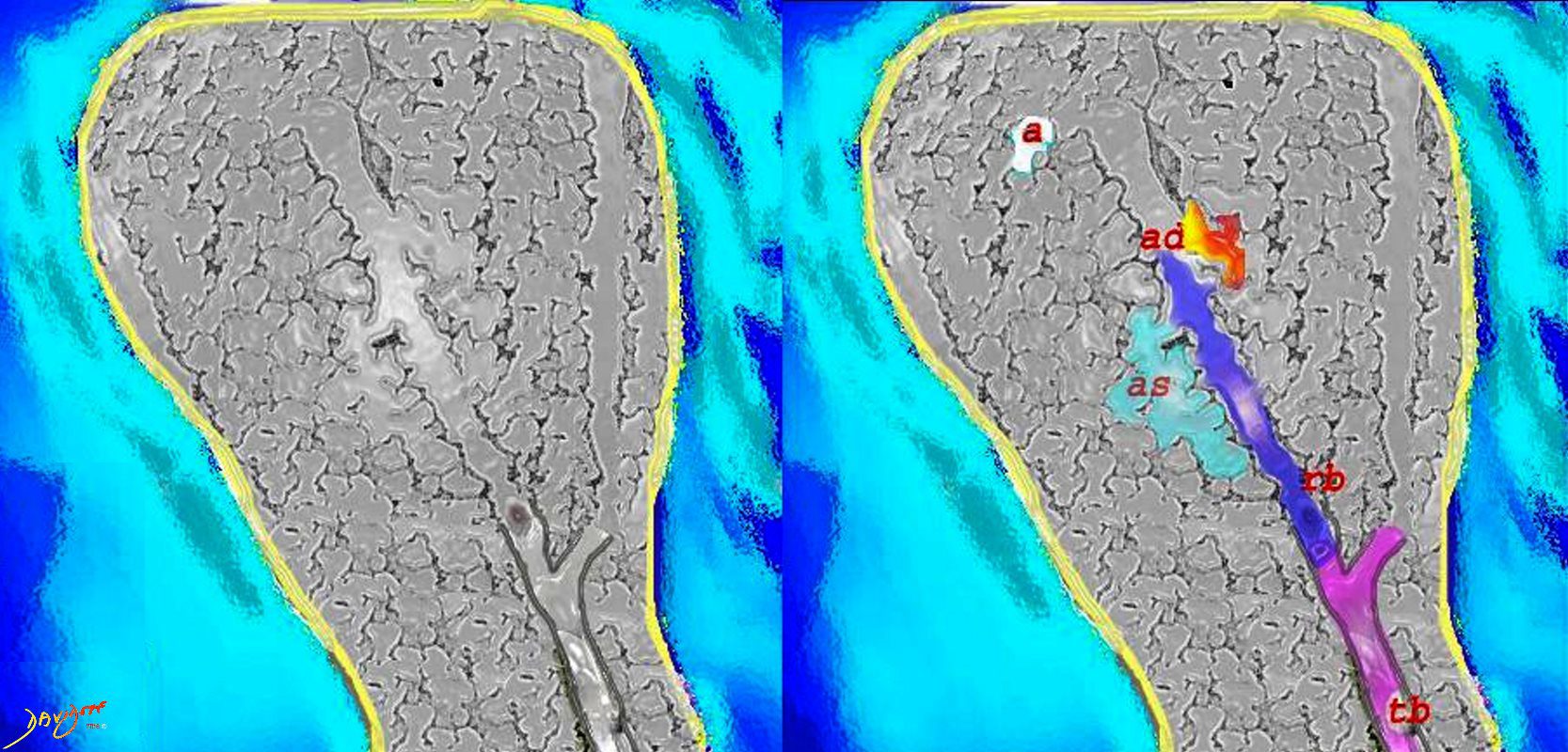
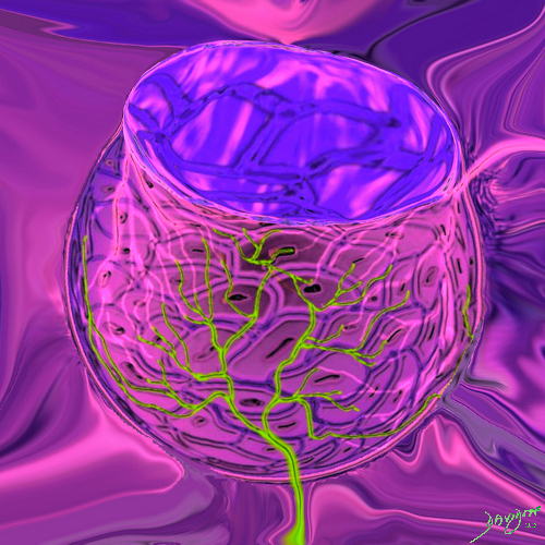
The acinus is defined as a unit of lung consisting of a single first order respiratory bronchiole that subtending a cluster of alveoli reminiscent of a bunch of grapes or berries (acinus in Latin means berry) . The lobular bronchiole (lb) branches into the terminal bronchiole (tb), which then branches into the first order respiratory bronchiole (rb). Subsequent branching after the respiratory bronchiole, includes in order, the alveolar duct (ad), alveolar sac (as), and then finally the berry like alveoli.
Courtesy Ashley Davidoff 2019
lungs-0030-low res

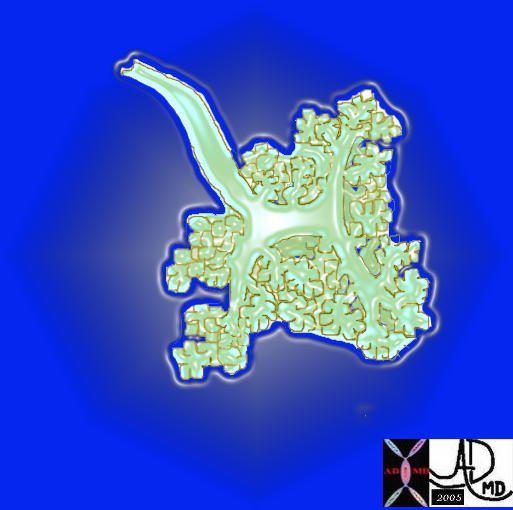
This artistic rendition of the small parts of the lung shows the beginning of the peripheral system just before it enters the acinus. This duct is called the terminal duct and it is the last part of the ductal system that has no ability for gas exchange. After its first division, the bronchioles become the respiratory bronchioles, and they are the first in the system to have an ability to both transport the gases as well as enable gas exchange.
Ashley Davidoff
TheCommonVein.net
32645b04b05.8s
The acinus with its arborizations is shaped more like a bunch of grapes.
Courtesy of: Ashley Davidoff, M.D 42650
TheCommonVein.net
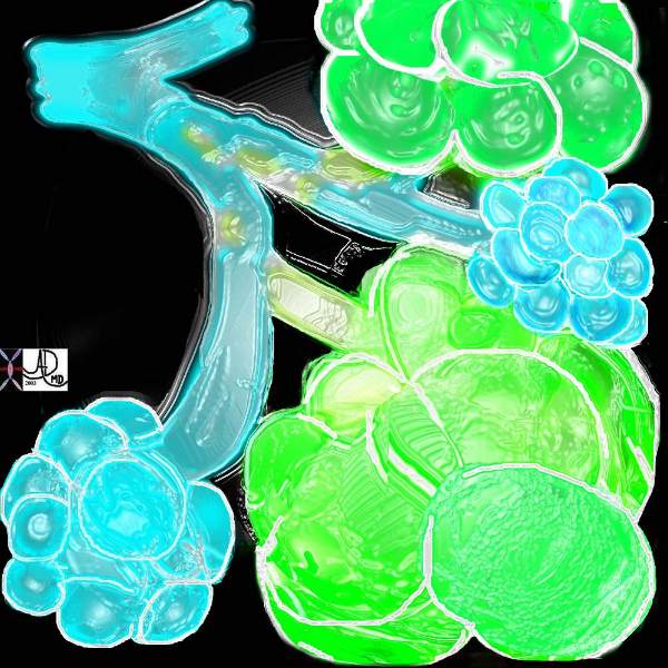
A drawing showing the normal acinus in teal and the abnormal emphysematous acinus in green characterised by destruction of the septal walls, enlargement of the alveoli, and loss of elasticity. The absence of involvement of the respiratory bronchiole makes the pathological diagnosis of centrilobular emphysema. Ashley Davidoff MD
TheCommonVein.net 32645
The Alveolus
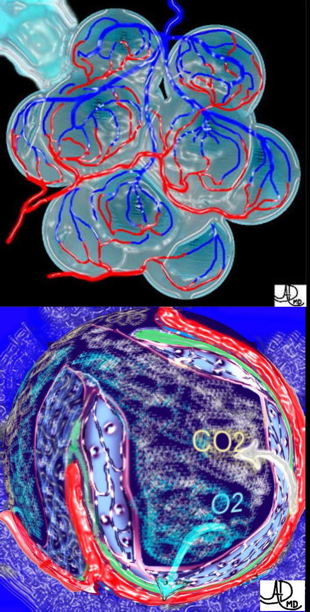
This is a drawing of a cluster of alveoli surrounded by the capillary network, fed by an arteriole in blue, and drained by a venule in red. The second image shows the exchange of life giving oxygen for the by product of metabolic activity – carbon dioxide
Ashley Davidoff MD
TheCommonVein.net
32165c
The five major layers that keep the air moving include the outer bony cage, the muscular layer represented in maroon, the pleural complex (orange yellow orange) the lung (blue) and surfactant within the alveolus. (pink) 42530b05b09b01a08
Ashley Davidoff art
by Ashley Davidoff MD

“Alveolus Cells and Capillaries of the Lung” shows an alveolus with single cell lining and associated arteriole, capillary and venous circulation. The cool fresh air flows into the alveolus, and oxygen flows into the blue blooded arteriole converting into a red blooded venule. A breeze of carbon dioxide flows through the single celled alveolus and into the airways for expiration
by Ashley Davidoff MD
by Ashley Davidoff MD
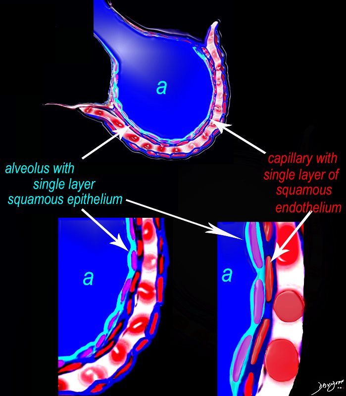 Alveolus at a Cytologic Level
The diagram shows an alveolus (a) above, lined by a single layer of squamous cells, surrounded by a capillary with red cells which is also lined by a single layer of squamous endothelial cells . The images below show progressive magnification of the alveolar wall demonstrating the two thin layer of the alveolar membrane .
Courtesy Ashley Davidoff 2019
lungs-0028-low res
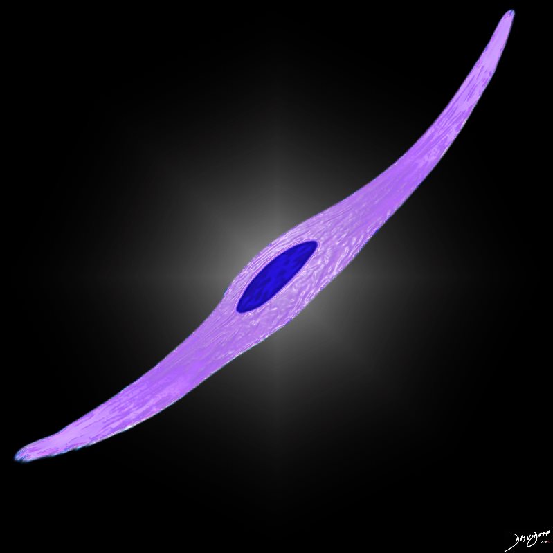
Squamous Cell
Ashley Davidoff MD

Ashley Davidoff MD
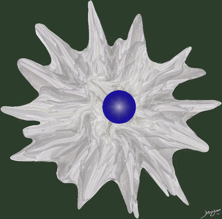 Macrophage
Ashley Davidoff MD