
The Common Vein Copyright 2007
The lungs are closely connected to the immediate environment and hence they are directly exposed to any and all pathogenic entities in the environment, including biological and non biological elements.
Bacteria and viruses are universal and therefore are common pathogens and affect everybody. Immune status is key to survival from these infections, and so the young and the old, and those with immune deficiencies are particularly vulnerable to these unseen enemies. Other diseases such as the dust diseases and pneumoconioses, occur in isolated work related environments, but there are some diseases, like bird fancier’s diseasea that have both acute and chronic manifestations due to exposure to the antigen which is found in the droppings of birds – most commonly pigeon’s but also from domestic birds like budgerigars (small parakeets).
Diseases of the Airways
Diseases
Principles
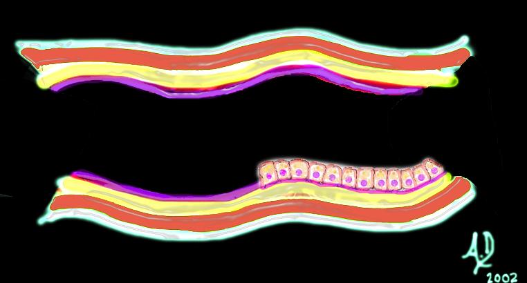 Basic Strcture of Tubular Systems Basic Strcture of Tubular Systems |
| 32347 tube colon small bowel lung bronchus bronchi esophagus stomach large bowel bile duct ureter tube principles Courtesy Ashley DAvidoff MD Davidoff art mucosa submucosa muscularis adventitia serosa histology |
 Mucosal Lesion Mucosal Lesion |
| 32347d01 mucosa submucosa muscularis adventitia serosa mucosal mass polyp neoplasm carcinoma acute angles with the lumen histopathology imaging diagnosis Davidoff art Davidoff MD |
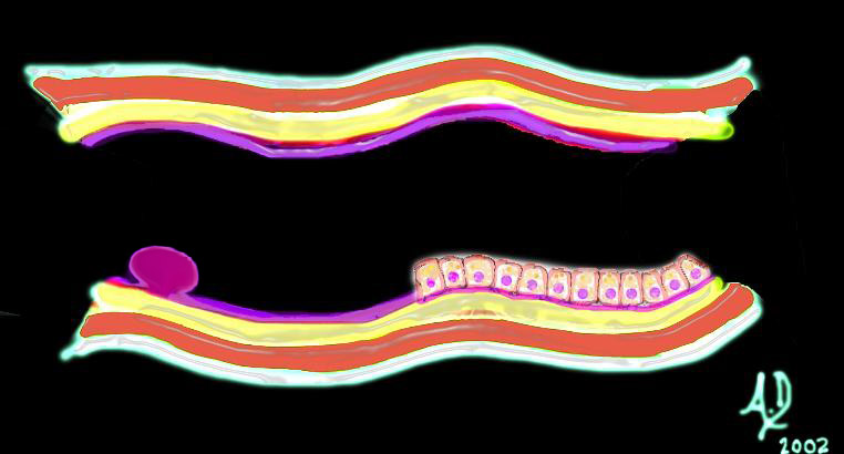
 Submucosal Lesion Submucosal Lesion |
| 32347d02 mucosa submucosa muscularis adventitia serosa submucosal mass edema hemorrhage obtuse angles or right angle 90 degree ninety degree angle with the lumen histopathology imaging diagnosis Davidoff art Davidoff MD |
 Submucosal Lesion Submucosal Lesion |
| 32347d03 mucosa submucosa muscularis adventitia serosa submucosal mass edema hemorrhage obtuse angles or right with the lumen histopathology imaging diagnosis Davidoff art Davidoff MD |
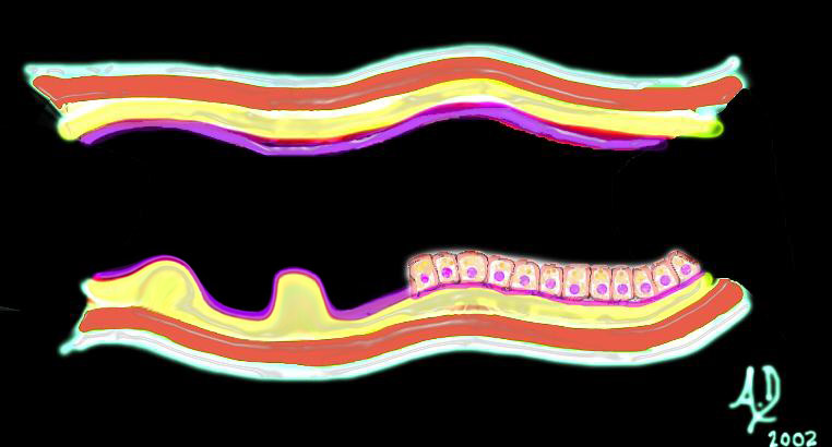
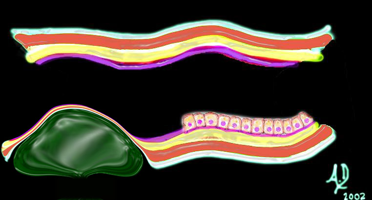 Extrinsic Lesion Extrinsic Lesion |
| 32347d04 mucosa submucosa muscularis adventitia serosa submucosal mass edema hemorrhage neoplasm malignancy benign obtuse angles with the lumen histopathology imaging diagnosis Davidoff art Davidoff MD |
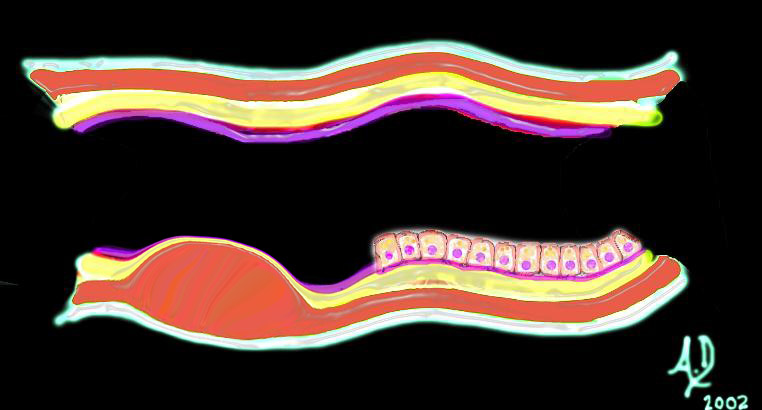
 Circumferential Lesion Circumferential Lesion |
| 32347d06 mucosa submucosa muscularis adventitia serosa submucosal mass edema hemorrhage neoplasm malignancy benign obtuse angles with the lumen circumferential narrowing constriction obstruction histopathology imaging diagnosis Davidoff art Davidoff MD |
Inflammation
Acute
 Acute Pneumonitis Secondary to Chemotherapy – Drug Reaction Acute Pneumonitis Secondary to Chemotherapy – Drug Reaction |
| 47095 lung chest fx lobar infiltrates fx air bronchogram dx chemotherapy related pneumonitis acute inflammation CTscan Davidoff MD |
Infection – Pneumonia
Infections to the lungs are caused by a host of organisms and most commonly are viral or bacterial in origin. Anatomically they can affect the airways causing epiglottitis, pharyngitis, tracheitis, bronchitis or bronchiolitis, The infection, on the other hand may affect the alveolar components of the lungs causing lobar or segmental pneumonia, or the interstium of the lungs causing an interstitial pneumonitis. Since the airways and alveoli by definition are connected the involvement of one part does not preclude the involvement of the other, though certain organisms have a prediliection for certain parts of the respiratory epithelium. Additionally an infection may start out as viral and progress and become complicated by secondary bacterial infection. Within the lung, interstitial infections (usually viral), will appear on chest X-ray with a linear pattern while the classical bacterial pneumonia will present as a consolidation.
The most common form of pneumonia in the population is viral in origin while the most common bacterial pneumonia is streptococcal in origin.
 Hemorrhagic Lobar Pneumonia Hemorrhagic Lobar Pneumonia |
| A gross pathoogy specimen showing a hemorrhagic bacterial lobar pneumonia (red hepatasisation) Courtesy Jeffrey Pierce and Ashley Davidoff MD 32320 |
Bacterial Pneumonia
There are some characteristic features about the bacterial pneumonias that are sometimes helpful in the differential diagnosis, though it is often difficult to make a organism specific diagnosis based on imaging. In general the bronchopneumonias affect infants young children and the elderly are caused by streptococcus pneumoniae and affect airways and alveoli contiguous to the larger bronchioles. Bronchopneumonia is characterized by peribronchial thickening, lobular nodules of patchy airspace disease, which may progress to lobar disease. Lobar pneumonia affects young adults and presents as a consolidated mass affecting segments, lobes, singly or at multiple locations. Lobar pneumonia is also usually caused by Streptococcus pneumoniae aka pneumococcal pneumonia (Todar’s On Line Text of Bacteriology), and Klebsiella. It is primarily a disorder of the alveoli resulting in segmental or lobar consolidation with the air bronchogram being chatacteristic.
Position
 Bronchopneumonia- Centrilobular Bronchopneumonia- Centrilobular |
| 47614c01 lung axial interstitium bronchioles connective tissue fx bronchial plugging peribronchial halo peribronchial thickening dx bronchopneumonia CTscan Davidoff MD |
 MAI – Surgical Proof MAI – Surgical Proof |
| 83065c.8 lung mass cavitating calcification eccentric air cavitation MAI infection atypical mycobacterium CTscan Courtesy Ashley Davidoff MD copyright 2008 |
Klebsiella infection tends to occur in the upper lobes and causes significant swelling of the lung so that bulging fissures may be seen. It also can cause cavitation.
Legionella has a tendency to affect the lower lobes
Mycoplasma has a tendency to affect the lower lobes
Chronic Inflammation
 Necrotizing Granuloma Necrotizing Granuloma |
| 72372c03 60 female with long history of smoking lung nodule slow growth SUV 1.3 right lower lobe dx necrotizing granuloma Most necrotizing granulomas are related to infection. Wegener’s granulomatosis Churg-Strauss syndrome – vasculitis sarcoidosis usually non necrotizing CTscan PETscan Davidoff MD |
Immune
| Acute Pneumonitis Secondary to Chemotherapy – Drug Reaction |
| 47095 lung chest fx lobar infiltrates fx air bronchogram dx chemotherapy related pneumonitis acute inflammation CTscan Davidoff MD |
 Scleroderma Scleroderma |
| This coronally reformatted CT of the posterior aspect of the chest shows interstitial lung disease within the lower lobes in this patient with scleroderma. The upper lobes were relatively spared. Courtesy Ashley Davidoff MD. 30476 code lung pulmonary interstitium interstial disease thickening lower lobes chronic inflammation fibrosis scleroderma collagen vascular disease imaging radiology CTscan |
Character
Air bronchograms tends to occur with stapphylococcal infections.
Cavitation occurs with Klebsiella, Staphylococcus, anerobic, gram negative, and tuberculous infections.
Atelectasis
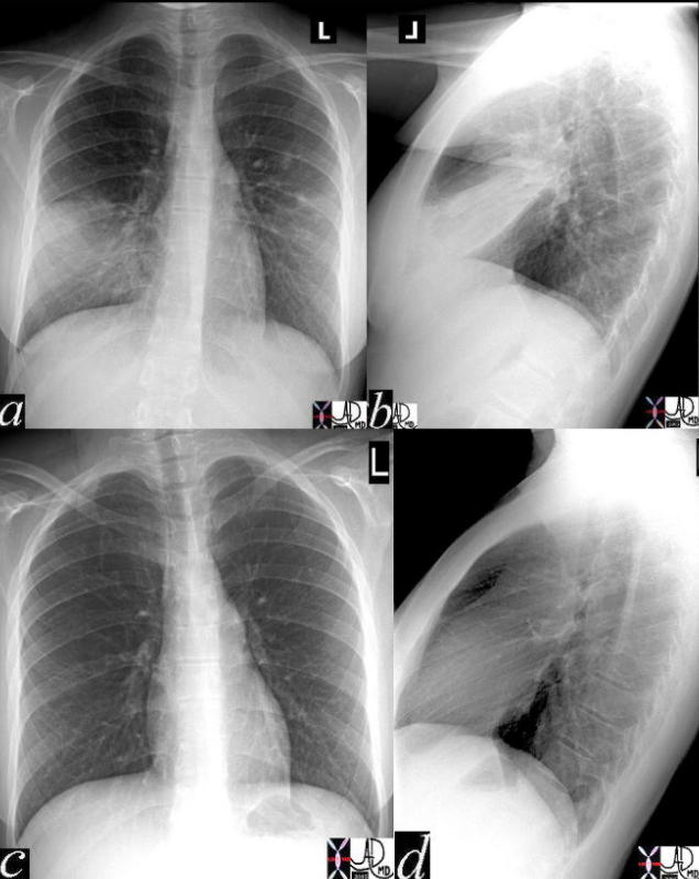 Classical RML pneumonia Classical RML pneumonia |
| The CXR in lobar pneumonia usually shows a consolidation exemplified by an air bronchogram. In this case an infiltrate is seen in the right middle lobe(a,b) which resolved 4 weeks later, after antibiotic therapy (c,d).
41819c02 Courtesy Ashley Davidoff MD medical students code chest CXR imaging lung plain film pneumonia radiology resolution |
 Active Tuberculosis Calcification and Air – Power of CT Active Tuberculosis Calcification and Air – Power of CT |
| 49812c01 50 yr F Korean fevers weight loss night sweats cough chest lung apex pleura fx calcified granulomas parenchyma pleural granulomas calcification cavitating nodules dx active tuberculosis active TB acute infection chronic infection infectious isolation Davidoff MD 49812c02 |
Viral Pneumonia
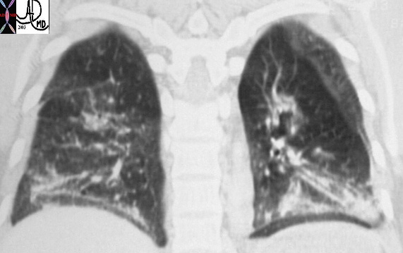
 Interstitial Pneumonia Interstitial Pneumonia |
| This plain film of the chest (CXR) shows a classicial reticular interstitial process pneumonia. Note this pattern is more linear and represents an interstitial pneumonitis.
Courtesy Priscilla Slanetz MD 30662 |
The most common agents causing viral pneumonia are influenza A, respiratory syncitialk virus (RSV) and paraifluenza 1,2,3.
Radiological findings are classically of a linear interstitial nature but they may also show alveolar changes such as consolidation. Pleural effusion and peribronchial thickening are associated findings.
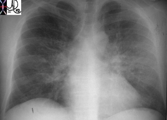
Pneumonia in the Immunocompromised Host
There is a high incidence of pneumocystis pneumonia (PCP) in patients with AIDS. Other organisms such as gram negative bacilli, staphylococcus, aspergillus and cytomegalovirus are also well known pathogenic organisms in this population of patients. (eMedicine – Fernandes). Up to 45% of all HIV respiratory infections are bacterial in origin occuring at any CD4 count. As the CD4 count decreases the incidence increases, particulalrly when the CD4 count is less than 200 cells/mms3
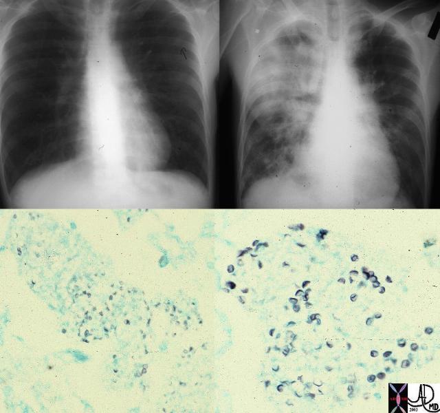 Pneumocystis Pneumocystis |
| These images are from a patient with immune deficiency and they show a progression of disease from near normal CXR to a consolidated right lung dominating in the right upper lobe. Pneumocystis (GMS stain reveals the capsule of the organism lower images – ) was identified as the incriminating organism. The most frequently described appearance on chest radiograph is a diffuse, bilateral interstitial infiltrate, consisting of fine-to-medium reticular interstitial change.
Courtesy Ashley Davidoff MD. a87-212 32236c |
PCP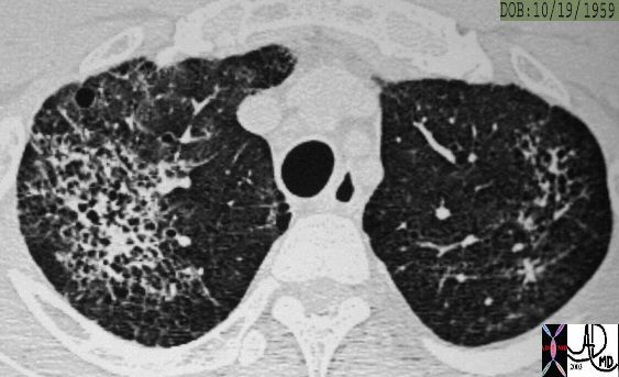
PCP
This is a CT of a 44 year old patient with HIV and with PCP infection. In this instance there is diffuse involvement of thick walled irregular cavities associated with a thin walled cyst seen anteriorly in the left lung. The most frequently described appearance on chest radiograph is a diffuse, bilateral interstitial infiltrate, consisting of fine-to-medium reticular interstitial change.
Courtesy Ashley Davidoff MD. 30048 30049 30050 code lung pulmonary infiltrate cystic cavity infection pneumocystis PCP imaging radiologyMycoplasma Pneumonia
Mycoplasma is a genus of bacteria that lacks cell walls. When it affests the respiratory system it presents with fever headache malaise and anorexia. The white cell count is usually normal but may show a left shift. Cold agglutinins are detected in 50-70% of patients and the diagnosis is made with a PCR assay of a nasopharyngeal swab or serologic testing.. The CXR has a variable appearance and can present both with airspace disease in ssegmental or lobar fashion (45-60%) or with an interstitial pattern of nodular or reticular type (15-50%). Pleural effusons are relatively uncommon (<20%). On CT they may present with a bronchioloitis with centrilobular nodules, alveolar consolidation interstital thickening of the axial interstitium or of the interlobular septa.
Most infections resolve and the morphology of the lung returns to normal. With repeated infection and or untreated disease, there may be permanent and ongoing structural damage to the lung or the airways and entities such as bronchiectasis may result.
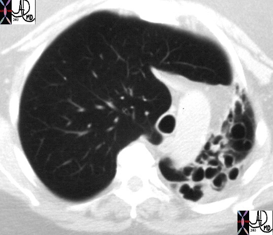 Atelectasis, bronchiectasis, hyperinflation. Atelectasis, bronchiectasis, hyperinflation. |
| This CT image shows a hyperinflated and large right lung, with volume loss and cystic change in the left lung. Note the trachea is pulled to the left by the contracted left lung, and is pushed by the hyperinflated right lung. The disease may be as a result of previous severe or untreated pulmonary infection
Courtesy Ashley Davidoff MD 30084 |
Inflammatory Diseases
Inflammatory diseases such as the collagen vascular diseases, interstitial pulmonary fibrosis, sarcoidosis are idiopathic slowly advancing understanding of the causes of these disease. The underlying pathology is one of an inflammatory process affecting the airways, vessels connective tissues or the parenchyma. In the acute phases exudation hyperemia swelling are characteristic while in long standing disease fibrosis is the hallmark.
 Acute Bronchovascular inflammatory Disease Acute Bronchovascular inflammatory Disease |
| This patient demonstrates significant patchy bilateral lung disease that dominates in the right lung. (a) In image b, there are ground glass changes (areas of increased density) areas of mosaic perfusion (areas of decreased density), and peribronchial halos of edema. In c there are small air bronchograms suggesting alveolar involvement. These findings are characteristic of an acute inflammatory process such as an acute allergic pneumonitis.
47170c01.800 Davidoff MD |
 PH probe PH probe |
| 49462 chest esophagus PH probe intraoperative monitoring for aspiration of stomach contents CXR plain X-Ray of the chest Davidoff MD |
 Chronic Inflammation – post XRT Chronic Inflammation – post XRT |
| The straight line of transition between the involved mediastinal side of the right lung and the relativeluy spared lung on the lateral side of the right lung is classical of radiation induced pneumonitis. The black holes in the dense parenchyma are areas of bronchiectasis – secondaryty to the effect of the radiatio on the smooth muscle indicating the chronic nature of the disease.
46968c01 Davidoff MD |
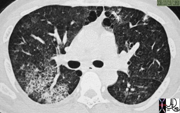
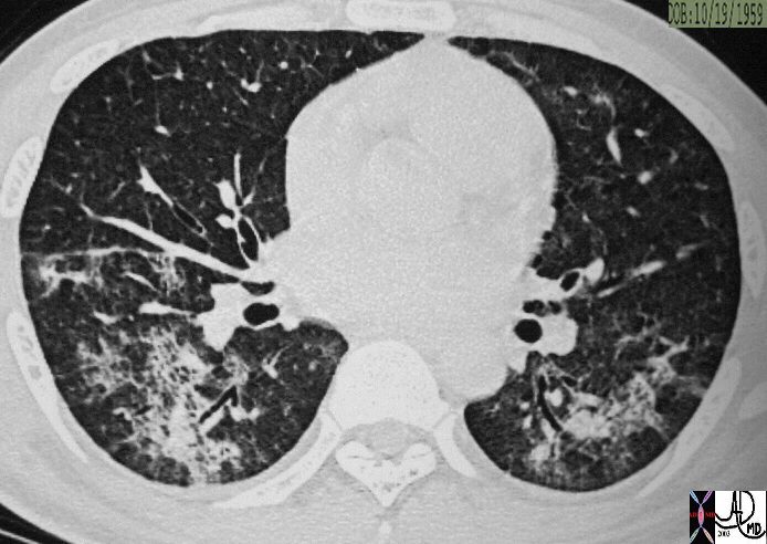
 End Stage Fibrosis – IPF End Stage Fibrosis – IPF |
| Ongoing inflammatory disease of the intertstitium will result in pulmonary fibrosis typified by the interstitial and reticular (net like changes) seen on the plain film (a) and the “honeycombing effect exemplified in the high resolution images (b,c) and conventional image in (d).
47145c01 Davidoff MD |
 Asbestosis Asbestosis |
| Chronic exposure to asbestos results in asbestos related disease which is characterised by pleural plaquesseen in images a and b. Some patients develop interstitial fibrosis and tthen the disease is called asbestosis. The “shaggy” heart border, (a) architectural distortion and honeycombing (c,d) particulalrly affecting the lingula and RML is reminiscent of a patient with asbestosis.
47060c01 Davidoff MD |
Malignant Disease and Other Growth Abnormalities
The malignant diseases of the lungs commonly have a cigarette smoking habit to blame, and the aggressive, space occupying, spreading nature of the disease usually has a negative outcome as we struggle with different and new therapeutic regimens to prolong life. The small cell carcinomas of the lung are usually bulky and aggressive, respond dramatically to therapy but then tend to recur. The non small cell cancers have surgical options and a potential cure. The potential for cure rests on the staging of the disease.
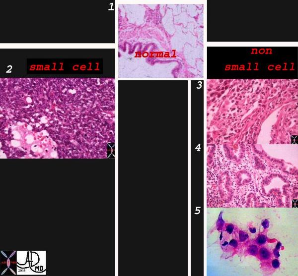 Classification of Lung Carcinoma Classification of Lung Carcinoma |
| Classification system of carcinoma of the lung based on histological subtype. This collage of histopatholgy images of lung carcinoma shows part of a normal bronchiole and a few alveoli (1), followed by a small cell carcinoma (left lower 2) and non small cell carcinoma grouped together on the right side with squamous cell(3), adenocarcinoma with glandular formation (4), and undifferentiated large cell derived from pleural aspirate (5). 1-4 Courtesy Dr Armando Fraire MD, and image 6 courtesy of tumorboard.com 32501cL02.jpg code lungs pulmonary neoplasm primary malignant malignancy small cell carcinoma non small cell NSCC classification histopathology |
 Malignant Neoplasm Malignant Neoplasm |
| In this patient a malignant mass is seen abutting the posterior mediastinum (a) is characterised as a spiculated mass in the superior segment of the left lower lobe (b,c), and is associated with A-P window nodes in c.
46218c01 Davidoff MD |
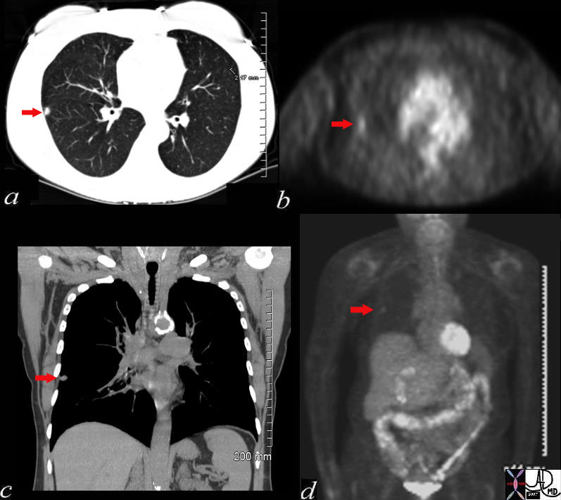
Malignant Disease in the RUL with PET positive Left Adrenal Gland – Stage 4 |
| This is a patient with lung carcinoma presenting with a large mass in the right upper lobe with a lymphangitic pattern, adrenal metastasis and a PET positive scan for the mass and for the left adrenal gland.
Courtesy Ashley Davidoff MD 32269b
|
Congenital Disease
Congenital and genetic disease of the lungs are not common in the adult population but sporadic cases do occur. Progress in the management of patients with cystic fibrosis have resulted in an enlarging adult population with this disease which was previously fatal in childhood.
Mechanical Diseases
Emphysema can be considered a mechanical disease of the lungs since the loss of elasticity of the lung tissue that occurs together with the destruction of tissue results in mechanical disadvantage. As a result air cannot be moved efficiently and there is an increase in air that is trapped in “dead” space and which cannot be efficiently moved to the capillaries for exchange nor efficiently removed. Emphysema is a major cause morbidity and mortality in the smoking population.
Asthma, another common disorder is an obstructive disease that inhibits the transport air to and from the lungs as well.
Mechanical Disease – Air Trapping
 Air Trapping Due to Small airway Disease and Obstruction Air Trapping Due to Small airway Disease and Obstruction |
| In this patient the lower lobes and particulalrly the LLL is affected by air trapping probably related to bronchioler disease rwith esulting ball valve effect. In this entity, air can enter and cannot leave the alveoli and hence there is progressive in flation of the affected segments. The entity called Swyer James effect , is a disease usually of the RML whih has bronchiolar stenosis with ball valve effect caused by childhood infection.
47057c01 CTscan Davidoff MD |
Mechanical Disease – Atelectasis
Atelectasis is a common disorder and has many causes including asiration, impaction of mucus, compression by tumor, or by enlarged lymph nodes.
 Atelectasis Atelectasis |
| Both lower lobes in this case are densely consolidated with no air seen in the atelectatic airways suggesting that they are filled with fluid and completely devoid of air. There are bilateral effusions as well (a,b,c). While the left mainstem bronchus contains air, the right contains bubbly aspirated fluid (b) so that the reconstruction (d) reveals a total blockage of the right mainstem.
46206c01 chest lung fx consolidation bronchus bronchi fx occluded pleural effusion dx aspiration CTscan Davidoff MD dx lobar atelectasis dx atelectatic RLL and LLL dx collapse due to bronchial obstruction 46198 46200 46201 46202 46204 46206 46206c01 46196b02 |
 Left Upper Lobe Obstruction Collapse and Compensatory Forces Left Upper Lobe Obstruction Collapse and Compensatory Forces |
| 49460c03 respiratory system lung left upper lobe fx atelecatsis compensation mediastinal shift lung shift volume loss plain CXR chest X-ray popcorn calcification Davidoff MD 49460c02.800 |
Trauma
Traumatic injury to the lungs is common manifesting with pneumothorax, pneumatoceles, and hemothorax.
Metabolic Diseases
Metabolic disease as a result of endocrine disease is not common, but the lungs are a vital organ in acid base metabolism. Most of these diseases revolve around functional rather than structural aberrance.
Circulatory Diseases
The circulatory abnormalities of the lung are a major source of morbidity and mortality, with particular reference to pulmonary embolism. The advent of multidetector CT has been a major advance in the diagnosis of this disease. Pulmonary hypertension secondary to left heart disease is common, as well as a result of chronic lung disease. The latter entity is called cor pulmonale.
From an imaging point of view it is key to have an understanding of the anatomy of the bronchi, the segments of the lungs, and the relative position of the segments. Knowledge of the secondary lobule, the location of the pulmonary arterial radicals, bronchiolar radicals, the lymphatics, the venules.
Pulmonary Embolism (PE)
The advent of multidetector CT has brought about a revolution in our ability to diagnose this relatively common and often fatal disorder. Our ability to diagnose PE with CT reqiures meticulous care to the technique and bolus timing since contrast volume limitations precludes a second injection because of the nephrotoxicity of the contrast.
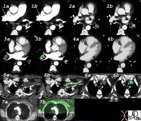
Pulmonary Emboli
This elderly woman presented with acute respiratory difficulty with swelling of her left upper limb. CT shows extensive pulmonary embolic disease to the lower lobes from second order branches to the tertiary branches (1-4 with green overlay) and evidence of thrombosis of the left subclavian vein. (5,6). Swelling over the anterior chest including the pectoralis muscles is evident in 7. Courtesy Priscilla Slanetz MD. 32538cLW
PE with Pulmonary Infarction
Although pulmonary embolism is common pulmonary infarction is distinctly uncommon because the lung receives blood supply from the bronchial as well as the pulmonary artery and can also be oxygenated directly from the alveoli.
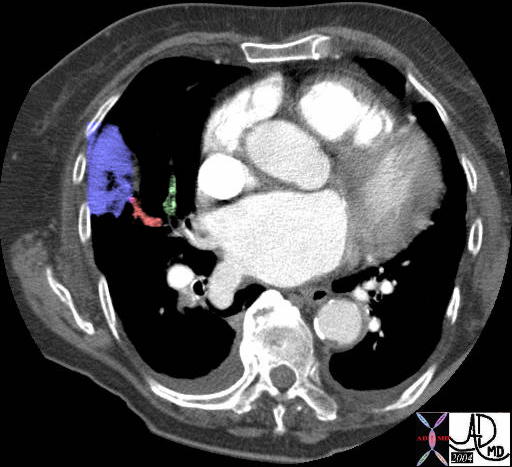 Pulmonary Embolus with Pulmonary Infarction Pulmonary Embolus with Pulmonary Infarction |
| In the case above a wedge shaped infarction is seen in the periphery of the right lung (purple) Both vessels coursing toward it are occluded by thrombus (red and green). The patient is in heart failure with cardiomegaly and small bilateral effusions noted.
39665b01 39665b02 Courtesy Ashley Davidoff MD |
Heart Failure
Heart failure is a very common entity and despite the progress with MDCT, the CXR from 1896 is still the most sensitive imaging technique for this entity.
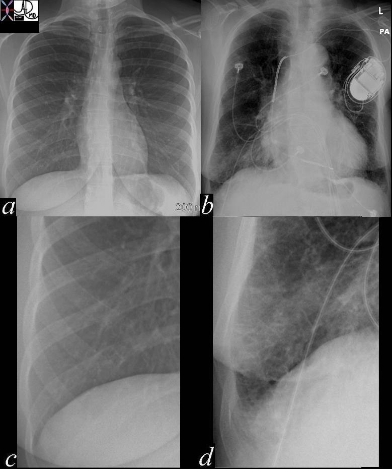 Heart Failure Heart Failure |
| In the images above the normal example is noted in image a with the normal costophrenic angle noted in c. Image b reveals a patient in congestive heart failure with an enlarged heart and prominent interstitial changes in the costophrenic angles called Kerley B lines that characterise thickened and congested interlobular septa (d)
42545c01 Davidoff MD |
Functional Disorders
Functional disorders often relate to poor muscular function including Guillain Barre syndrome, diaphragmatic paresis, and myasthenia gravis. Although not strictly a primary pulmonary functional disoder, aspiration and aspiration pneumonia from incoordinated swallowing mechanisms is common and often a cause of death in the elderly.
 Aspiration Aspiration |
| In images b,c and d contrast is seen in the trachea representing aspiration during a barium swallow.
46508c01.800 Davidoff MD |
References
e Medicine Bacterial Pneumonia
eMedicine Viral Pneumonia