
Source
Signs in Thoracic Imaging
Journal of Thoracic Imaging 21(1):76-90, March 2006.
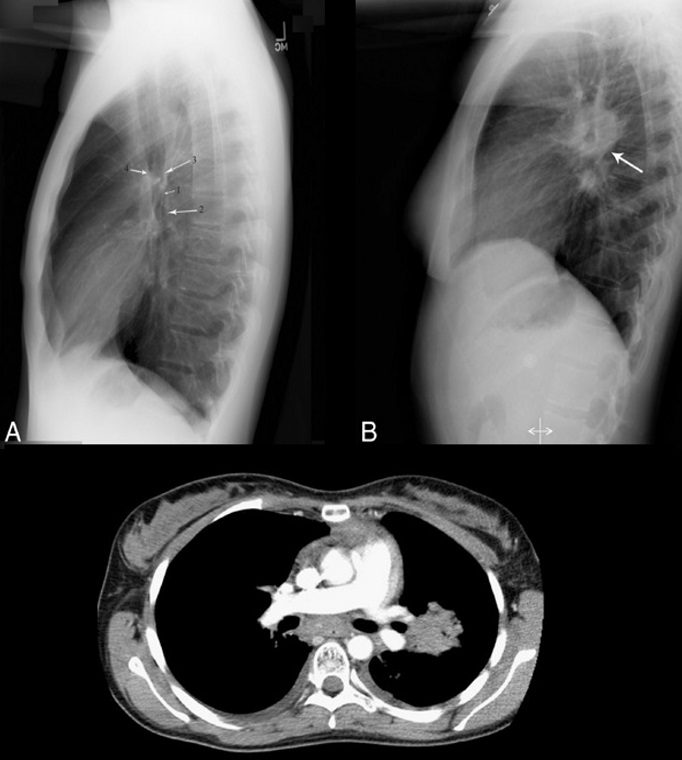
Although CT is widely accepted as the primary modality for detecting mediastinal lymphadenopathy, chest radiography is generally the initial radiographic investigation performed. On a normal lateral chest radiograph (Fig. 10A), the aortic arch and right and left pulmonary arteries are visualized in an “inverted horseshoe” configuration.34 In the presence of subcarinal lymphadenopathy (Figs. 10B, C), the inferior portion of the “horseshoe” fills in.34 The lymphadenopathy appears as a mass posterior to the bronchus intermedius and inferior to the tracheal bifurcation, completing the rounded hilar “doughnut” density.34,35