- Immunotherapy and XRT

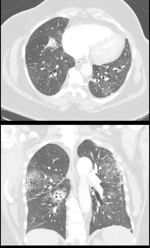
Drug-induced pneumonitis. (a) Axial and (b) coronal CT images in lung windows in a 61-year-old woman with history of stage IIIA non–small cell lung cancer. The patient had undergone chemoradiation followed by immunotherapy (durvalumab). Her course was complicated by pneumonitis. The patient improved with steroid treatment. Incidental note is made of dilated right side of heart. The patient has a recent history of acute pulmonary embolism with right-sided heart strain.
Parekh, M et al Review of the Chest CT Differential Diagnosis of Ground-Glass Opacities in the COVID Era Radiology Vol. 297, No. 3 July 2020 - Methotrexate
-
Methotrexate-induced NSIP in a 41-year-old woman with rheumatoid arthritis who presented with dyspnea and decreased diffusing capacity of the lungs for carbon monoxide (Dlco). (a) High-resolution CT scan shows
scattered ground-glass attenuation and thickened inter- and intralobular lines (arrow). (b) Photomicrograph (original
magnification, 400; hematoxylin-eosin stain) of a specimen from lung biopsy shows patchy interstitial fibrosis, expansion of the interstitium by chronic inflammatory infiltrates, and reactive hyperplastic type II pneumonocytes (arrow), findings consistent with NSIP induced by the pulmonary toxic effects of methotrexate.
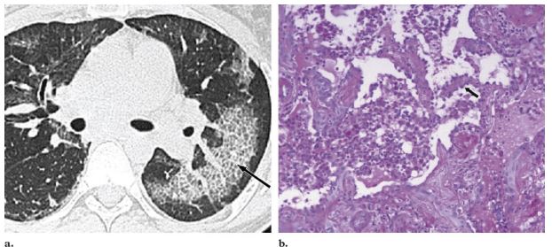
Diffuse mucinous bronchioloalveolar carcinoma in a 78-year-old man. (a) High-resolution CT scan
shows a bilateral crazy-paving pattern and centrilobular nodules. (b) Photomicrograph (original magnification,
400; hematoxylin-eosin stain) of a specimen from open lung biopsy shows replacement of the alveolar epithelium
by epithelial neoplastic cells with abundant intracytoplasmic mucin (arrows).
Rossi, S.E et al “Crazy-Paving” Pattern at Thin-Section CT of the Lungs: RadiologicPathologic Overview Radiographics Volume 23 – Number 6, 2003