
Embryonic Phase
Around the fourth week of gestation, the beginnings of the future respiratory system can be seen. A ridge of endodermal tissue forms in the ventral foregut. As the ridge elongates and separates, it defines what will become the trachea and the esophagus. Bronchial buds form caudally from the distal end of the newly formed trachea. Within the following days the buds will divide vigorously, resembling the dichotomous branching of the fully formed lung. As the trachea continues to elongate, the bronchial buds are carried into the thorax and pushed into the surrounding mesenchymal tissue. This interaction allows the growth of connective tissue and the smooth muscle of the tracheal wall. The epithelium lining the lungs originates from the endodermal tissue of the trachea. The dichotomous division of the airways continues throughout this phase.
Pseudoglandular Phase
This phase lasts from 52 days gestation to week 16. The bronchi continue to grow, as well as spread and elongate. They are lined with a high columnar epithelium that decreases in size and approach a cuboidal shape in the terminal branches. This gives the lung a tubular, glandular shape. This phase is characterized by the maturation of the conductive airways. The right main bronchus splits to form three secondary bronchi. These form the upper, middle, and lower lobes of the right lung. The left main bronchus divides once, forming the bronchus of the upper lobe. The lower lobe is the terminus of the main stem bronchus. The majority of branching occurs during weeks 10-16. A total of 18 tertiary branches results from dichotomous branching, forming the basis for the bronchopulmonary segments.
Lung buds from a human embryo of about four weeks, showing commencing lobulations. www.bartleby.com
Gray’s Anatomy, 2000
Lungs of a human embryo more advanced in development. www.bartleby.com
Gray’s Anatomy, 2000
Canalicular Phase
By the end of the pseudoglandular phase, all conductive elements of the airway tree have been established. This phase, comprising weeks 16 through 26 of gestation, will see a dramatic change in lung morphology, specifically in the future gas exchange region. So much so that the lung will have the potential to function, if the fetus is born prematurely. This change is due primarily to the differentiation of the pulmonary epithelium, which also aids in the synthesis of surfactant as well as the capillarization of the lung parenchyma. Interstitial tissue thins around the enlarged air spaces. Capillaries begin to insert themselves near the bronchial lumen, in between the cuboidal epithelium, with an intervening layer of cytoplasm. As this occurs, the acinar endothelium begins to differentiate into type I and II pneumocytes.
Saccular Phase
During this phase all airspaces located distally to the terminal bronchioles widen and elongate, expanding the future gas exchange region. This area continues to expand and mature until the formation of the alveoli in the next phase. The lung gains elasticity as elastic fibers from interstitial cells forms tissue around the air spaces. This defines each saccular unit as well as the acinus. Fibroblastic cells will begin to differentiate during this phase. They interact with the epithelium as well as play a role in the control of surfactant secretion. The vascular system increases in length and diameter. New generations of arteries form to keep up with the expansion of the gas exchange region.
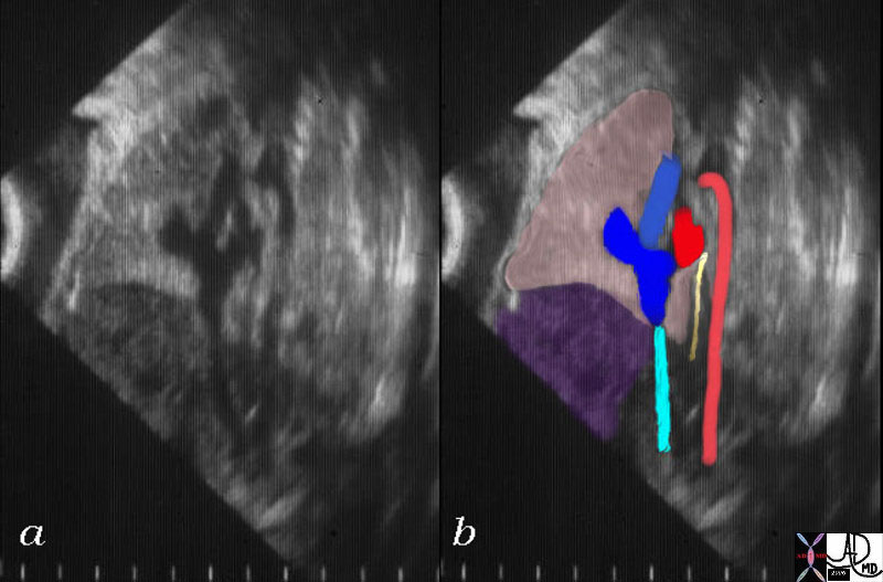 Fetal Lungs Fetal Lungs |
| 06401c01 heart cardiac fetus right atrium superior vena cava SVC right atrial appendage RAA inferior vena cava IVC left atrium LA esophagus aorta |
 24 week old Lungs 24 week old Lungs |
| 47755 24 week pregnancy heart lungs normal OB fetus amniotic sac placenta placental vessels uterus gallbladder T2 weighted MRI scan Davidoff MD concepts |
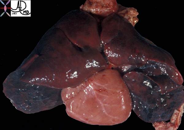 Atelectasis, bronchiectasis, hyperinflation. Atelectasis, bronchiectasis, hyperinflation. |
| This post mortem specimen of the lungs and heart of a child showing normal lobar pattern of the lungs with 3 lobes on the right and two on the left. The lungs appear hemorrhagic with the interlobular septa and secondary lobules are prominent. A small piece of glistening pleura are seen at the lower portion of the left lung.
Courtesy Ashley Davidoff MD 32558 |