
Tension Pneumothorax
30 year old female presents with tension pneumothorax 1 month prior to current admission
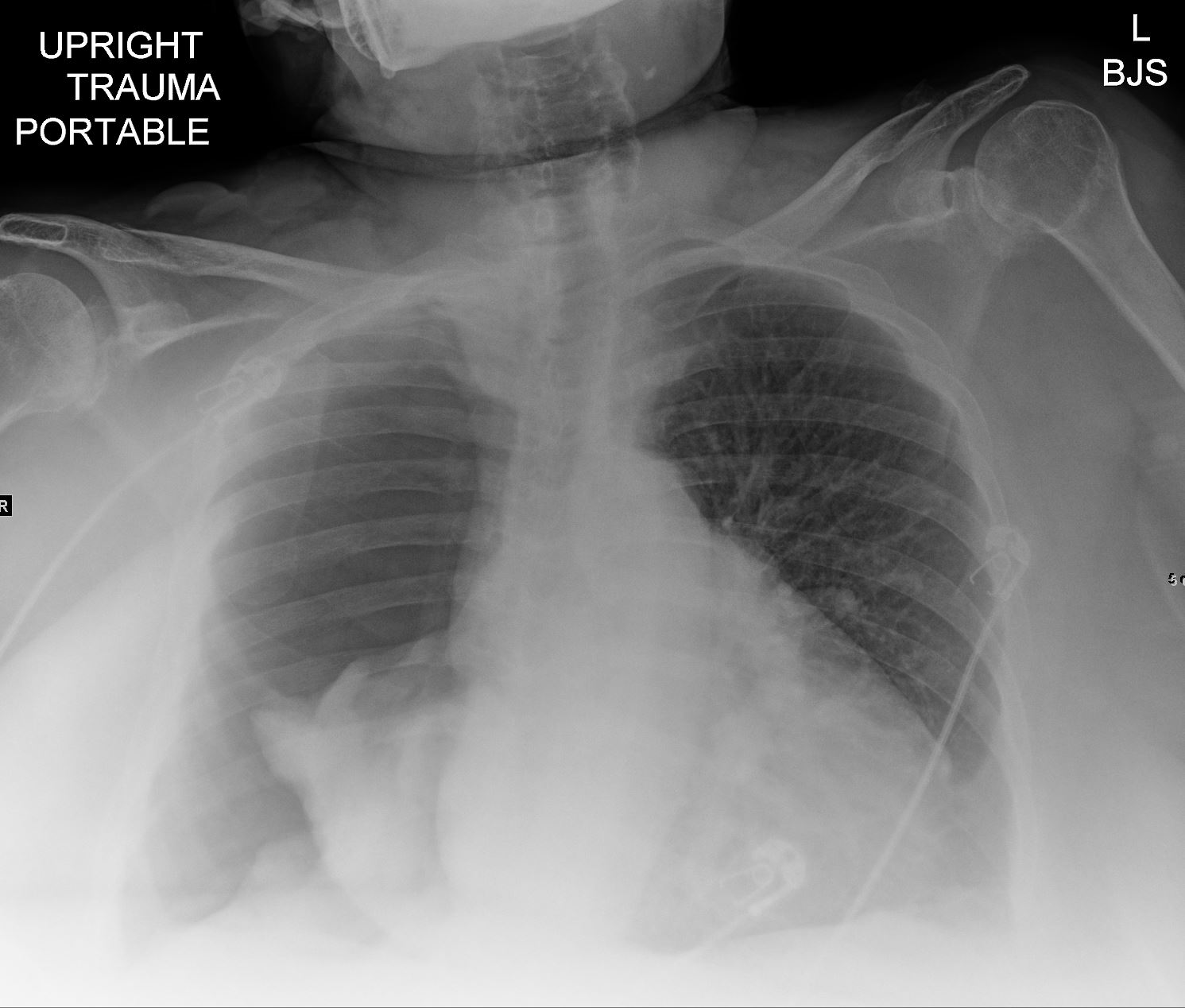
Ashley Davidoff MD TheCommonVein.net
Ashley Davidoff MD TheCommonVein.net
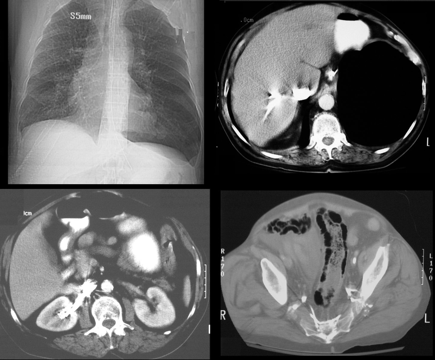
65 year old male s/p MVA presents in shock. Scout film (top left) shows left sided tension pneumothorax with rightward mediastinal shift. Axial CT through the liver (top right) shows expanded pneumothorax at the left lung base with reflux of contrast into the IVC.. Contrast also refluxes into the right renal vein (bottom left) and into the internal iliac veins (bottom right) Associated pneumatosis intestinalis in the sigmoid colon is present as well and likely secondary to the tension pneumothorax
Ashley Davidoff MD TheCommonVein.net
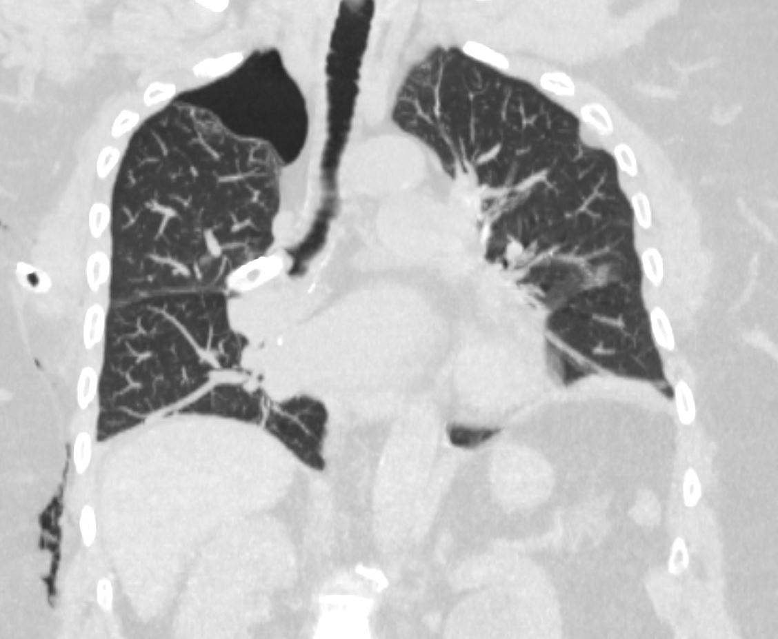
Tension Hydro/Hemothpneumothorax
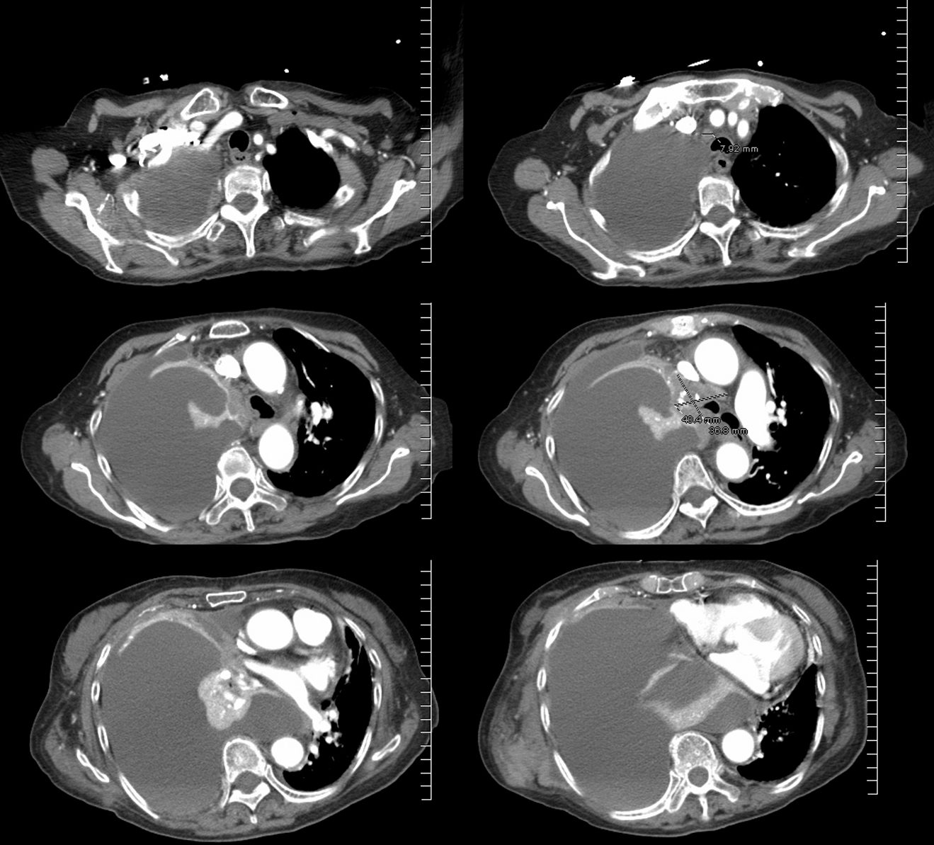
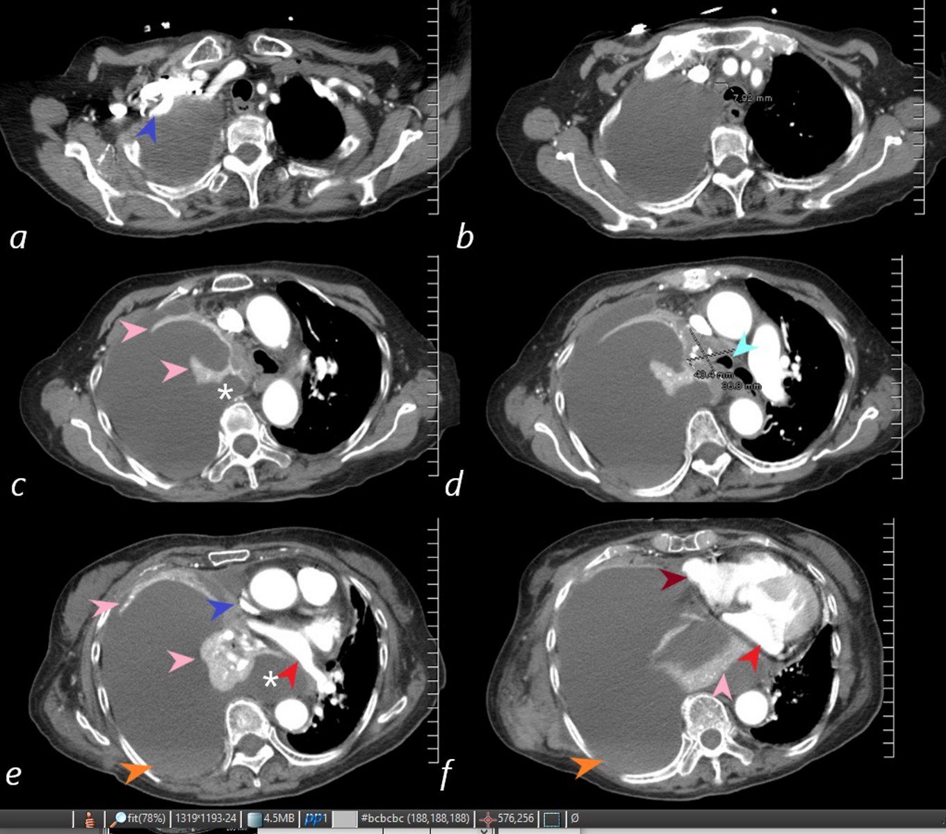
85-year-old female with a history of lung cancer, presents with a dyspnea and hypotension. CT scan shows a large right pleural effusion under pressure, suggestion of blood in the pleural cavity (orange arrowheads e, f) and mediastinal shift to the right. In addition, there is compression of the heart with back up of venous return due the pressure effect on the heart and vascular structures. Among the structures showing compressive effects are the SVC (blue arrowhead, c) with venous distension on right sided upper limb veins (blue arrowhead a). There is also pressure effect on the trachea with narrowing (light blue arrow d) The effusion in the right pleural cavity with atelectatic lung herniates into the left hemithorax, (white asterisk c, e). There is a dense sediment in the pleural fluid (orange arrowheads, e and f) suggesting blood in the pleural cavity. The left atrium is compressed (red arrowhead, e, f), and the right atrium is compressed (maroon arrow f).
Ashley Davidoff MD TheCommonVein.net 106Lu 118450c

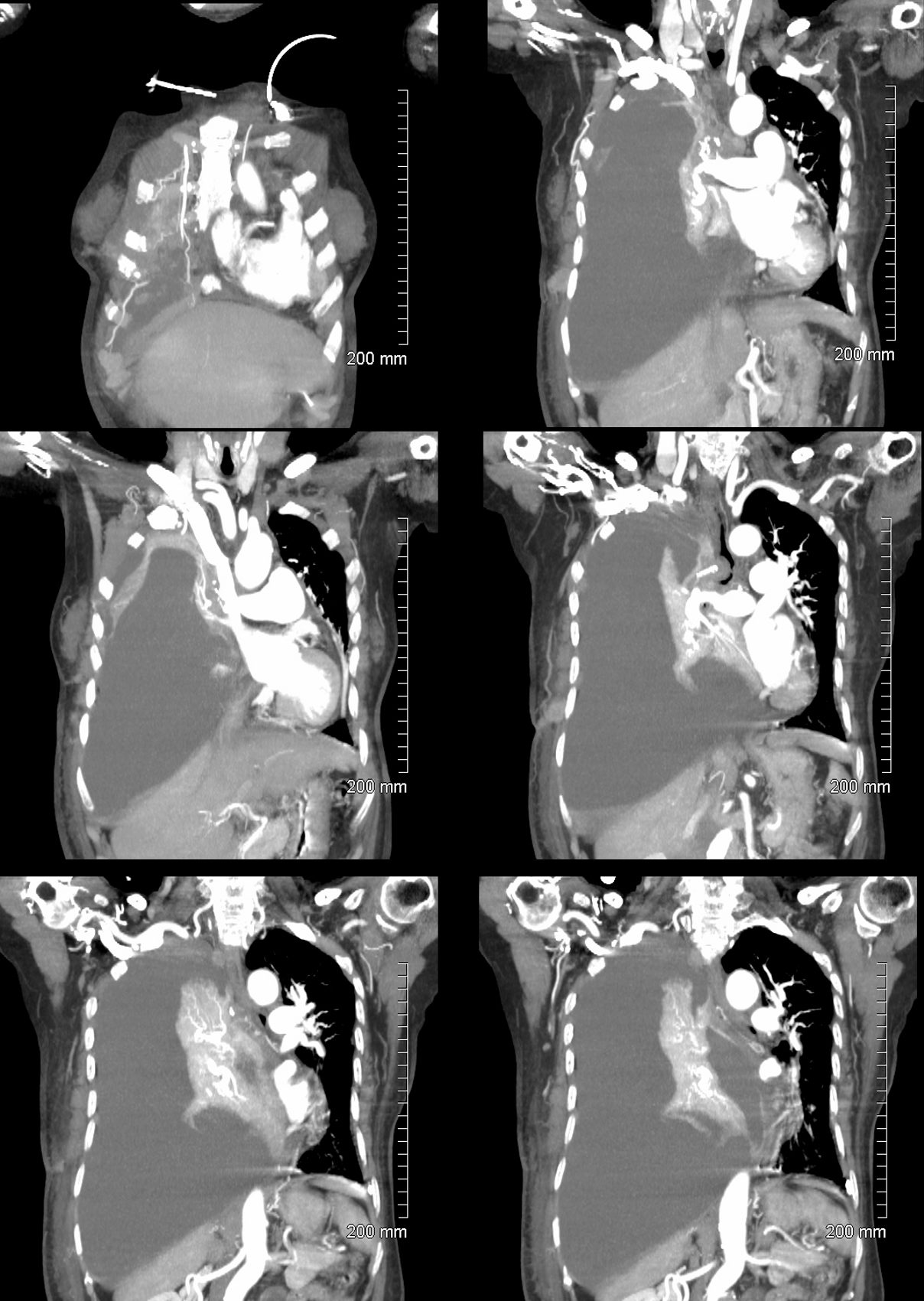
85-year-old female with a history of lung cancer, presents with a dyspnea and hypotension. Reconstruction of the CT scan in the coronal plane, shows a large right pleural effusion under pressure with herniation into the left chest (white asterisk e,and f) , with mediastinal shift to the left (yellow arrowhead b, c, d). In addition, there is compression of the heart with back up of venous return due the pressure effect on the heart and vascular structures. Among the structures showing venous distension are the SVC (blue arrowhead, c) right sided upper limb veins (blue arrowhead d) and the left upper pulmonary veins (red arrowhead, d and f). The density of the systemic venous abd arterial systems is similar, but vascular structures as noted by the green arrowhead in a could represent venous collaterals.
Ashley Davidoff MD TheCommonVein.ne 106Lu 118467cL
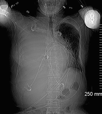
85-year-old female with a history of lung cancer, presents with a dyspnea and hypotension. Scout film prior to the CT scan shows “white out” of the right hemithorax (white asterisk) with pressure effect characterised by narrowing of the trachea (blue arrow) mediastinal shift yellow arrows) and herniation into the left chest characterised by a leftward shift of the azygo-esophageal junction line (white arrow).
Ashley Davidoff MD TheCommonVein.net106 Lu 118463
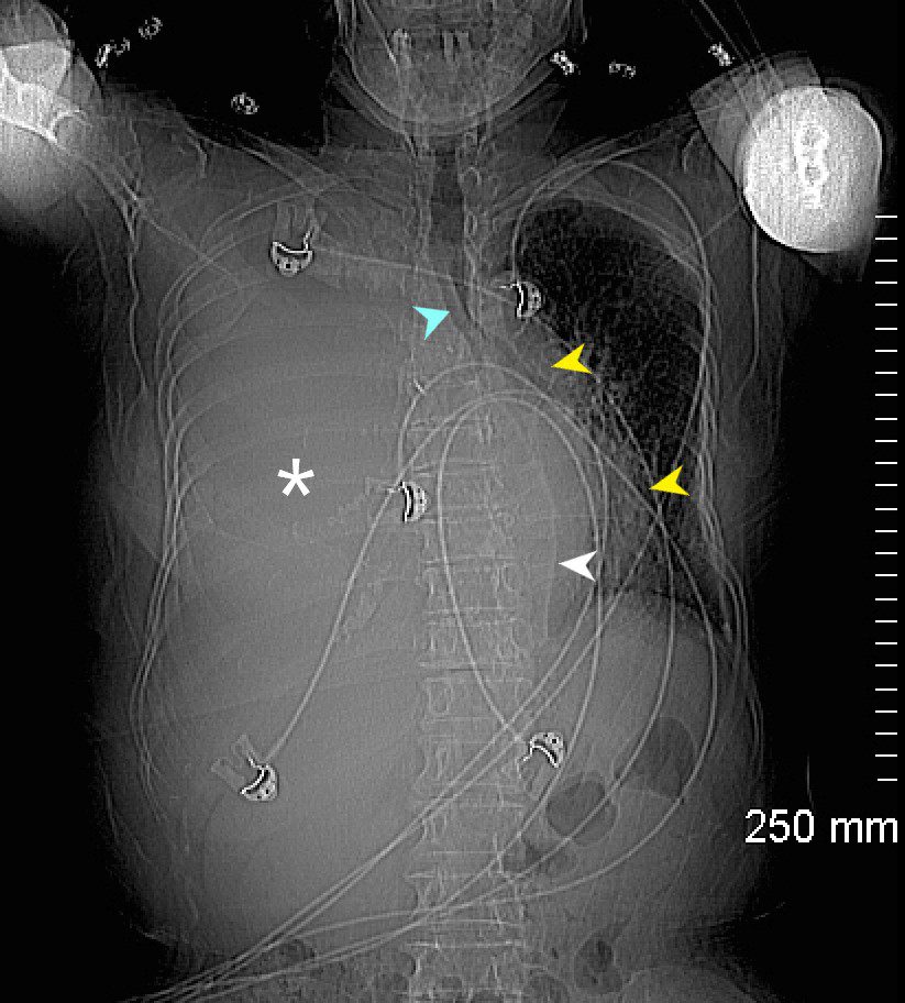
85-year-old female with a history of lung cancer, presents with a dyspnea and hypotension. Scout film prior to the CT scan shows “white out” of the right hemithorax (white asterisk) with pressure effect characterised by narrowing of the trachea (blue arrow) mediastinal shift yellow arrows) and herniation into the left chest characterised by a leftward shift of the azygo-esophageal junction line (white arrow).
Ashley Davidoff MD TheCommonVein.ne 106Lu 118463L
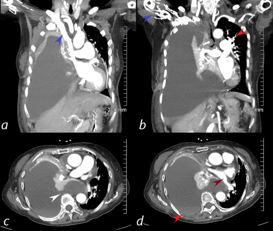
85-year-old female with a history of lung cancer, presents with a dyspnea and hypotension. CT scan shows a large right pleural effusion under pressure, suggestion of blood in the pleural cavity (orange arrowheads e, f) and mediastinal shift to the right. In addition, there is compression of the heart with back up of venous return due the pressure effect on the heart and vascular structures. Among the structures showing compressive effects are the SVC (blue arrowhead, c) with venous distension on right sided upper limb veins (blue arrowhead a). There is also pressure effect on the trachea with narrowing (light blue arrow d) The effusion in the right pleural cavity with atelectatic lung herniates into the left hemithorax, (white asterisk c, e). There is a dense sediment in the pleural fluid (orange arrowheads, e and f) suggesting blood in the pleural cavity. The left atrium is compressed (red arrowhead, e, f), and the right atrium is compressed (maroon arrow f).
Ashley Davidoff MD TheCommonVein.net 106Lu 118450cL
85-year-old female with a history of lung cancer, presents with a dyspnea and hypotension. CT scan shows a large right pleural effusion under pressure, with mediastinal shift to the right. In addition, there is compression of the heart with back up of venous return due the pressure effect on the heart and vascular structures. Among the structures showing venous distension are the SVC (blue arrowhead,a) right sided upper limb veins (blue arrowhead b) and the left upper pulmonary veins (red arrowhead, b. The effusion in the right pleural cavity with atelectatic lung herniates into the left hemithorax, (white arrowhead, c). There is a dense sediment in the pleural fluid (red arrowhead, d) suggesting blood in the pleural cavity. The left atrium is compressed (maroon arrowhead, d)
Ashley Davidoff MD TheCommonVein.net