
Ashley Davidoff TheCommonVein.net
Ashley Davidoff TheCommonVein.net
Ashley Davidoff TheCommonVein.net
Ashley Davidoff MD
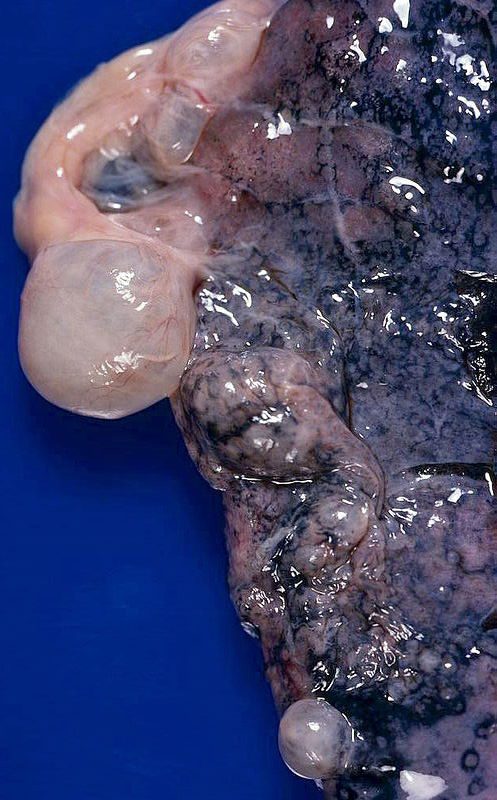
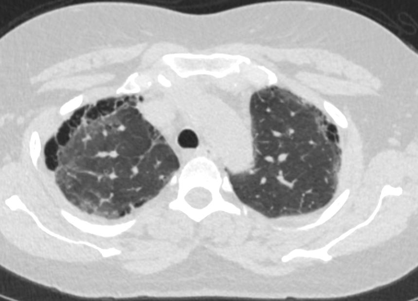
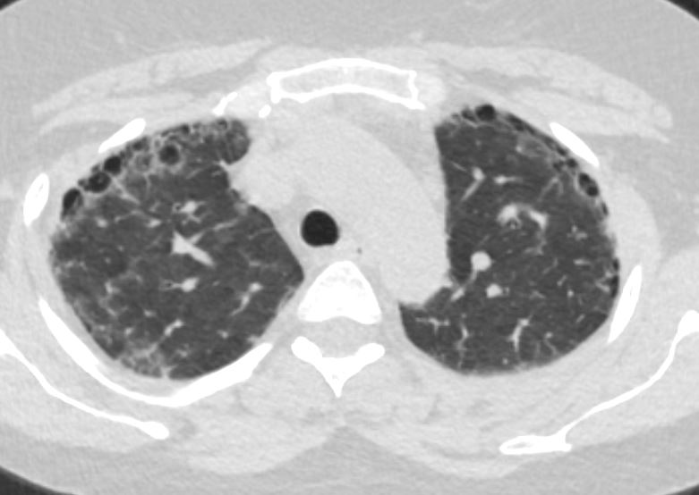
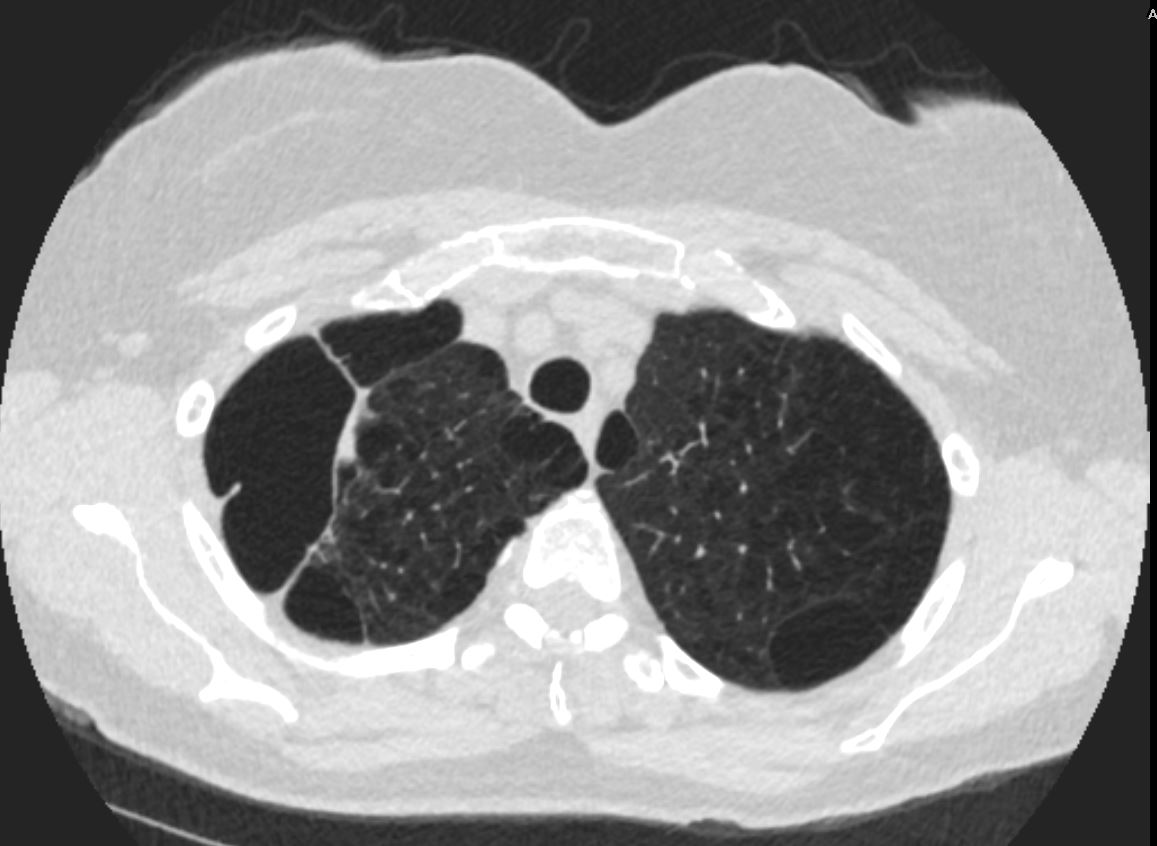
Large, prominent subpleural bullae. Spontaneous pneumothorax often results from rupture of such lesions.
Bullous (paraseptal) emphysema limited to lung periphery. Usually not smoking-related. Rupture is most common cause of spontaneous penumothorax…mostly young adults.
Courtesy Yale Rosen MD
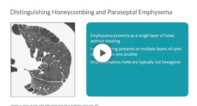
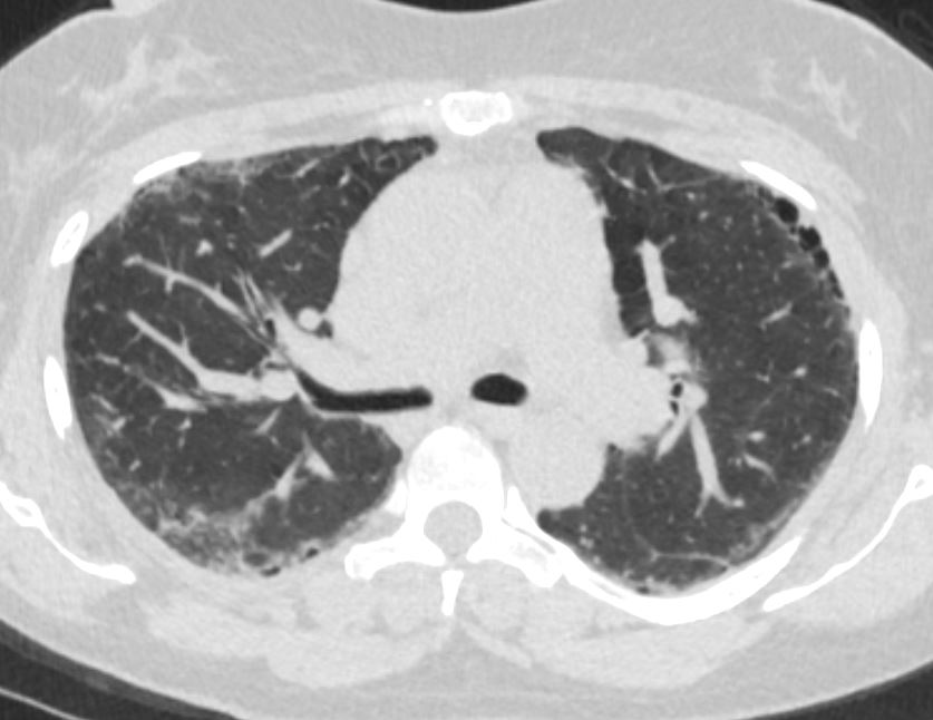
Distinguishing between honeycombing and paraseptal emphysema may be difficult, especially when coexisting on a single scan. As compared with honeycombing, which may present as multiple layers of cysts stacked upon one another, emphysema presents as a single layer of holes without stacking.7 Furthermore, emphysematous holes are typically not hexagonal; therefore, the shape of the cysts and their propensity to stack can help to distinguish one from the other.7
as relatively reduced CT attenuation at subpleural or peribronchovascular areas with or without intact interlobular septa
Paraseptal emphysema is a
- chronic lung condition
- characterized by the destruction and enlargement of the alveoli
- involves the peripheral areas of the lungs,
- Alveolar Destruction:
- the walls of the alveoli in the peripheral areas of the lungs
- become damaged and
- lose their elasticity.
- Air Trapping:
- As the alveoli lose their elasticity, t
- become less efficient at expelling air during exhalation.
- leads to air becoming trapped in these distal alveolar spaces, resulting in localized overinflation.
- Tension of the pleura
- pulling on the periphery of the lung during inspiration
- contributes to greater mechanical stress on the peripheral alveoli.
- Stress Distribution:
- The tension on the pleura is
- transmitted to the adjacent alveoli.
- Stretching and Shearing
- The mechanical forces
- can cause stretching and shearing forces on the alveoli.
- result in damage to the alveolar walls and
- more so if smoking or disease
- have caused decreased elasticity
- Stress Distribution:
-
- Bullae Formation:
- Prolonged air trapping and
- overinflation can cause the
- formation of large, bullae.
- can exert pressure on surrounding lung tissue and interfere with normal lung function.
- Associated Conditions:
- often associated with other forms of emphysema,
- Smoking is a significant risk factor
- as well as other lung conditions.
- Why does paraseptal emphysema affect the periphery of the lungs?
- due to differences in ventilation and anatomical structure within the lung tissue.
- Ventilation Distribution:
- The lungs are not uniformly ventilated throughout their structure.
- Peripheral lung regions, which are further away from the main airways,
- receive less ventilation compared to the central regions.
- Alveolar Structure: The alveoli, in the peripheral or subpleural areas
- have thinner walls
- Pressure Differences:
- alveoli near the periphery have
- relatively higher pressures during exhalation
- due to the increased resistance
- lead to stress and strain on the alveolar walls
- alveoli near the periphery have
- Air Trapping:
- When air becomes trapped in the alveoli due to reduced elasticity of the alveolar walls,
- tends to accumulate more in the peripheral regions