
Fibrotic HP
Upper lobe predominance
- reticulation,
- reaction bronchiectasis and
- volume loss,
- with or without evidence of honeycombing, [26,28]. Patients
- often an
- upper lobe predominant distribution of fibrosis,
- but diffuse and lower lobe predominant changes also.
- bronchocentricity to the fibrosis can be observed at the lung apices.
- reticulation in fibrotic HP i
- predominantly subpleural or
- peri-bronchovascular distribution.
Normal Small Airways
The Duct, and the Artery
The pulmonary arteriole accompanies the airway as it carries oxygen from the trachea to the alveoli. They part ways at the alveoli where the pulmonary venule then takes the oxygenated blood from capillary network around the alveoli back to the left atrium.
The intimate relationship of the airways and the pulmonary artery and their close approximation in size, is helpful in radiology, firstly to identify theese structures and secondly to define disease such as heart failure and bronchiectasis.
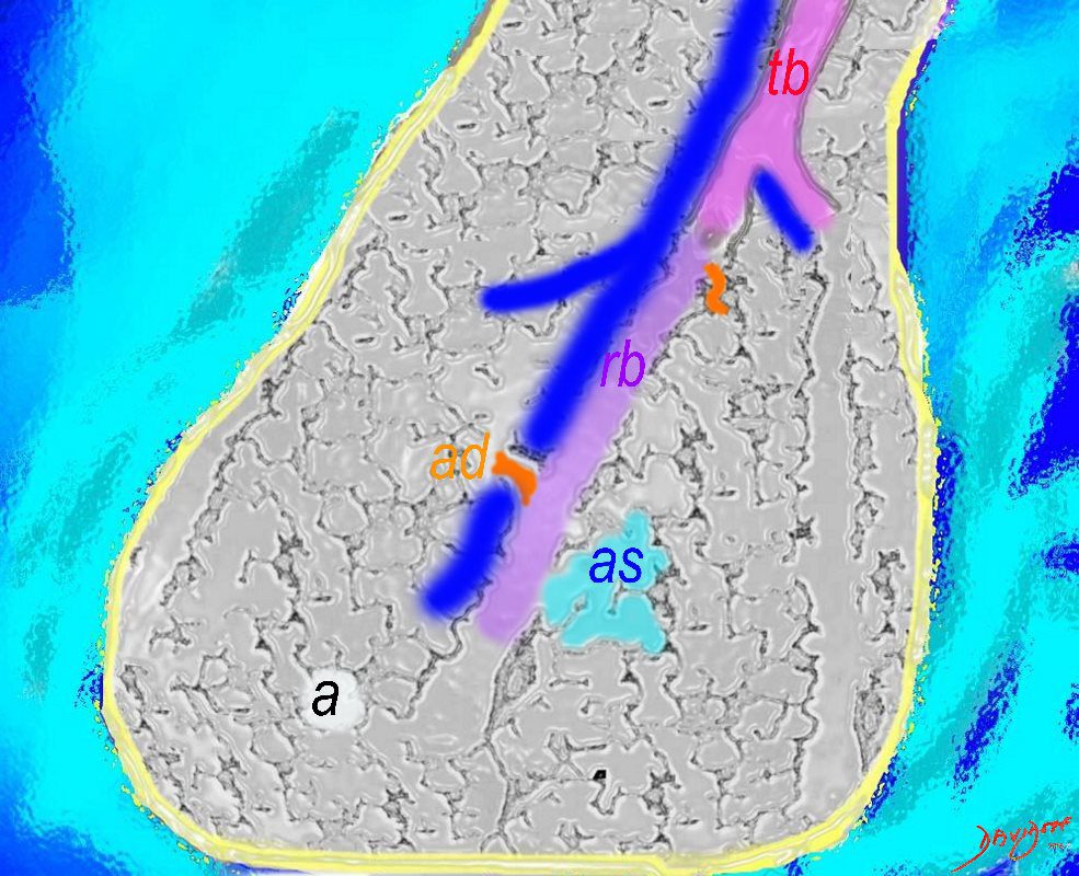
The acinus as shown in this image is defined as a unit of lung consisting of a single first order respiratory bronchiole that subtending a cluster of alveoli reminiscent of a bunch of grapes or berries (acinus in Latin means berry) . The lobular bronchiole (lb) branches into the terminal bronchiole (tb), which then branches into the first order respiratory bronchiole (rb). Subsequent branching after the respiratory bronchiole, includes in order, the alveolar duct (ad), alveolar sac (as), and then finally the berry like alveoli.
Courtesy Ashley Davidoff 2019
lungs-0033-low res
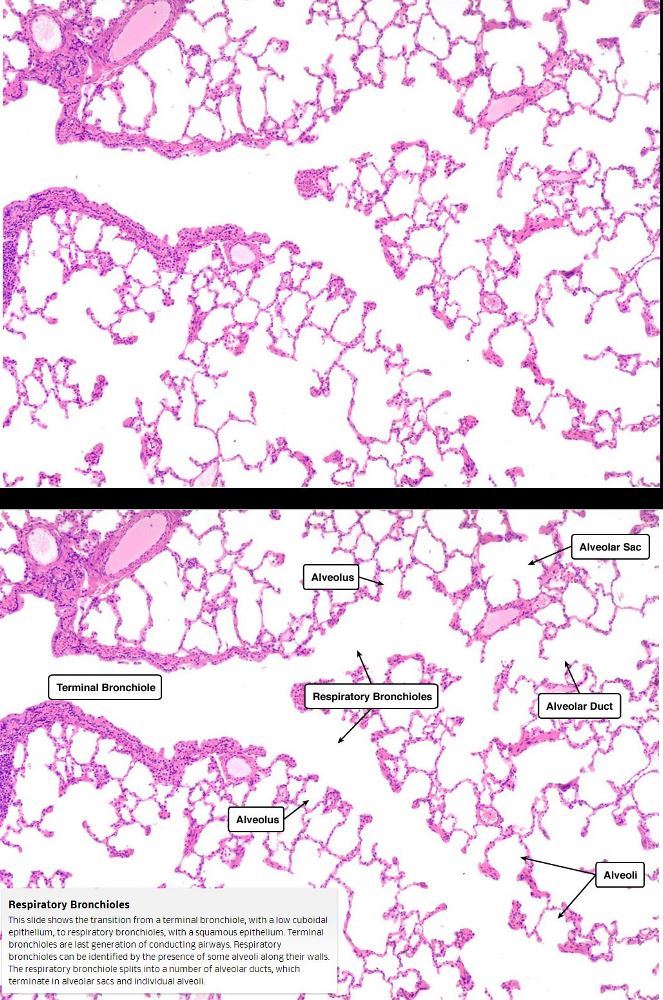
This slide shows the transition from a terminal bronchiole, with a low cuboidal epithelium, to respiratory bronchioles, with a squamous epithelium. Terminal bronchioles are last generation of conducting airways.
Respiratory bronchioles can be identified by the presence of some alveoli along their walls. The respiratory bronchiole splits into a number of alveolar ducts, which terminate in alveolar sacs and individual alveoli
Courtesy medcell.med.Yale.edu
The Buck Ends Here
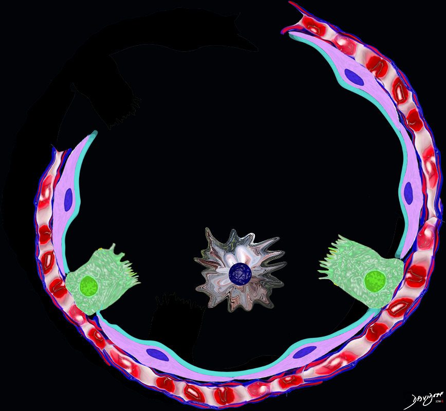
The alveolus is lined by a simple epithelium – one cell layer thick. There are two types of lining cells; Type 1 pneumocytes are squamous cells that cover 90% of the surface of the inner lining of the lung , and type II cuboidal pneumocytes that are in fact much more numerous than Type I. They are involved in the production of surfactant . In the lumen there are resident macrophages which play a crucial role in the immune system. The mucosa is grounded by a basement membrane and a lamina propria, and connected to the lamina propria and basement membrane of the surrounding capillary. The alveolus is lined by a thin layer of surfactant. (teal blue)
Ashley Davidoff
TheCommonVein.net

Lower magnification of the lung with H and E stain shows cup-shaped alveolar spaces outlined by delicate thin alveolar capillary membrane.
key words
lung, pulmonary, normal alveolus, alveoli, histology, interstitium, interstitial
Courtesy Armando Fraire MD. 32819
Small Airway Disease Terminal Bronchiole

Courtesy Dr Yale Rosen
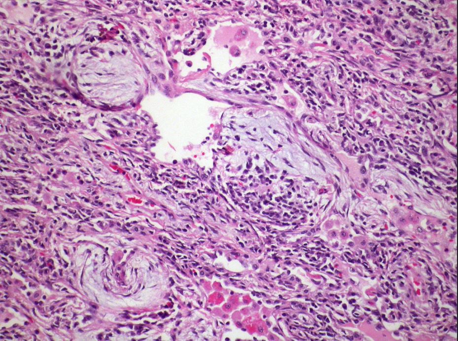
Small Airway Disease Giant Cell Around the Terminal Bronchiole

Multinucleate giant cells within a terminal bronchiole.
Courtesy Dr Yale Rosen
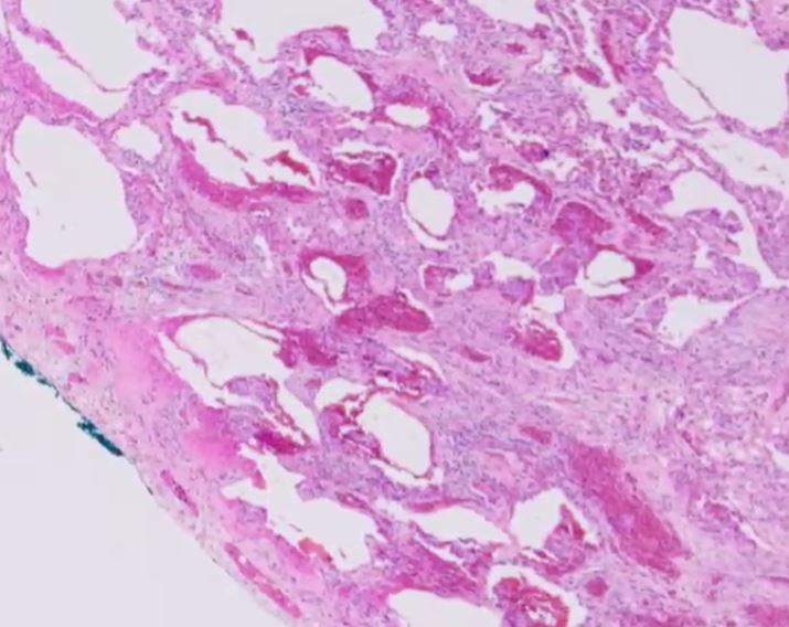
Small Airway Disease – Organizing Pneumonia – Terminal Bronchiole
Areas of organizing pneumonia are frequently seen in hypersensitivity pneumonitis.
Courtesy Dr Yale Rosen
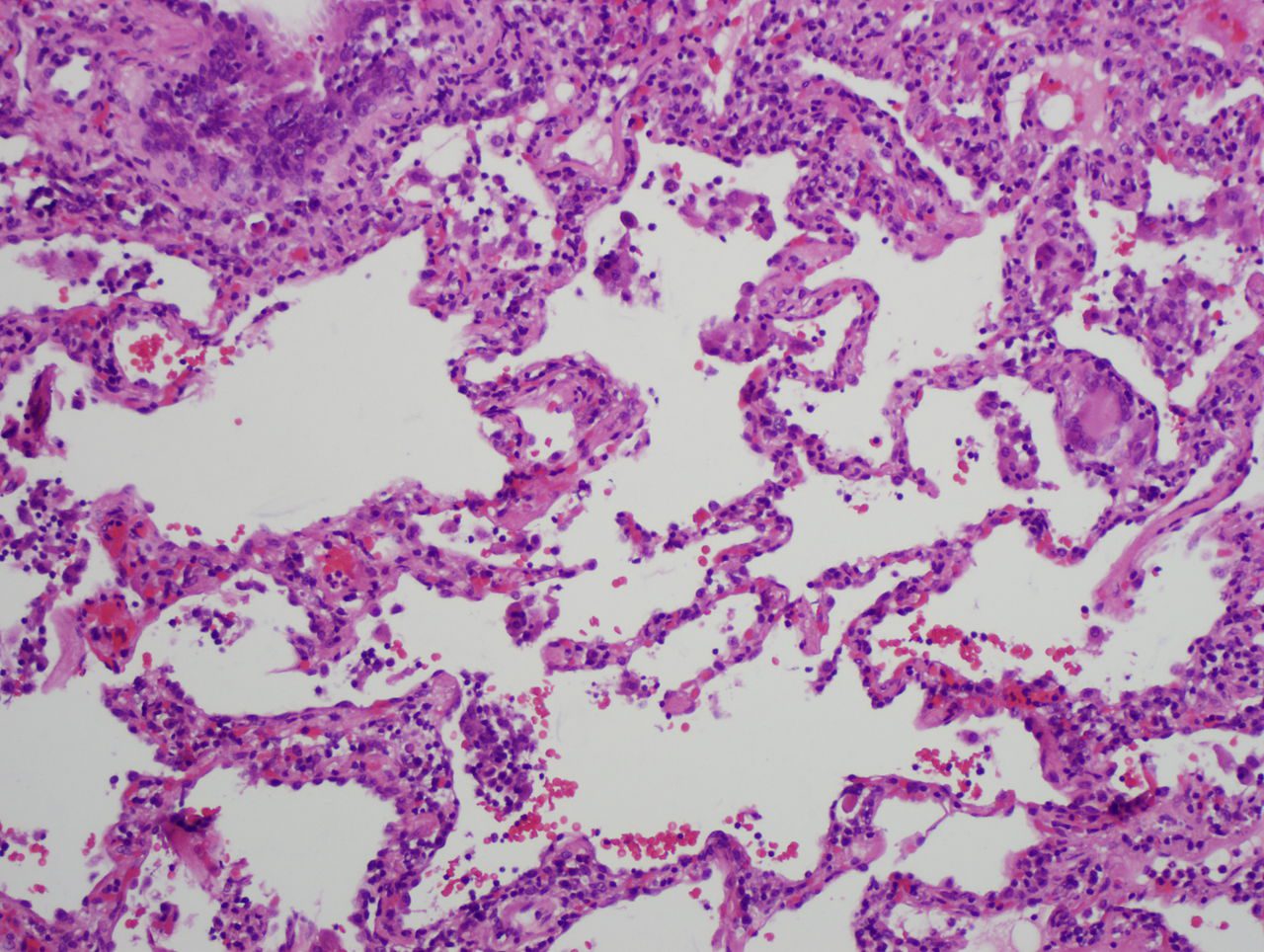
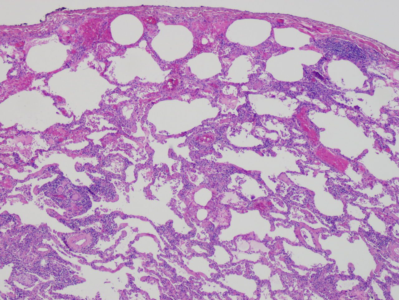
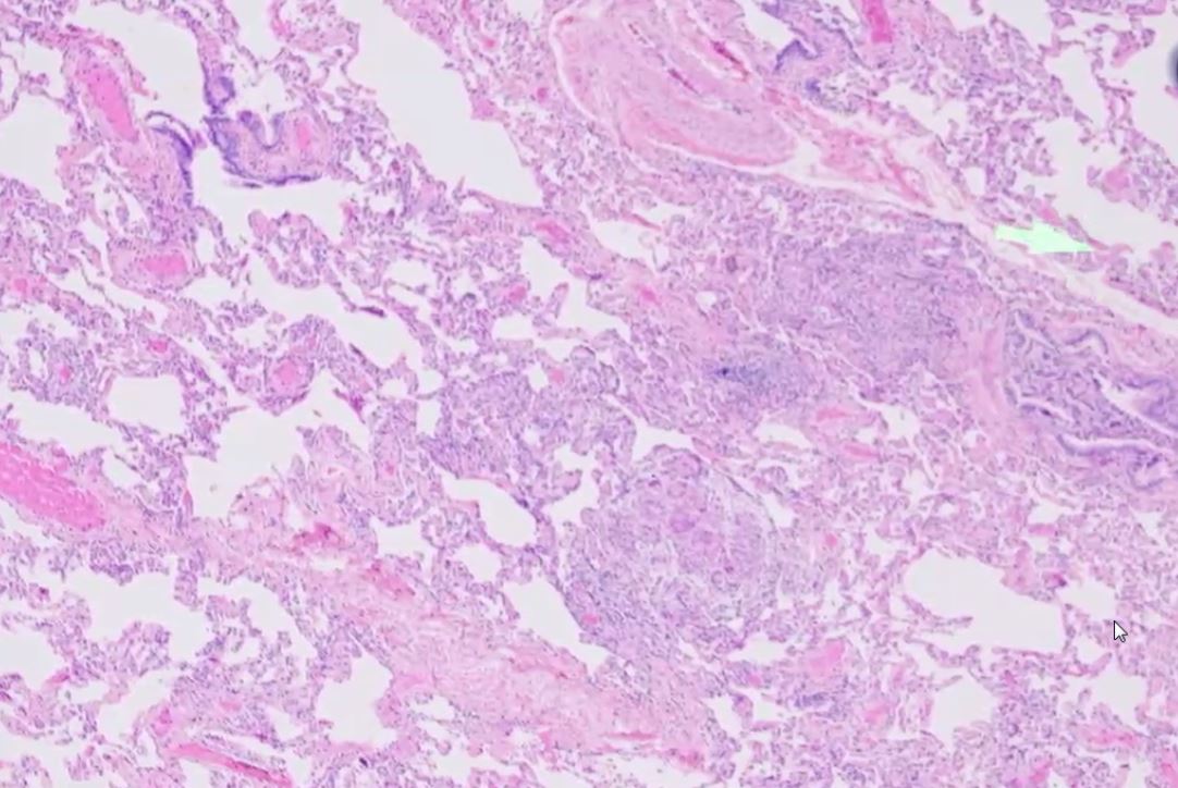
Interstitial Infiltrates
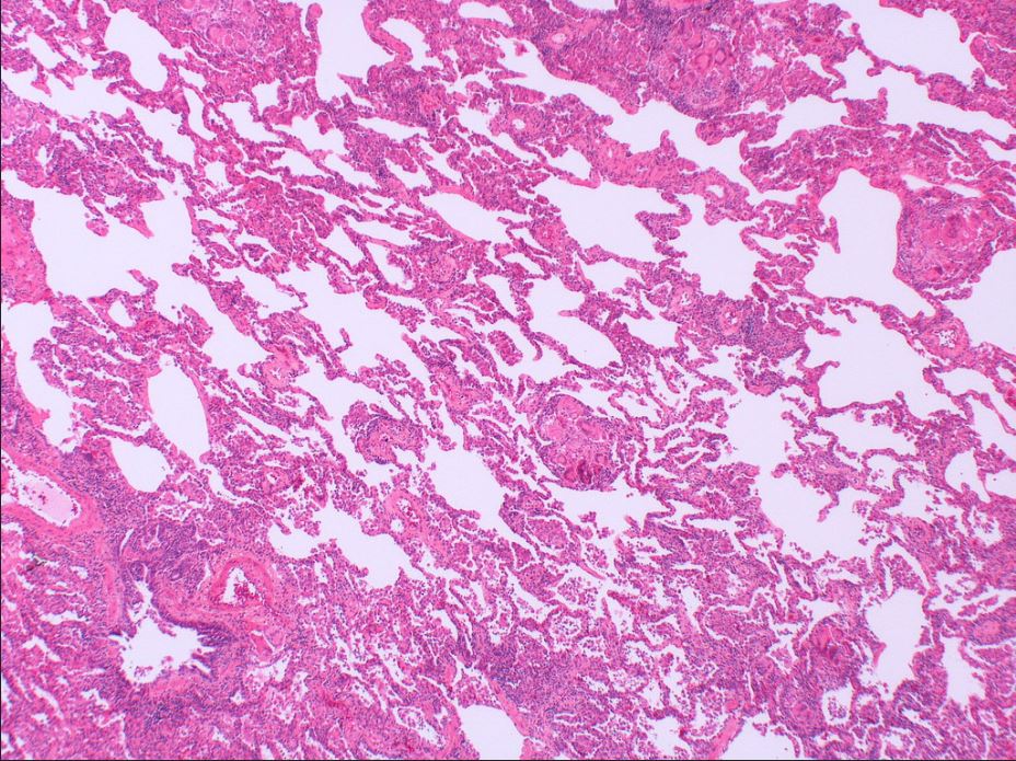
Mild interstital inflammation and multiple granulomas.
Courtesy Dr Yale Rosen
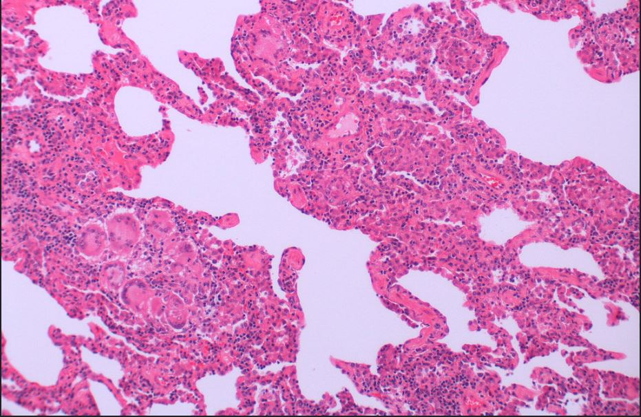
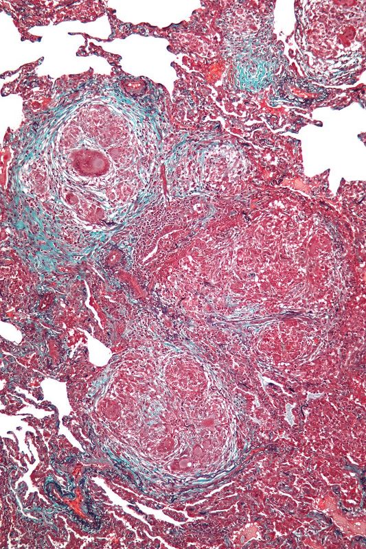
Interstitial chronic inflammation and a granuloma composed of multinucleate giant cells.
Courtesy Dr Yale Rosen
Mild interstital inflammation and multiple granulomas.
Courtesy Dr Yale Rosen

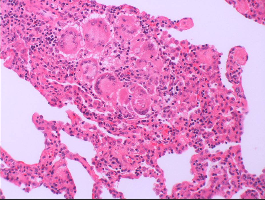
An interstitial granuloma composed of multinucleate giant cells.
Courtesy Dr Yale Rosen
Patchy chronic interstitial inflammation and a granuloma composed of multinucleate giant cells.
Courtesy Dr Yale Rosen
Courtesy Wikiwand
web lungs 435
Courtesy Mutleysmith WikipediaL
Courtesy Nephron
Ashley Davidoff MD TheCommonVein.net
Ashley Davidoff MD TheCommonVein.net
Links and References
- Videos