
NB
IPF in the upper lobes
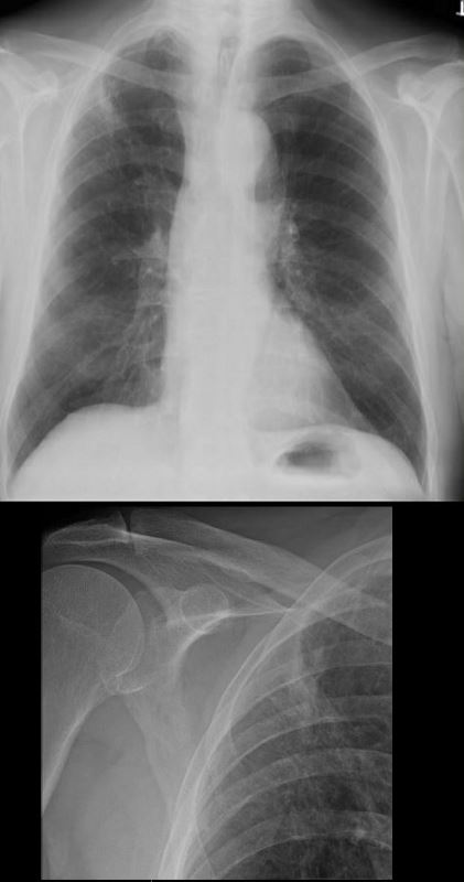
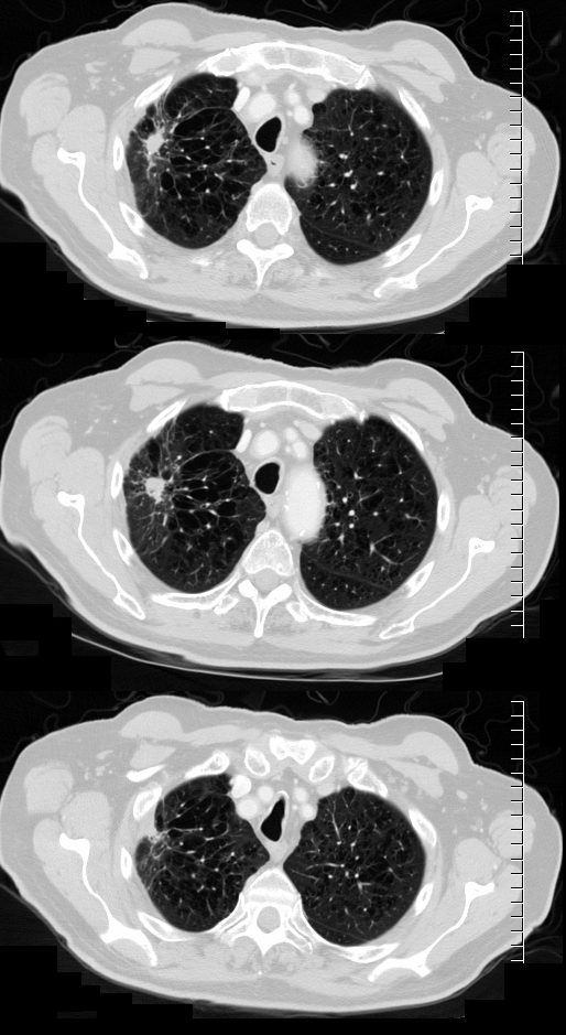
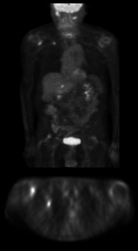
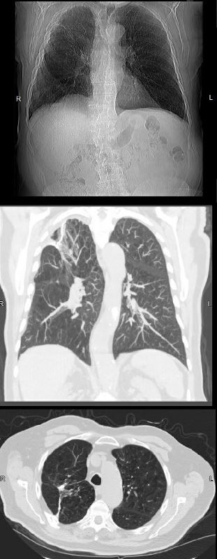
83 year old man with COPD and emphysema presented with bilateral shoulder pain. X-ray examination of his shoulders revealed a stellate nodule in his LUL. CT showed evidence of emphysema with a 15x14mm nodule with evidence of surrounding scarring and retraction.
On PET scan the nodule was PET avid with an SUV of 4.5 consistent with a malignant neoplasm
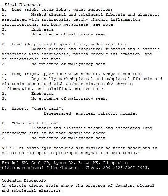
Surgery was performed and the pathology was consistent with idiopathic pleuroparenchymal fibroelastosis
Follow up CTs 5 years following surgery have shown stability
Ashley Davidoff MD
83 year old man with COPD and emphysema presented with bilateral shoulder pain. X-ray examination of his shoulders revealed a stellate nodule in his LUL. CT showed evidence of emphysema with a 15x14mm nodule with evidence of surrounding scarring and retraction.
On PET scan the nodule was PET avid with an SUV of 4.5 consistent with a malignant neoplasm
Surgery was performed and the pathology was consistent with idiopathic pleuroparenchymal fibroelastosis
Follow up CTs 5 years following surgery have shown stability
Ashley Davidoff MD
5 YEARS POST OP
83 year old man with COPD and emphysema presented with bilateral shoulder pain. X-ray examination of his shoulders revealed a stellate nodule in his LUL. CT showed evidence of emphysema with a 15x14mm nodule with evidence of surrounding scarring and retraction.
On PET scan the nodule was PET avid with an SUV of 4.5 consistent with a malignant neoplasm
Surgery was performed and the pathology was consistent with idiopathic pleuroparenchymal fibroelastosis
Follow up CTs 5 years following surgery have shown stability
Ashley Davidoff MD
Courtesy EPOS from the European Society of RadiologyReferences: RADIODIAGNÓSTICO , HOSPITAL CLÍNIC BARCELONA – Barcelona/ES
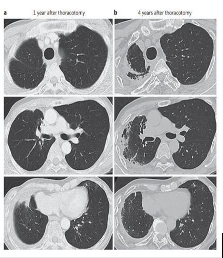
1 year after surgical resection of the right
upper lobectomy and middle lobe partial
resection. b A subpleural and parenchymal
lesion developed in the remaining right
lung 4 years after surgical lung resection.
Respiration 2017;94:431–441
References and Links
Current Respiratory Medicine Reviews
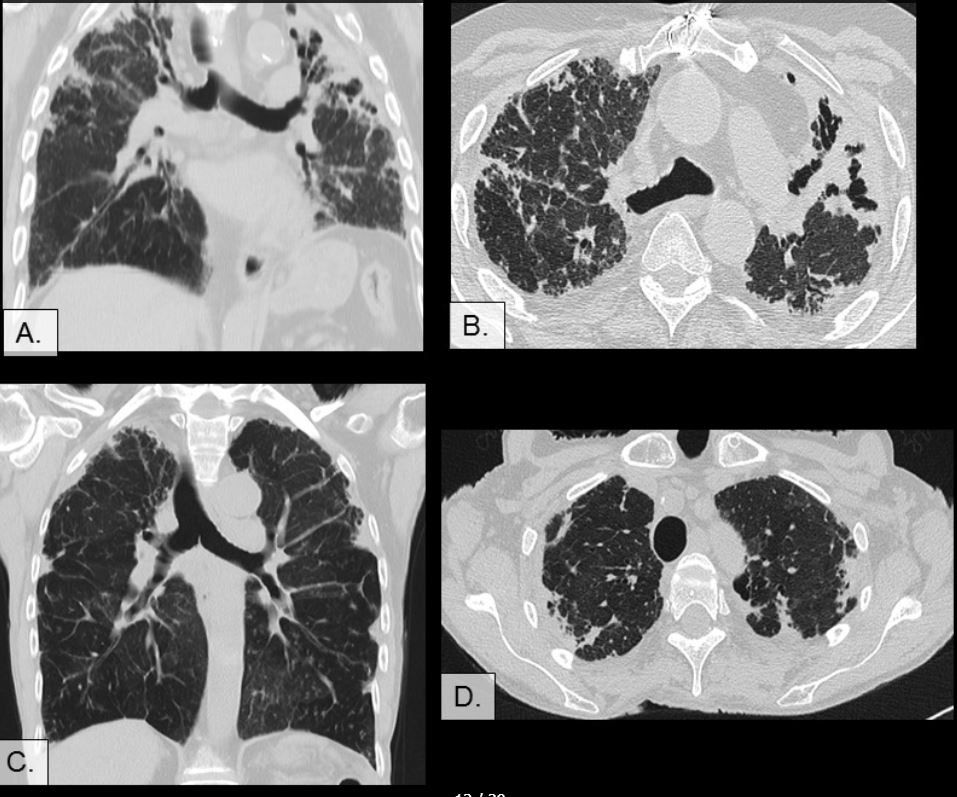
J Bronchology Interv Pulmonol. Case report with Spiculated nodule with PET positivity
Respiration – Unilateral Disease
-
- Usual interstitial pneumonia (UIP)
- Nonspecific interstitial pneumonia (NSIP)
- Cryptogenic organizing pneumonia (COP)
- Desquamative interstitial pneumonia (DIP)
- Respiratory bronchiolitis-interstitial lung disease (RB-ILD)
- Acute interstitial pneumonia (AIP)
- Lymphoid interstitial pneumonia (LIP)
- Idiopathic pleuroparenchymal fibroelastosis (PPFE)