
Classification
-
- Usual interstitial pneumonia (UIP)
- Nonspecific interstitial pneumonia (NSIP)
- Cryptogenic organizing pneumonia (COP)
- Desquamative interstitial pneumonia (DIP)
- Respiratory bronchiolitis-interstitial lung disease (RB-ILD)
- Acute interstitial pneumonia (AIP)
- Lymphoid interstitial pneumonia (LIP)
- Idiopathic pleuroparenchymal fibroelastosis (PPFE)
Practical Approach
- Chronically fibrosing (UIP and NSIP)
- Smoking related (DIP and RB-ILD)
- Acutely presenting (COP and AIP)
Based on Findings
Reticulations
UIP NSIP
Clinical Entities
asbestosis
collagen vascular disease
drug toxicities
chronic hypersensitivity pneumonitis
IPF
Distribution
upper mid lower
peripheral central
segmental diffuse
Homogeneous Heterogeneous
Subpleural Sparing
Ground Glass
Ancillary findings
plaques
esophagus
hyperdense liver
Nodules
Sarcoidosis
Hypersensitivity Pneumonia
Metastases
Cysts
Langerhans Cell Histiocytosis
LAM
Airspace Disease
OP COP DIP
RADIOLOGY
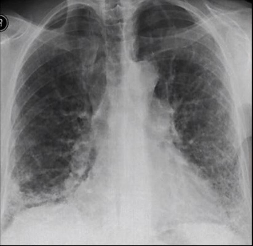
Frontal view exemplifies a diffuse reticular pattern of ILD
Courtesy You Tube on ILD
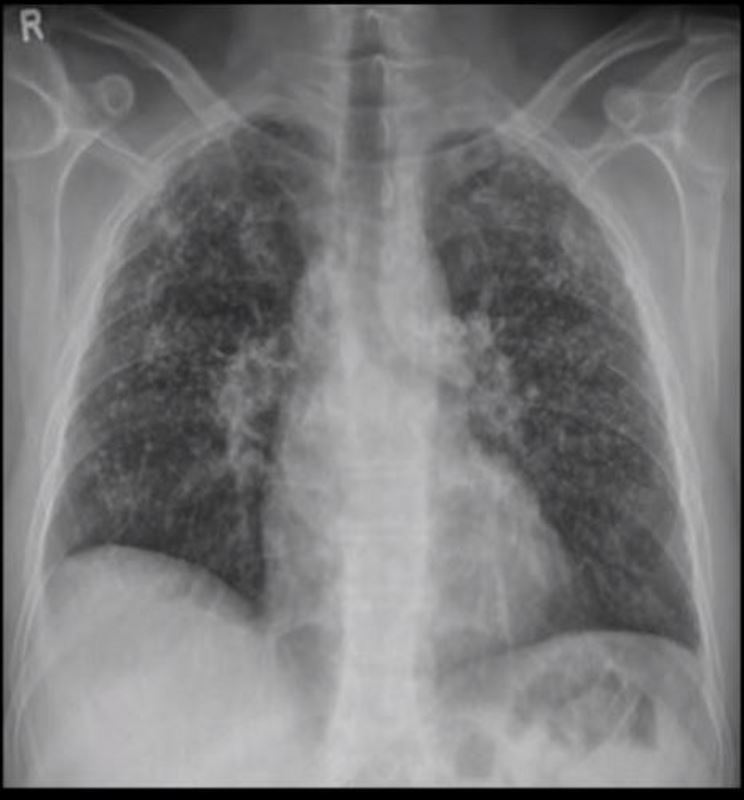
Frontal view exemplifies a diffuse nodular pattern of ILD such as is seen in silicosis and sarcoidosis
Courtesy You Tube on ILD
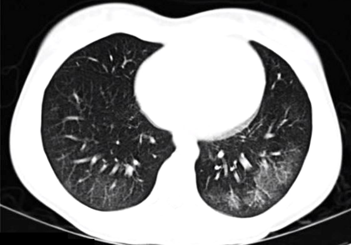
Ground glass infiltrates are one of the features of interstitial lung disease with increased density without obscuring the airways and vessels Courtesy You Tube on ILD
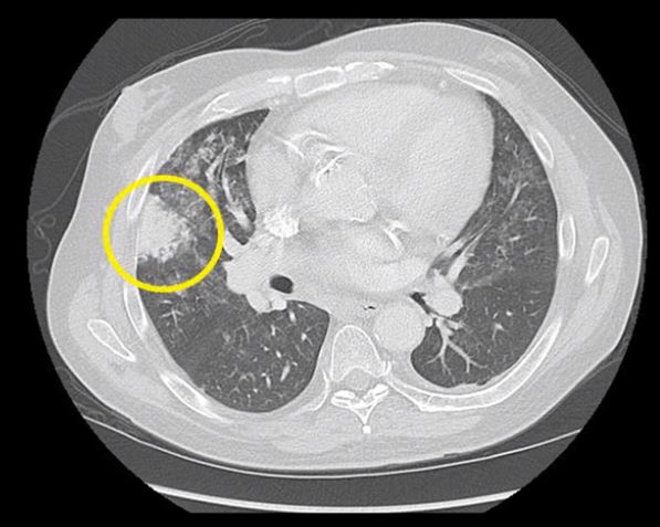
Focal consolidations or infiltrates are another feature of interstitial lung disease (yellow ring with obscuration of the airways and vessels Courtesy You Tube on ILD
Crazy paving in ILD is another another CT feature of interstitial lung disease and is characterised by diffuse ground glass caused by a combination of interlobular septal and intralobular septal thickening resulting well demarcated patchy densities in the lungs. Courtesy You Tube on ILD
Micronodules in ILD is another another CT feature of interstitial lung disease and is characterised by nodules of a variety of shapes and sizes and likely centrilobular in origin. Sometimes they are ill defined such as in this case. You Tube on ILD
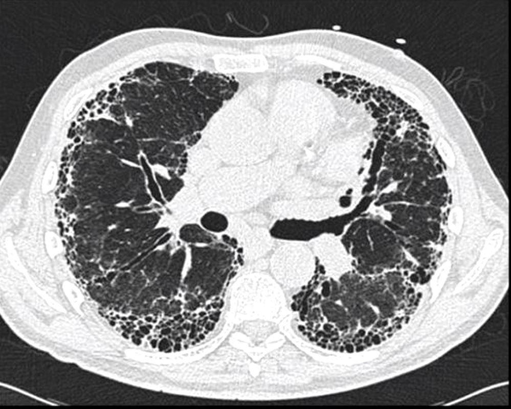
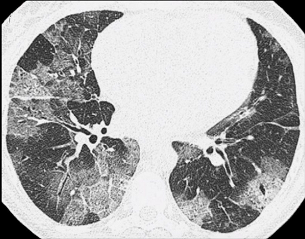
Honeycomb changes in ILD is a feature on CT of interstitial lung disease and is characterised by clusters of stacked cystic air spaces in the periphery and predominantly posteriorly at the bases of the lungs . They likely represent small airways that have been denuded of the their alveoli apparatus. It is most commonly associated with idiopathic pulmonary fibrosis (IPF) and its radiological equivalent UIP – usual interstitial pneumonia. You Tube on ILD
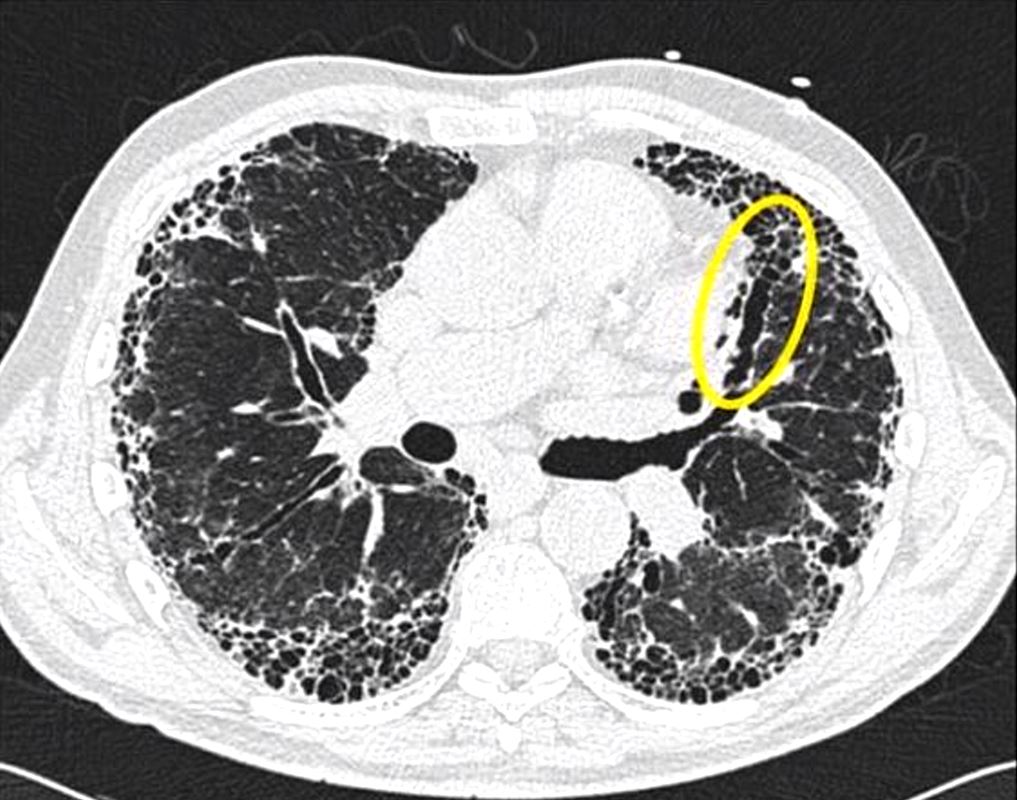
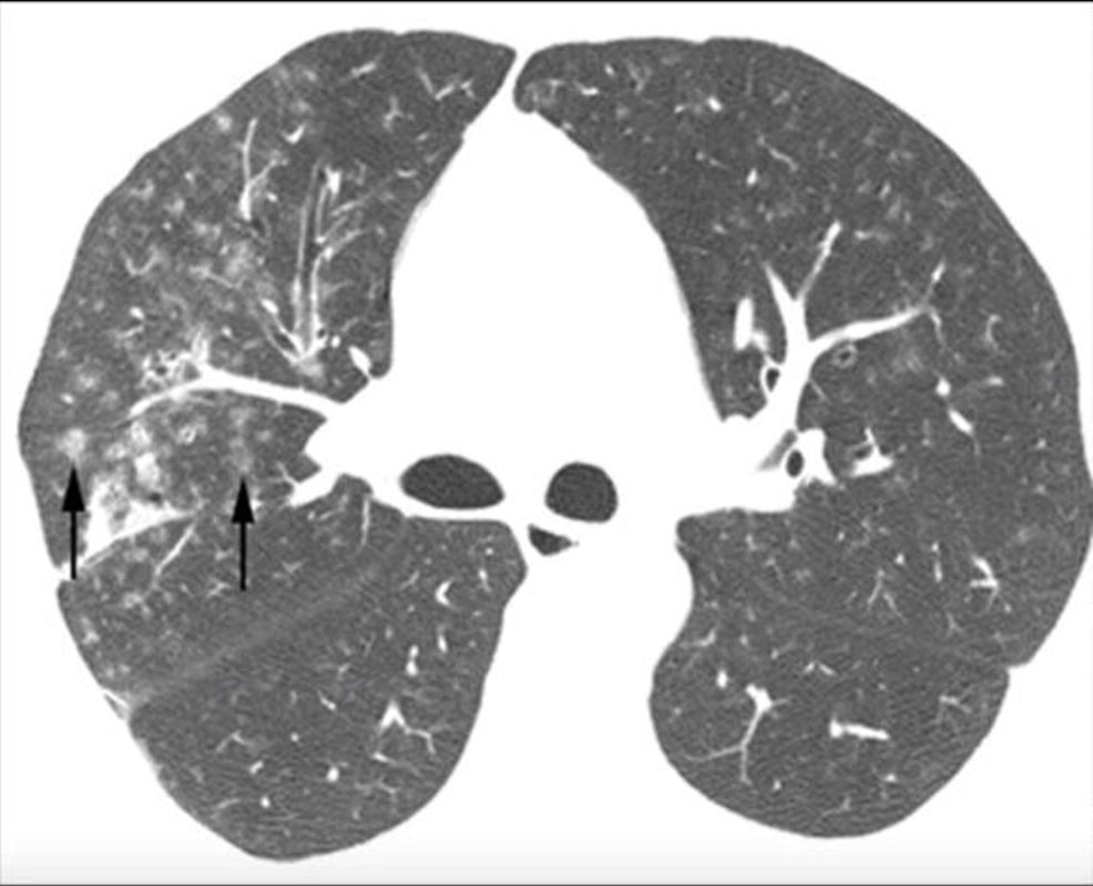
Traction bronchiectasis and bronchiolectasis (yellow ring) is as a result of fibrotic traction on the airways. You Tube on ILD
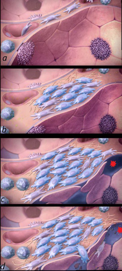
The first image shows the normal squamous cell lined alveolus with type 1 and 2 pneumocytes. An inciting agent causes white cells and myofibroblasts to enter into the interstitium with progressive fibrosis of the interstitium (b). The alveolar cells are also destroyed and the epithelium is denuded (c red asterisk) and are not regenerated allowing the white cells and myofibroblasts to enter the alveolus (d) and with continued fibrosis and destruction of the alveoli.
Idiopathic Pulmonary Fibrosis by Alan Sklar
Pathologically most of the ILD’s are characterized by inflammation and fibrosis resulting in injury and structural alteration of the airways and alveoli