
Source
Signs in Thoracic Imaging
Journal of Thoracic Imaging 21(1):76-90, March 2006.
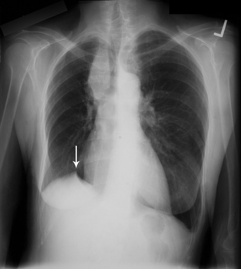
This sign, first documented by Kattan et al in 1980,49 is an ancillary sign of upper lobe collapse, depicted as a triangular opacity projecting superiorly over the medial half of the diaphragm, at or near its highest point (Fig. 16). It is most commonly related to the presence of an inferior accessory fissure.49,50 Although the mechanism is not certain, one theory suggests it is due to the negative pressure of upper lobe atelectasis causing upward retraction of the visceral pleura and extrapleural fat protruding into the recess of the fissure.49 A recent retrospective analysis of patients who have undergone upper lobectomies suggests that the prevalence of the sign increases in the ensuing weeks after intervention and documents its utility in specifically recognizing the type of surgery.51