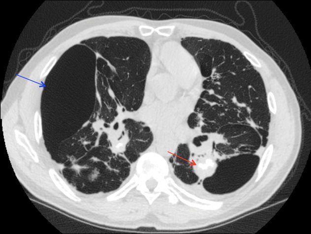
CXR (PA view) shows significant linear and reticular interstitial thickening (red arrow) in bilateral lungs. Several large bullae (blue arrow) scattered bilaterally, most notably in the RML. Increased opacity in left upper perihilar region (green arrow) consistent with a diagnosis of silicosis, complicated by progressive massive fibrosis. Differential diagnosis includes other ILDs, atelectasis, or pneumonia. Courtesy Maegan Lu, Jonathan Scalera, MDCT chest without contrast in the coronal projection at the level of the hilum shows eggshell calcifications in the hilar and mediastinal lymph nodes (red arrow) consistent with a diagnosis of silicosis, complicated by progressive massive fibrosis. Bullous disease (blue arrow) is also seen bilaterally, right greater than left. Differential diagnosis includes coal-worker’s pneumoconiosis, sarcoidosis, and blastomycosis. Courtesy Maegan Lu, Jonathan Scalera, MDCT chest without contrast in the axial projection at the level of the ascending aorta shows eggshell calcifications in the hilar and mediastinal lymph nodes (red arrow) consistent with a diagnosis of silicosis, complicated by progressive massive fibrosis. Bullous disease (blue arrow) is also seen bilaterally, right greater than left. Differential diagnosis includes coal-worker’s pneumoconiosis, sarcoidosis, and blastomycosis. Courtesy Maegan Lu, Jonathan Scalera, MD