
- Mosaic attenuation is an
- imaging pattern
- variable lung attenuation
- results in a heterogeneous appearance of the parenchyma.
- sometimes it is caused by air trapping
- sometimes by perfusion abnormalities
- sometimes normal
- imaging pattern
Nutshell Buzz
patchwork
differing pulmonary attenuation
Mosaic attenuation refers to a patchy or uneven pattern of lung attenuation seen on CT imaging. It appears as areas of increased lung density (hyperattenuation) adjacent to areas of decreased lung density (hypovascular or hypodense regions). This pattern can be caused by several different factors, such as:
- Small Airway Disease: Mosaic attenuation can be associated with diseases affecting the small airways, such as chronic obstructive pulmonary disease (COPD) or bronchiolitis obliterans.
- Air Trapping: In conditions like asthma or other obstructive lung diseases, air may be trapped in certain lung regions during exhalation, leading to areas of hyperinflation and hyperattenuation.
- Pulmonary Embolism: Blood clots in the pulmonary arteries can lead to mismatched lung perfusion and cause mosaic attenuation patterns.
- Pulmonary Edema: Fluid accumulation in the lungs due to heart failure or other causes can result in mosaic attenuation.
- Infection and Inflammation: Certain lung infections and inflammatory conditions can also contribute to mosaic attenuation.
Ashley Davidoff
TheCommonVein.net
https://pubs.rsna.org/doi/full/10.1148/radiol.13120908
Ashley Davidoff MD TheCommonVein.net
Mosaic Attenuation Caused by Obstruction of Small Airways
- Air trapping on the other hand
- is an imaging and physiologic term to
- retained air in a part or parts of the lung
- more easily identified during expiration
- caused by
- obstruction
Mosaic Attenuation of a Secondary Lobule
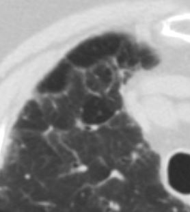
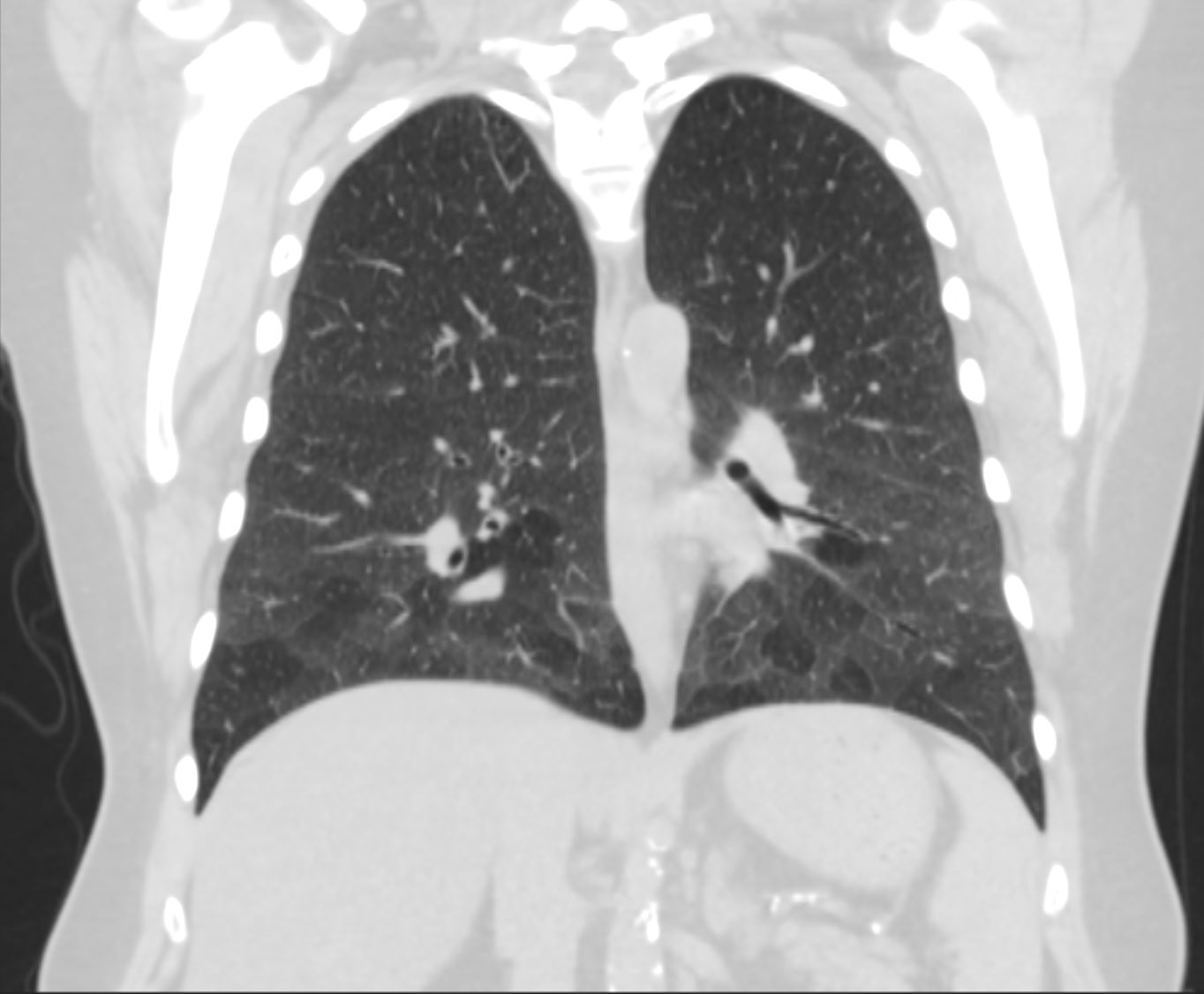
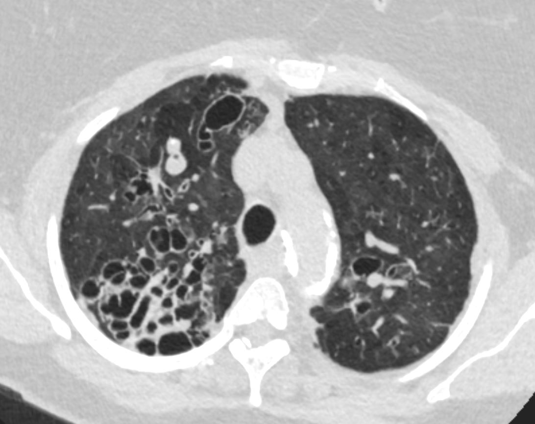
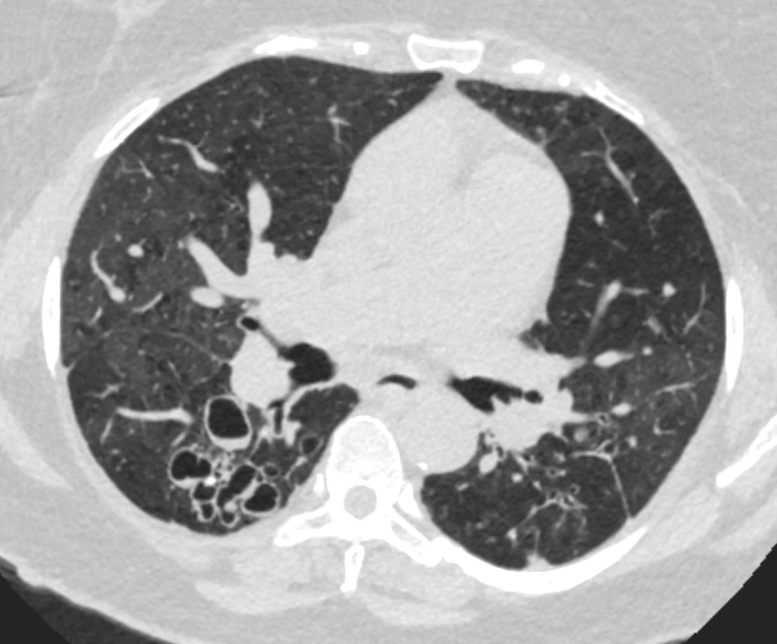
77F with long history of dyspnea and cough showing medium and small airway disease, centri-lobular nodules, para-septal nodules ground glass changes and mosaic attenuation Diagnosis includes Stage 3 sarcoidosis
Ashley Davidoff
TheCommonVein.net
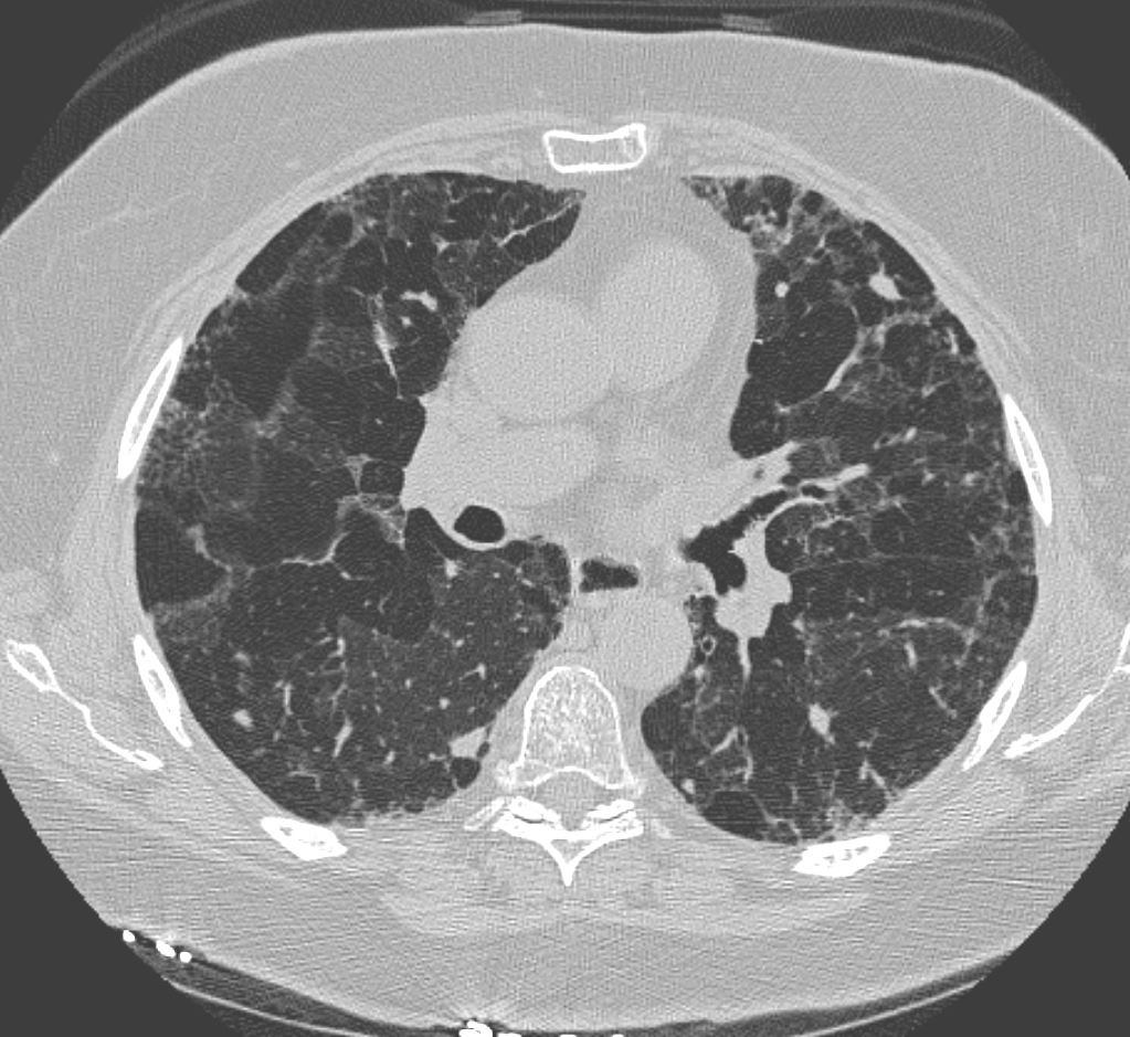
77F with long history of dyspnea and cough showing medium and small airway disease, centri-lobular nodules, para-septal nodules ground glass changes and mosaic attenuation Diagnosis includes Stage 3 sarcoidosis
Ashley Davidoff TheCommonVein.net
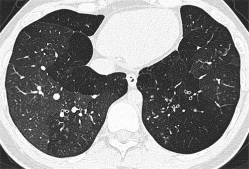
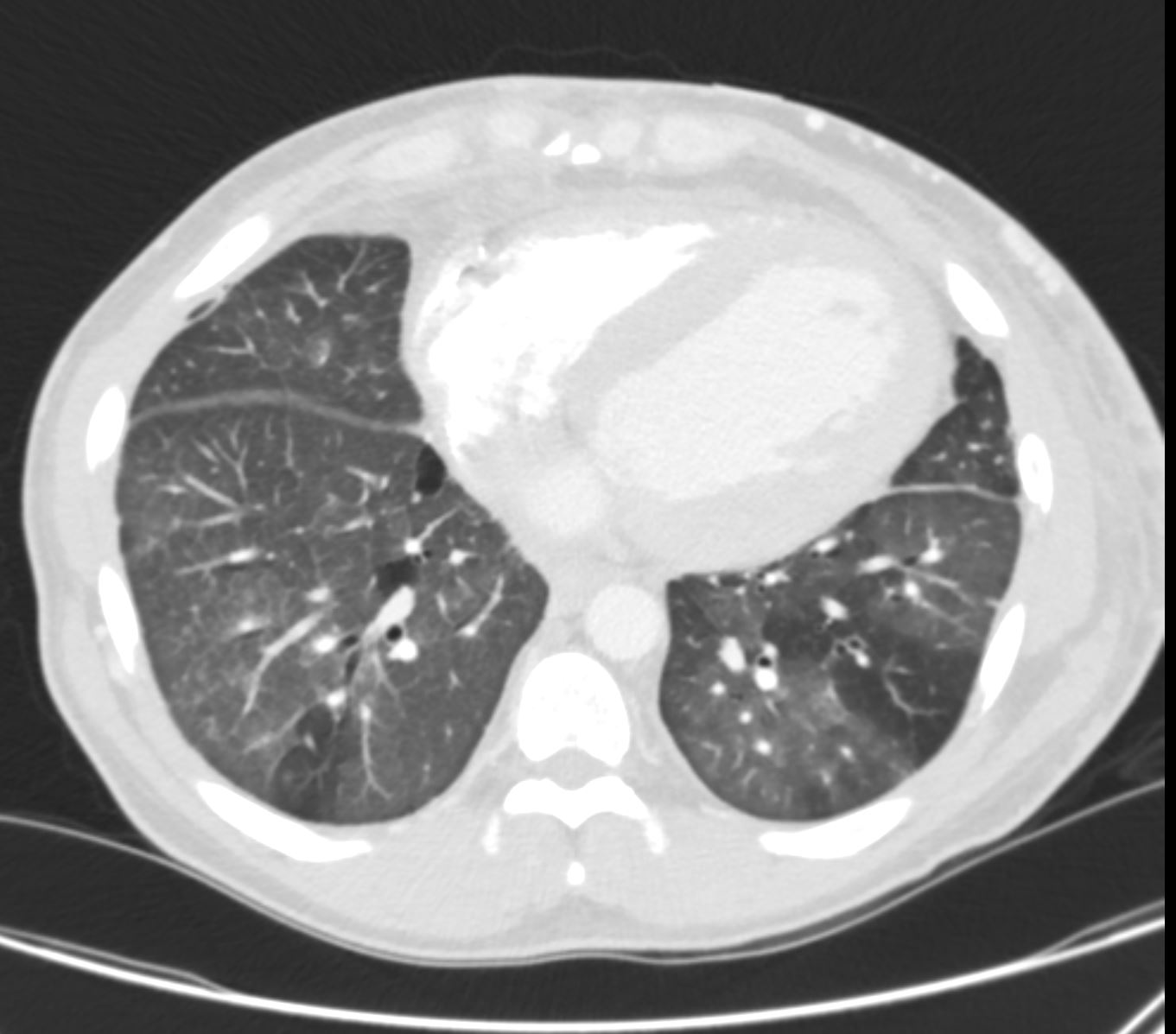
Mosaic Attenuation -Due to Mucoid Impaction COPD
Ashley Davidoff TheCommonVein.net bronchioles 004
Ashley Davidoff TheCommonVein.net bronchioles 003
in a patient with COPD – Small Airways are obstructed and air is trapped
Ashley Davidoff MD TheCommonVein.net bronchioles 002
Small Airways are filled with mucus in a patient with COPD – Note centrilobular impaction of mucus Small Airways are obstructed and air is trapped
Ashley Davidoff TheCommonVein.net bronchioles 001
Mosaic Attenuation with Bronchiectasis
Ashley Davidoff MD TheCommonVein.net bronchiectasis 006
Ashley Davidoff MD TheCommonVein.net bronchiectasis 009
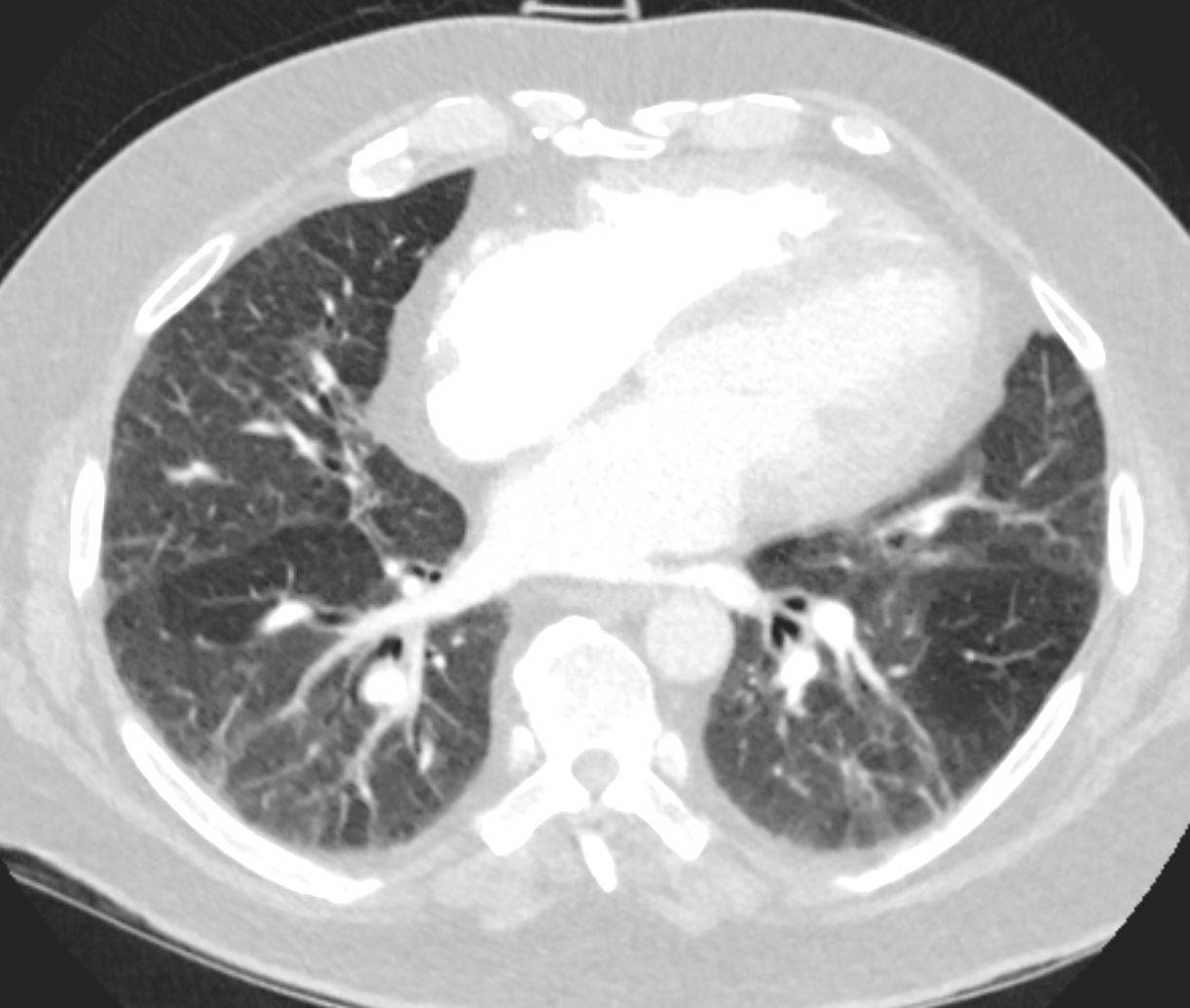
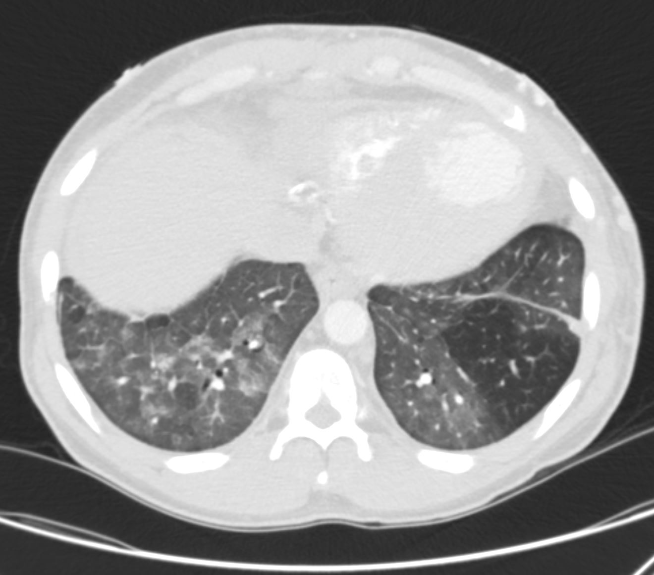
Mosaic Attenuation in CHF

Ashley Davidoff MD TheCommonvein.net 50-005-CT

Ashley Davidoff MD TheCommonvein.net 50-006-CT
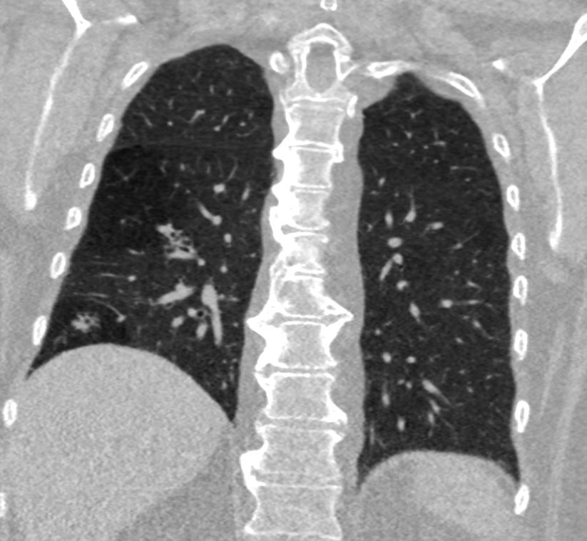
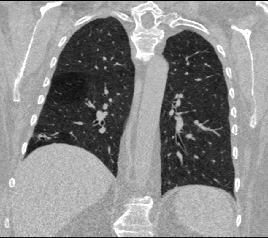
Mosaic Attenuation in a patient with SLE thought to represent small vessel disease
Ashley Davidoff TheCommonVein.net
Ashley Davidoff TheCommonVein.net
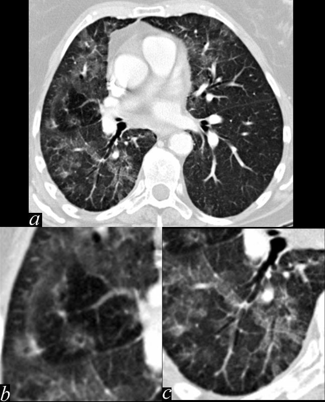
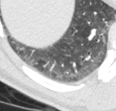
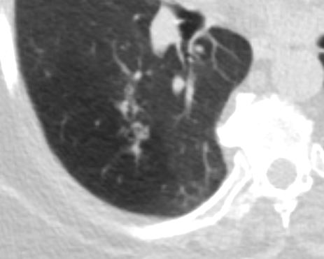
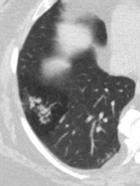
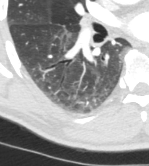
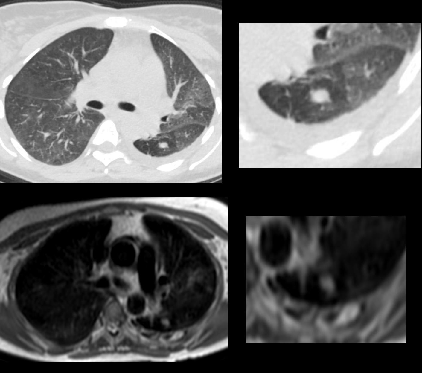
This series of images shows some subtle changes that reflect the local control of blood flow to a small segment of the right middle lobe. Note that in image a, there is a small area of increased lucency (blacker) in the right lung just lateral to the vessels of the right hilum. This region is highlighted in b. Note also that in b, the rapid diminution of the size of the blood vessel to that subsegment when compared to the size change of the vessels in the image in c. The lucent appearance of the lung suggests air trapping and the vasoconstriction reflects decreased perfusion – ie with decreased ventilation there is an associated consequent associated decrease in perfusion.
47170c01.800 bronchocentric inflammation lung bronchovascular bundle chest inflammation peribronchial halo air trapping mosaic perfusion ground glass changes alveolar change air bronchogram acute bronchovascular inflammation ddx allergic collagen vascular disease infection CTscan
Ashley Davidoff MD TheCommonVein.net
Ashley Davidoff MD TheCommonVein.net
Ashley Davidoff MD TheCommonVein.net
Ashley Davidoff MD TheCommonVein.net 002-CT-mucoid-impaction
Examples