
There are three types of nerve fibers that connect the lungs to the autonomic nervous system. Autonomic afferent fibers travel to the vagus nerve via the pulmonary plexuses, originating in the airways and the lungs. They transmit information from stretch receptors. Parasympathetic efferent fibers are all contained in the vagus nerve. They carry motor impulses to the airways and lungs which cause bronchiolar muscle contraction, gland secretion, and vasodilatation. The third group are the sympathetic efferent fibers which relax bronchial muscles, inhibit glandular secretion, and cause vasoconstriction.
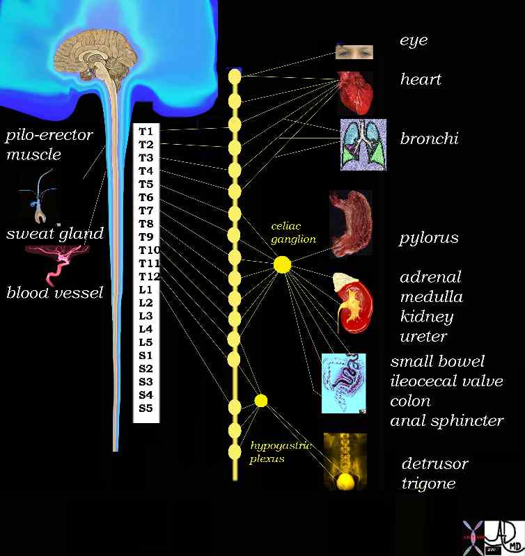 Sympathetic Efferent System Sympathetic Efferent System |
| 14798d03.800 sympathetic nervous sytem brain cerebral function autonomic nervous system efferent nerves eyes heart cardiac bronchi pylorus adrenal gland adrenal medulla kidney ureter small bowel ileocecal valve colon anal sphincter detrusor muscle of the urinary bladder trigone piloerector muscle sweat gland blood vessels arteries sympathetic ganglia sympathetic ganglion preganglionic sympathetic neurons nerve posganglionic sympathetic neurons Davidoff Art Davidoff MD |
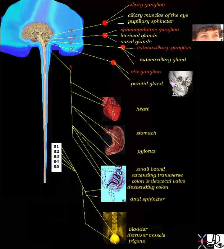 Parasympathetic Efferents Parasympathetic Efferents |
| 14798d06 brain control parasympathetic nervous system ciliary ganglion eyes ciliary muscle pupillary sphincter sphenopalatine ganglion lactrimal glands nasal glands submaxillary glands submaxillary ganglion otic ganglion parotid gland heart cardiac stomach pylorus small bowel colon anal sphincter bladder detrusor muscle trigone S1 S2 S3 S4 vagus nerve cranial nerves III oculomotor VII facial nerve V mandibular nerve IX glossophayngeal nerve control efferent system parasympathetic supply Davidoff Art Davidoff MD |
The walls of the bronchi and bronchioles are innervated by the autonomic nervous system. There are b2-adrenergic receptors in the bronchial epithelium and smooth muscle. The b2 receptors are responsible for bronchodilatation and secretion. The a1 adrenergic receptors, on the other hand inhibit secretion. VIP is another mediator that has been shown to cause vasodilatation.
Applied Anatomy
Bronchospasm classically seen in asthmatics is a failure of normal mechanisms to optimize air flow, and it is usually caused by bronchospastic behavior of the smooth muscle induced most commonly by allergens and exercise.
The nerves are not visible and their position in the chest is implied by their relationship to the other structures in the chest. For example the left recurrent laryngeal nerve curls around the left main stem bronchus. When the left atrium enlarges significantly, it can impinge on the left recurrent laryngeal nerve and cause hoarseness. The Pancoast tumor in the apex of the lung can affect the sympathetic system and result in Horner’s syndrome, with meiosis, anhidrosis, and hoarseness.
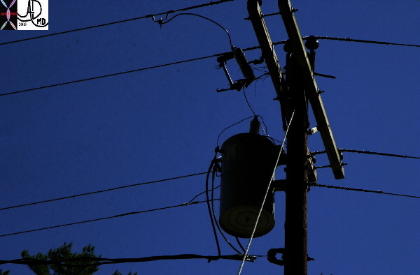 Contolling mechanisms Contolling mechanisms |
| This is a photograph of an overhead telephone wire with a control box – reminiscent of our nervous system with its central controlling ganglion. Courtesy Ashley Davidoff MD 70030 |