
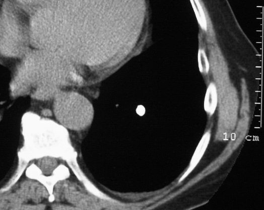
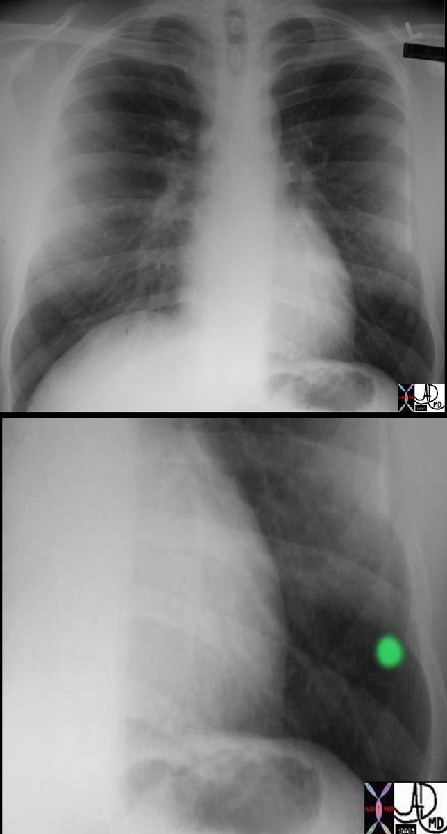
Classical Calcified Granuloma
Ashley Davidoff
TheCommonVein.net
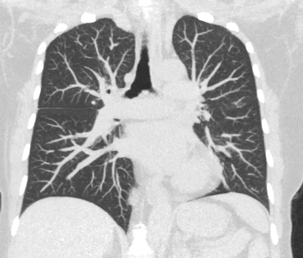
Granulomatous Tree in Bud – Likely TB possibly MAI
Ashley Davidoff
TheCommonVein.net
Ashley Davidoff
TheCommonVein.net
Ashley Davidoff
TheCommonVein.net
Ashley Davidoff
TheCommonVein.net
Central Calcification
Ashley Davidoff
The CommonVein.net
42244c01
Ashley Davidoff MD
TheCommonVein.net
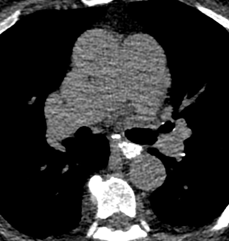
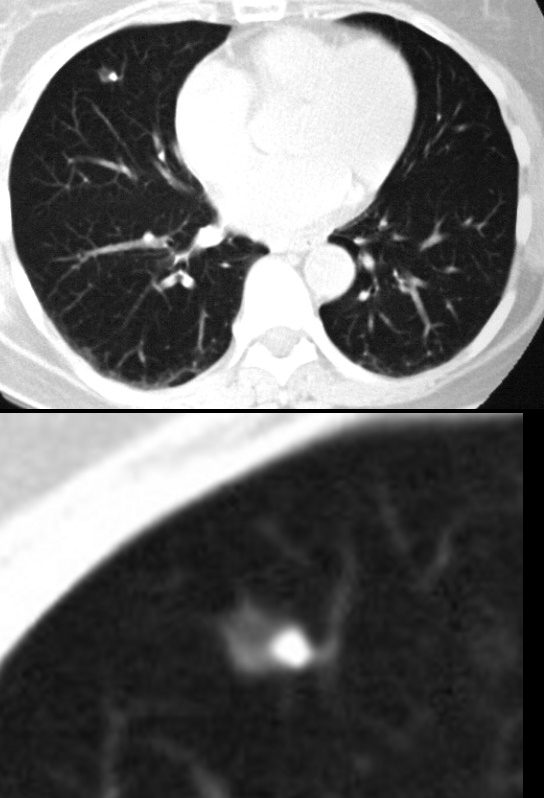
Central Calcification – Sarcoidosis
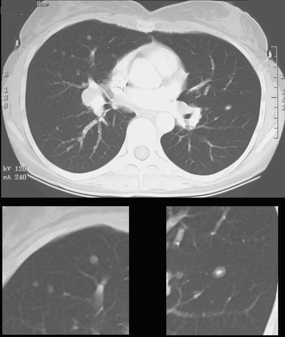
CT scan shows a 6mm nodule with central calcification in the ligula and ground glass nodules in the middle lobe
Ashley Davidoff
TheCommonVein.net
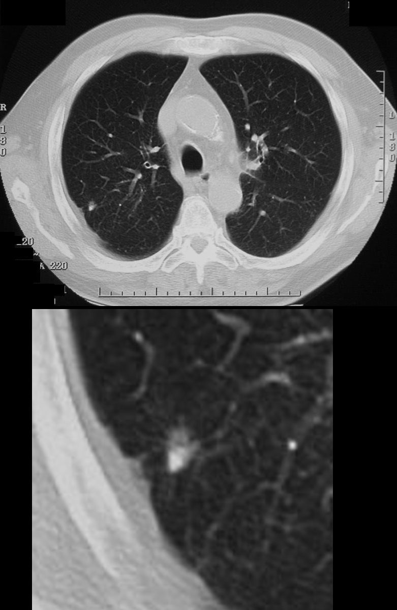
70060c
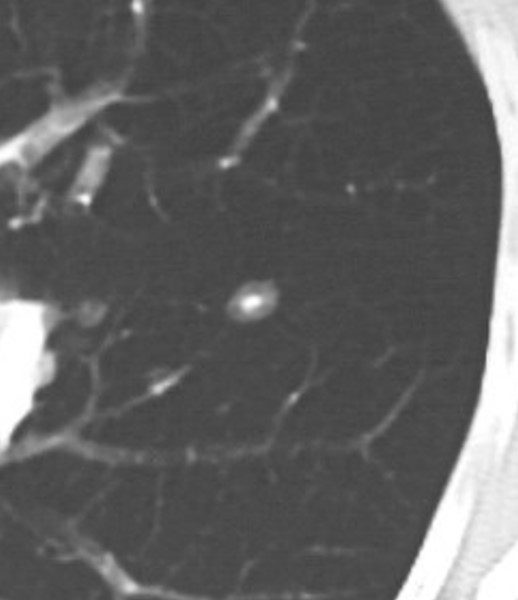
CT scan shows a 6mm nodule with central calcification
Ashley Davidoff
TheCommonVein.net
70060b
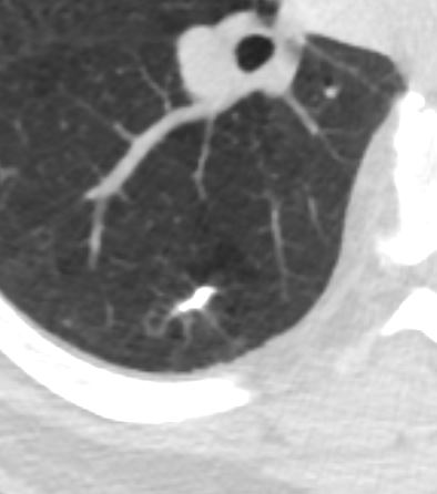
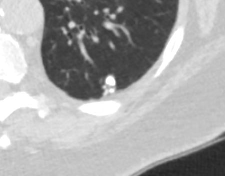
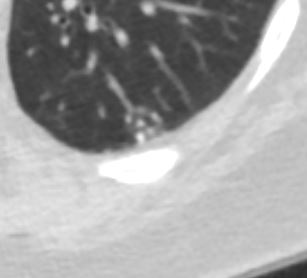
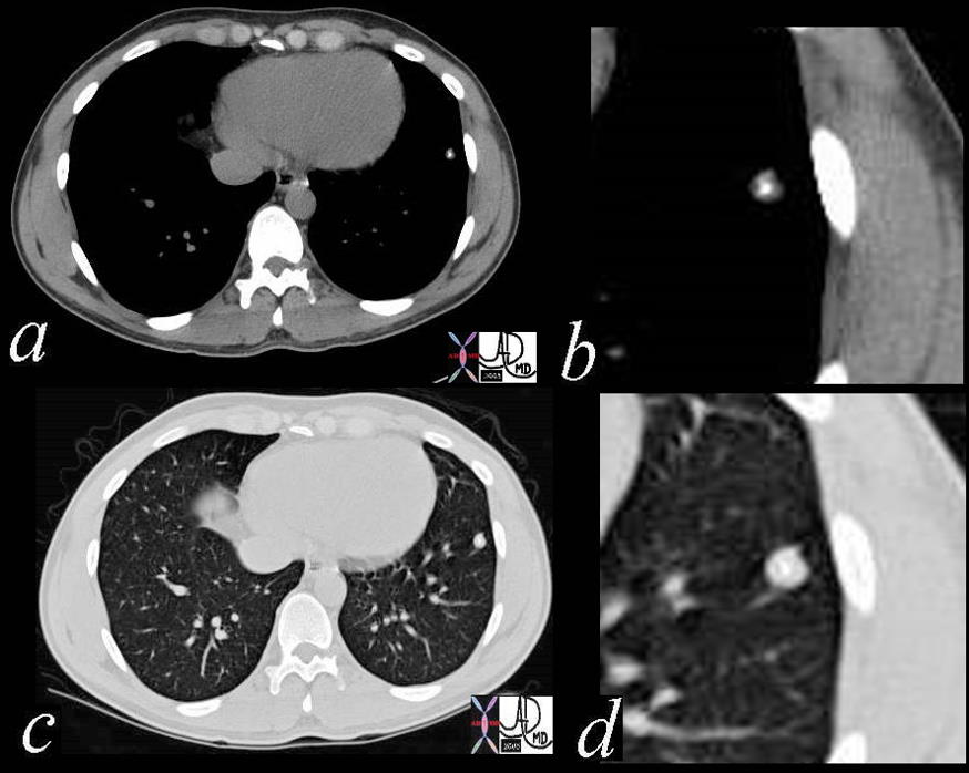
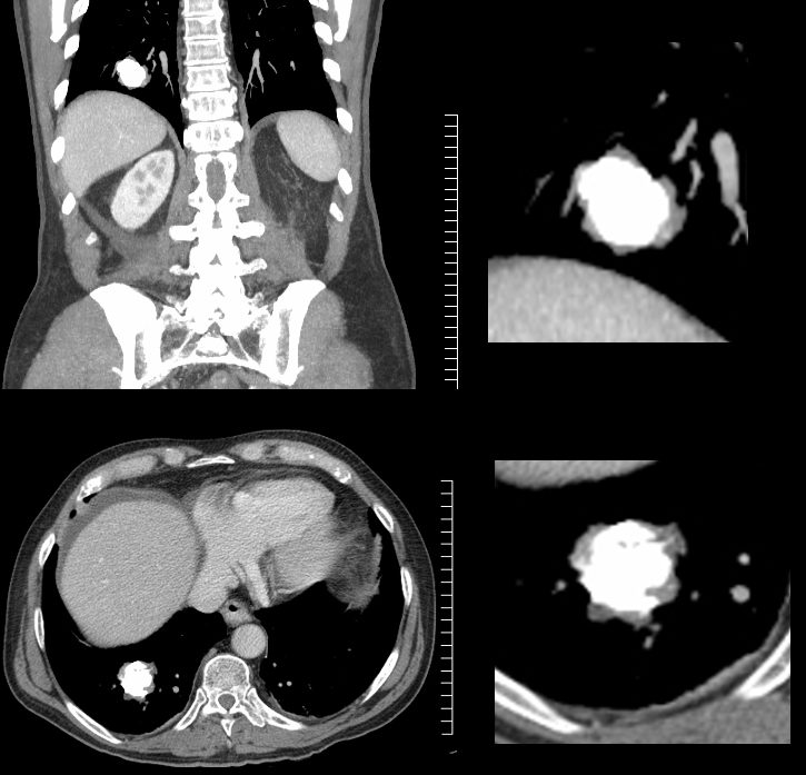
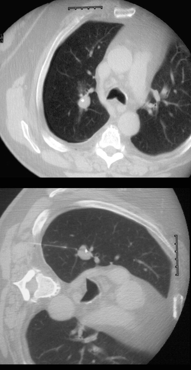
Central Calcification Amyloid
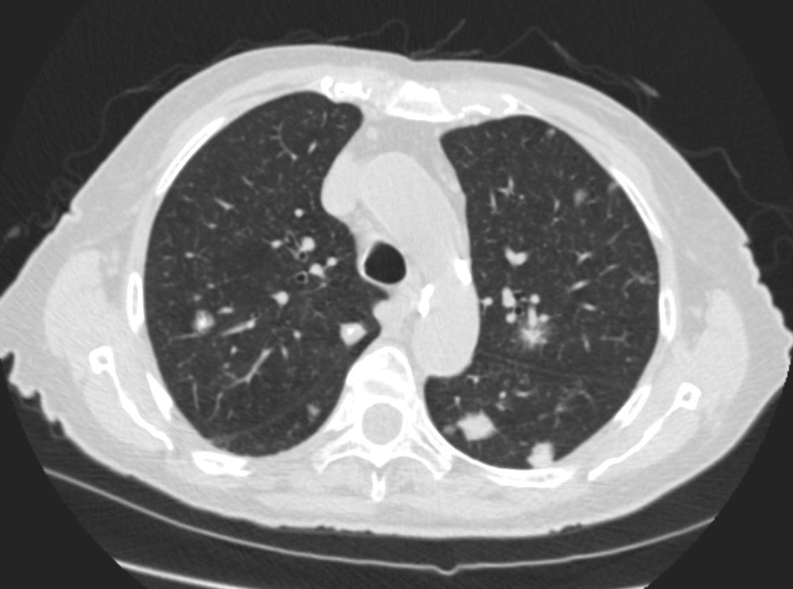
The nodule on the right close to the spine is fissural based (see next image)
Ashley Davidoff Boston Medical Center TheCommonvein.net LV-006
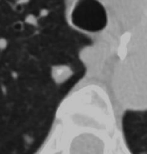
Axial CT images through the right upper lobe shows a solid amyloid nodule with central calcification abutting the major fissure. These features, although not pathognomonic are characteristic. Sarcoidosis would be a radiological consideration as well
Ashley Davidoff Boston Medical Center TheCommonvein.net LV-006
and amyloidoma
Ashley Davidoff TheCommonvein.net
hamartoma calcifications 004c stable
Eccentric Calcification
Unknown Diagnosis
Ashley Davidoff
TheCommonVein.net
Benign Eccentric Lobular Calcification Differential Hamartoma or Amyloid
Ashley Davidoff TheCommonvein.net
hamartoma 0001c01 86f
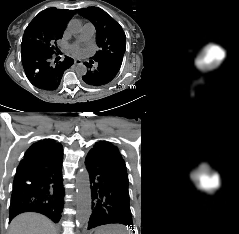
Malignant
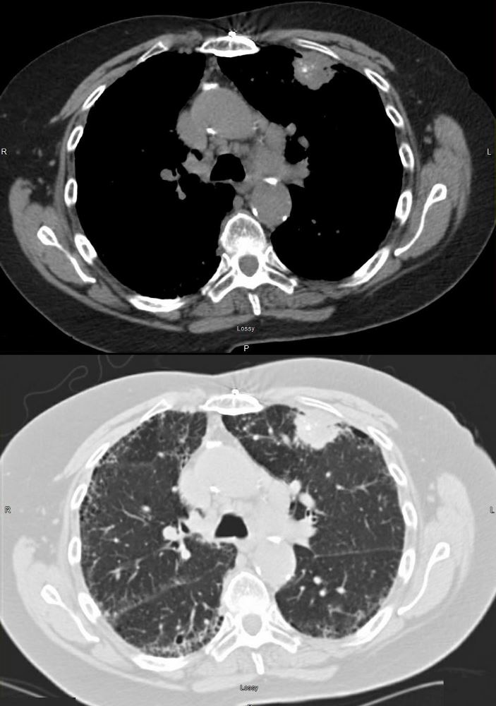
73 year old female with left mastectomy and IPF with new onset bilateral upper lobe masses. The LUL mass has scattered calcifications, and the right shows early cavitation.
Both lesions are PET positive
The LUL lesion was biopsied revealing squamous cell carcinoma with calcifications showing progressive growth and enlarging ipsilateral lymphadenopathy.
The right lesion shows progressive cavitation and enlargement likely a squamous cell carcinoma as well.
The ILD dominates in the periphery of the lower lobes but involves the upper lobes as well, reveals honeycombing in the right upper lobe, but shows no significant progression over the two years.
Ashley Davidoff MD TheCommonVein.net
Nodule with Eccentric Calcification
Biopsy showed Scar Carcinoma
Ashley Davidoff
TheCommonVein.net
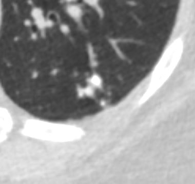
Eccentric Calcification
Benign
56 F with a middle lobe nodule with eccentric calcification which turned out to be granulomatous in origin
Ashley Davidoff
TheCommonVein.net
30252c
A Second Case
65 year male with lung nodule characterised by eccentric calcification that did not change over 2 year period
Ashley Davidoff
TheCommonVein.net
31211b
Psammomatous Calcification
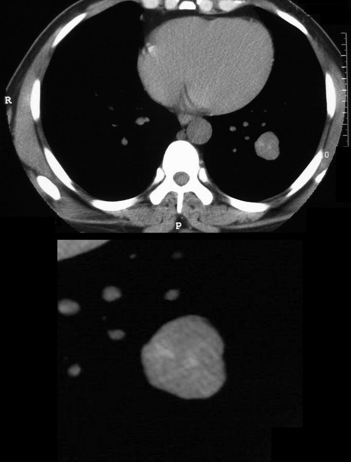
CT scan of the chest shows a metastatic nodule with psammomatous calcifications
Ashley Davidoff
TheCommonVein.net
31491c
Lung Nodules that are Outside the Chest
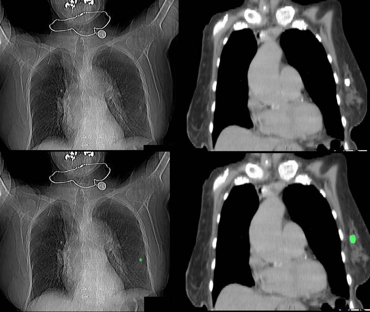
Ashley Davidoff
The CommonVein.net
-
Links and References
-
-
TCV
-
-
-