
-
Based on Size <3cms
- Single or Multiple
- Macronodules
- Micronodules
-
Based on Shape
- Round
- Stellate or Spiculated
-
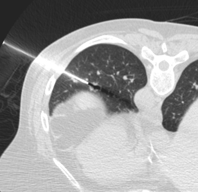
Based on Position
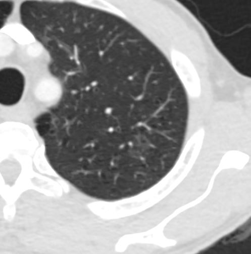
- Centrilobular
-

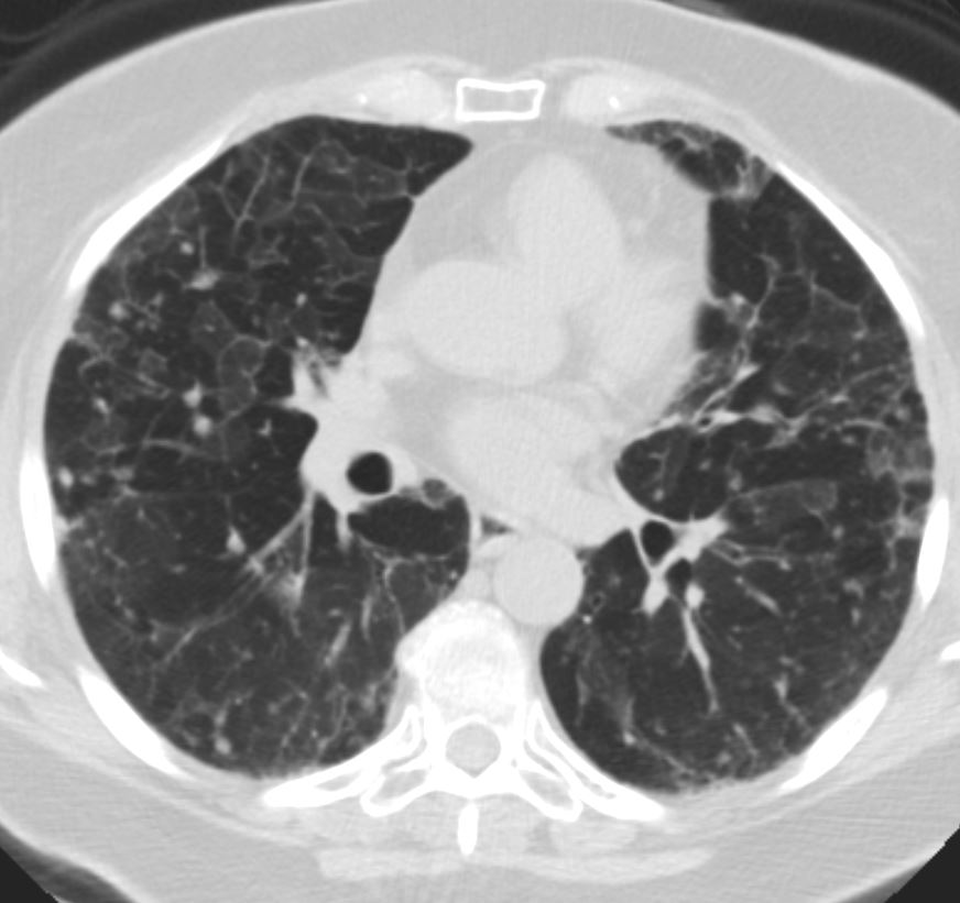
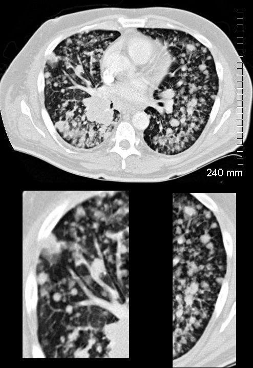
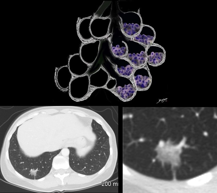
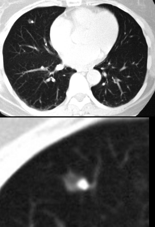
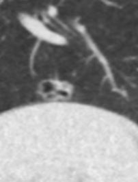
Centrilobular Nodules
77F with long history of dyspnea and cough showing medium and small airway disease, centrii-lobular nodules, paraseptal nodules ground glass changes and mosaic attenuation Diagnosis includes Stage 3 sarcoidosis
Ashley Davidoff
TheCommonVein.net
-
Based on Character
- Solid
- Subsolid
- Ground Glass
- Calcified
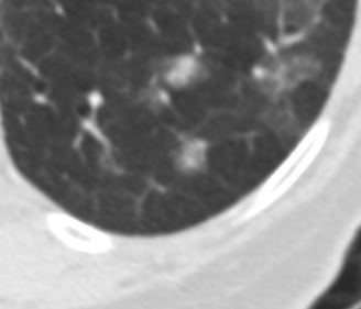
Lung nodules, by definition are less than 3 centimeters in diameter and are quite common. Lung nodules can be categorized based on their appearance, size, and other characteristics. The most common types of lung nodules seen on CT scans include:
Solitary pulmonary nodule (SPN): This refers to a single lung nodule that appears as a well-defined round or oval lesion in the lung tissue.
-
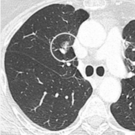
- Solid Pulmonary Nodule
Solid pulmonary nodule: These nodules appear as well-defined, dense lesions with a uniform density throughout. They can be benign or malignant.
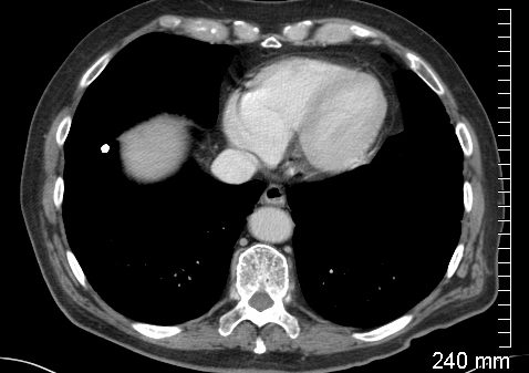
Multiple pulmonary nodules: Sometimes, more than one nodule can be present in the lungs. These can be caused by various conditions such as metastases from cancer or infectious diseases.
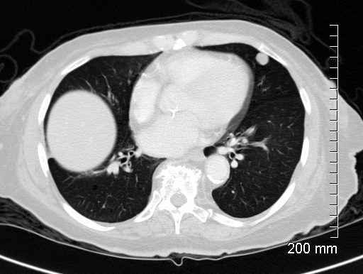
The lower panels demonstrate centrilobular distribution of someof the nodules
Ashley Davidoff MD
TheCommonVein.net
134336c
Ground-glass opacity (GGO): GGO appears as hazy, increased lung density that does not obscure the underlying bronchial structures. It can be a sign of various conditions, including early-stage lung cancer or inflammation.
1 year prior
Ashley Davidoff MD
TheCommonVein.net
-
- Ground Glass Opacity and Adenocarcinoma with Lepidic Growth
The Ground Glass Opacity (GGO) in this case is caused by partial filling of the alveolus with malignant cells Ground glass opacification may be caused by partial filling of the alveolus with cellular material resulting in partial replacement of air with solid material. The net density is gray rather than white in the situation where the alveolus is fully replaced with cells or fluid. There is blending of the black of the subtending airways and the white of the vessels with the gray density of the cellular infiltrate and hence the normal vessels are not visualized in ground glass opacities.
Part-solid nodule: Part-solid nodules have both solid and ground-glass components. These can also be associated with lung cancer, particularly adenocarcinoma.
Ashley Davidoff
TheCommonVein.net
-
- Part Solid Lung Nodule
- Courtesy Wiki Commons
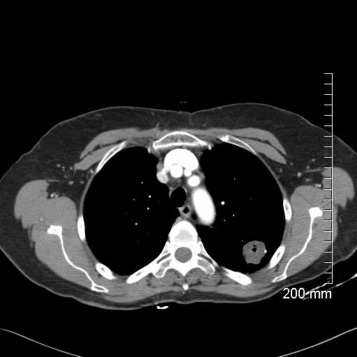
Calcified nodules:
Some lung nodules may contain calcium deposits, causing them to appear as dense spots on the CT scan. These are often benign and can result from previous infections or scarring.
-
- Calcified Granuloma CT scan
- Ashley Davidoff TheCommonVein.net
-
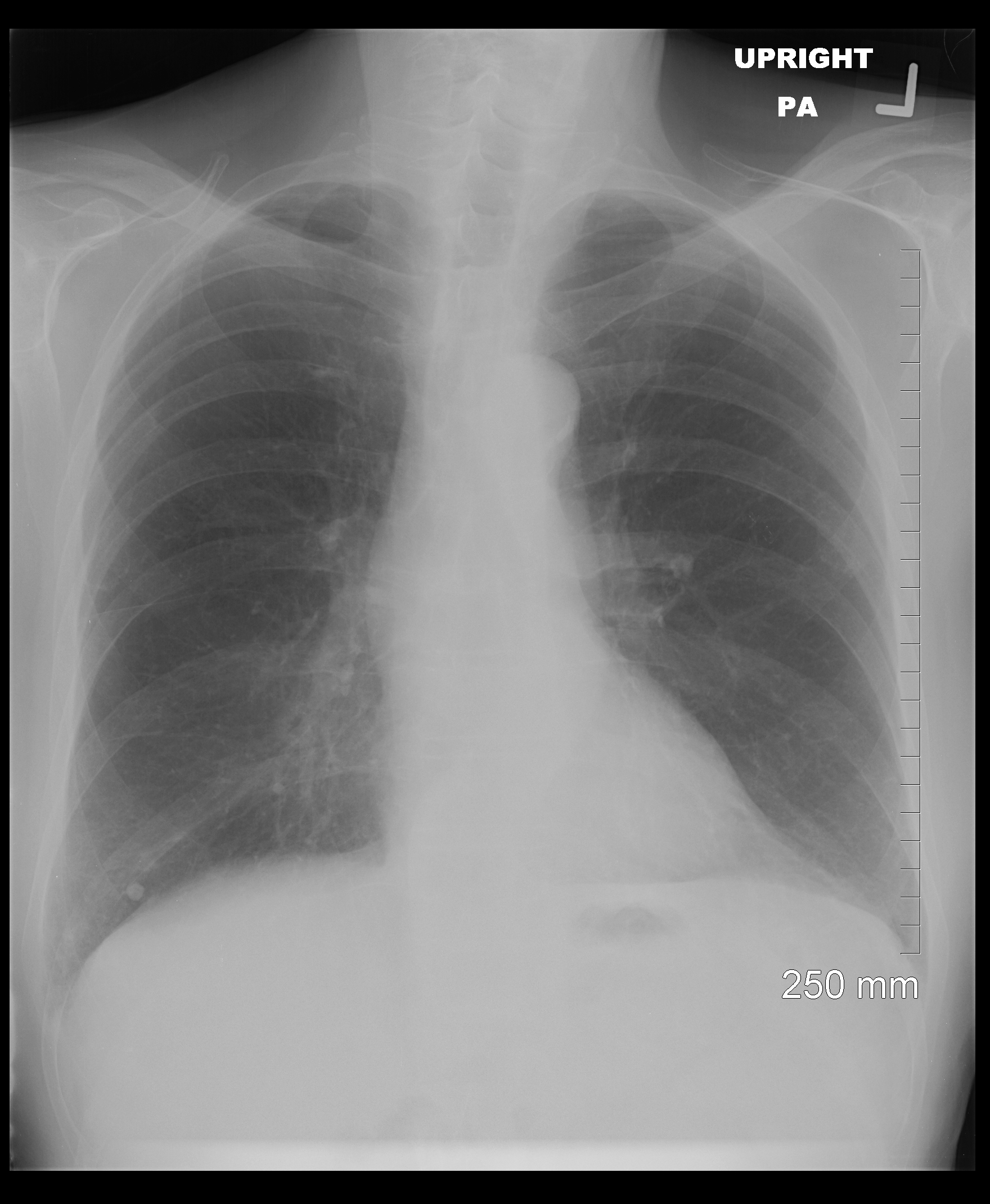
- Calcified Granuloma CXR scan
- Ashley Davidoff TheCommonVein.net
-
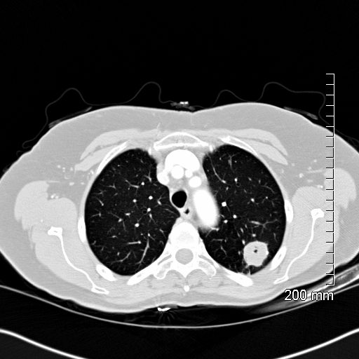
- Eccentric Calcification
-
- 56 F with a middle lobe nodule with eccentric calcification which turned out to be granulomatous in origin
-
- Ashley Davidoff
-
- TheCommonVein.net
- 30252c
Cavitating Nodule
-
- Cavitating Nodule
- Ashley Davidoff TheCommonVein.net
Pseudocavitation
Ashley Davidoff MD TheCommonVein.net
Ashley Davidoff MD TheCommonVein.net