
-
Cancer
-

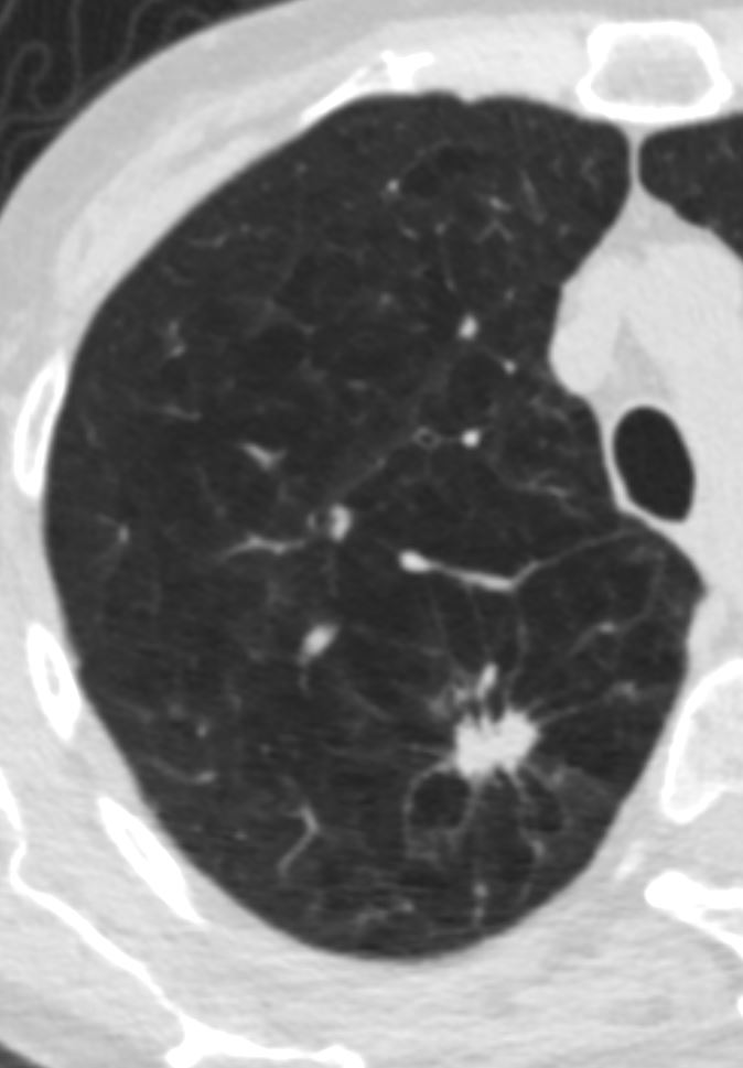
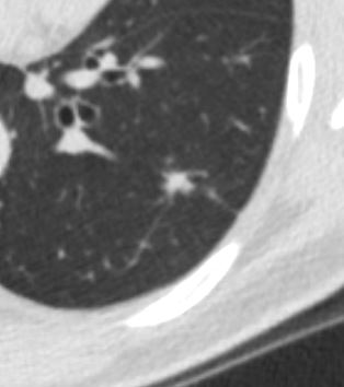
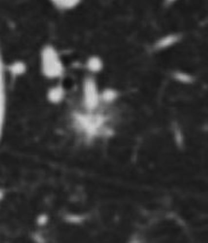
66F spiculated lesion with extension into the interlobular septa and background of severe centrilobular emphysema
Ashley DAvidoff
TheCommonVein.net -
Lung Cancer
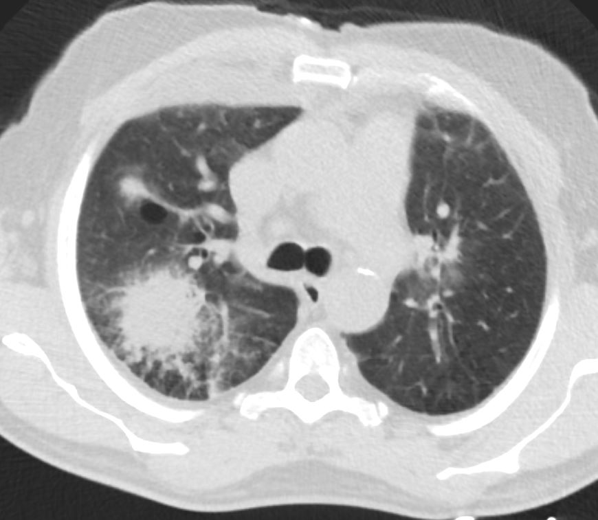
Spiculated and Cavitating Nodule
Ashley Davidoff
TheCommonVein.netSpiculaated PET positive Cancer with Satellite Nodules
Ashley Davidoff
TheCommonVein.net
28979c -
Inflammations
- Sarcoidosis
-
81 F with sarcoidosis
Irregular spiculated solid nodule
Ashley Davidoff
TheCommonVein.netSARCOIDOSIS with STELLATE NODULES
42 year old female with known history of sarcoidosis characterised by confluent granulomas, with spiculated nodules, retractile fibrosis and moderate adenopathy
Ashley Davidoff MD - Wegener’s Granulomatosis
-
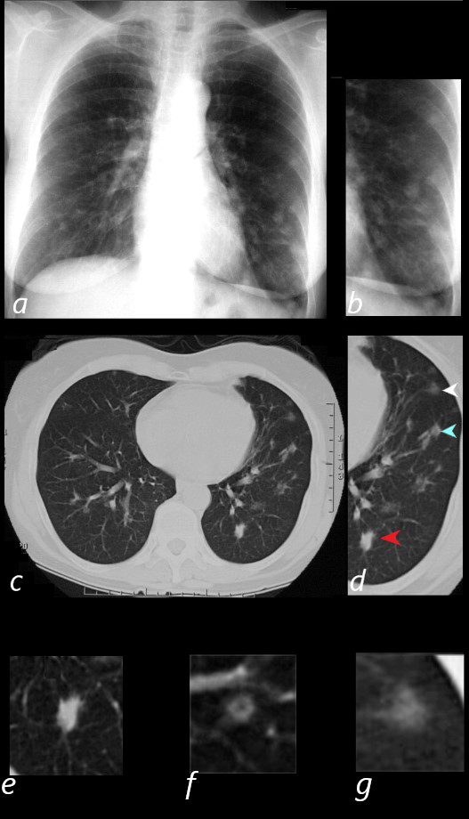
NODULES OF WEGENER”S GRANULOMATOSIS, aka GRANULOMATOSIS WITH POLYANGIITIS, GPA
65 year old female presents with epistaxis and with nodular changes on CXR (a) magnified in b.
CT scan in axial projection (c) and magnified in d, reveals 3 types of nodules.
A spiculated solid nodule (red arrow head) is magnified in e, a bronchocentric nodule (teal arrowhead) is magnified in e. This may represent a cavitating nodule or hemorrhagic change around a bronchiole (cheerio sign) A ground glass nodule (white arrowhead) is magnified in g.
Ashley Davidoff MD - Langerhans Cell Histiocytosis
-
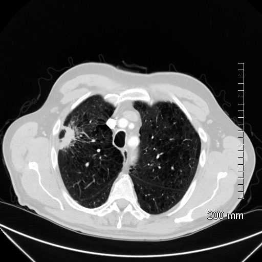
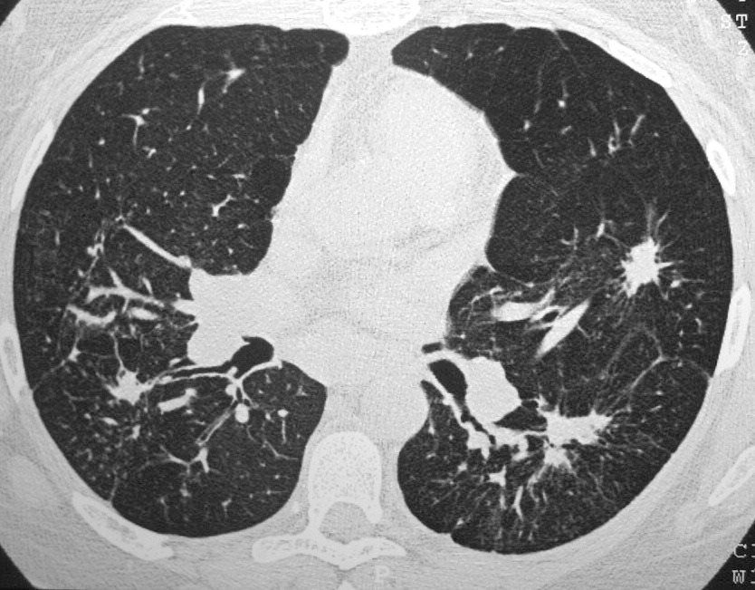
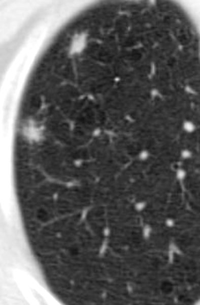
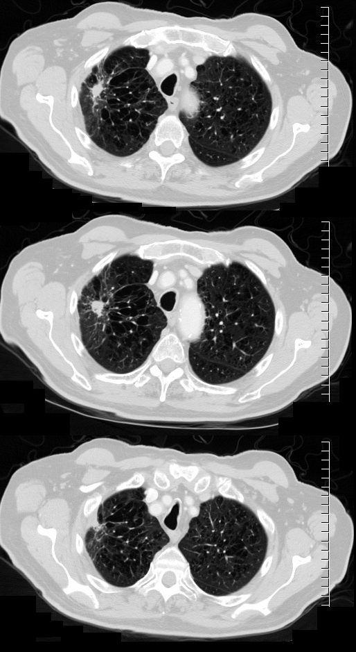
Spiculated Nodule of Langerhans Cell Histiocytosis in a background of Centrilobular Emphysema
TheCommonVein.netLangerhans Cell Histiocytosis
Langerhans Cell is a dendritic white cell with a wavy nucleus that creates granulomas and infiltrates the interstitium. It thus causes spiculated nodules that appear as spiculated nodules on CT
Ashley Davidoff
TheCommon Vein.netChronic inflammation giant cell reaction
44 year old male spiculated nodule bx showed chronic inflammation giant cell reaction
Ashley Davidoff MD TheCommonVein.net
-
Infections
- Candida
-
66 y/o m w/ hx of CLL presents
fever and chills
3 days later
IR guided CT biopsy of the right upper lobe lesion
progression of disease
neg for AFB
Ashley Davidoff TheCommonVein.net - Idiopathic Pleuro-parenchymal Fibroelastosis
-
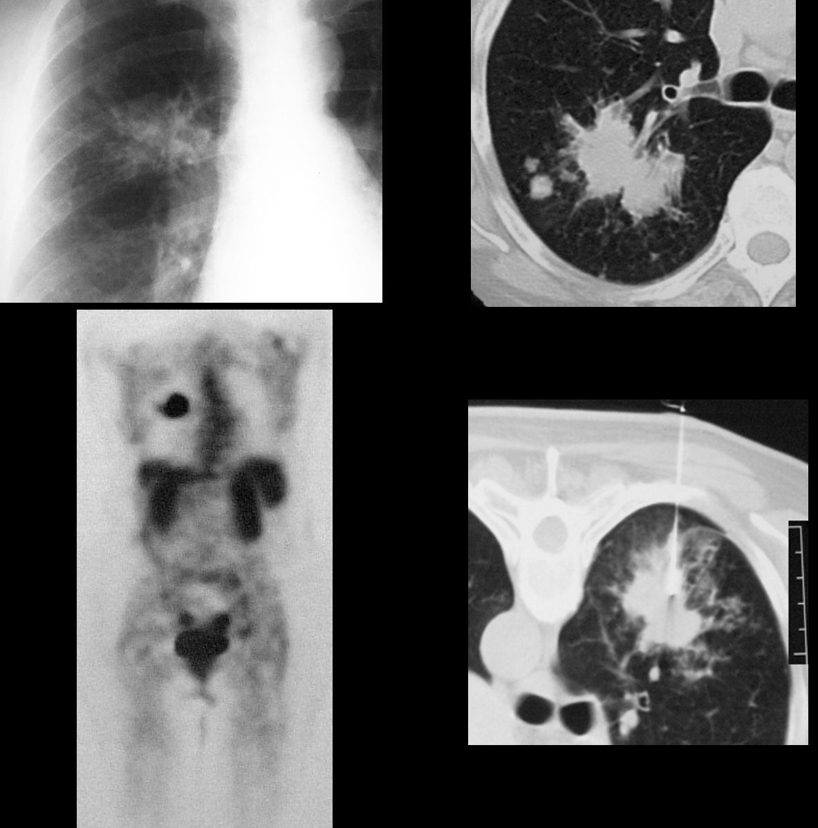
IDIOPATHIC PLEUROPARENCHYMAL FIBROELASTOSIS
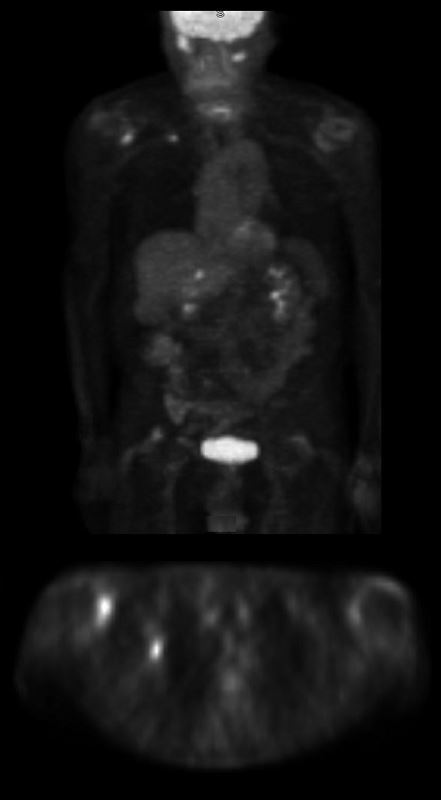
83 year old man with COPD and emphysema presented with bilateral shoulder pain. X-ray examination of his shoulders revealed a stellate nodule in his LUL. CT showed evidence of emphysema with a 15x14mm nodule with evidence of surrounding scarring and retraction.
On PET scan the nodule was PET avid with an SUV of 4.5 consistent with a malignant neoplasm
Surgery was performed and the pathology was consistent with idiopathic pleuroparenchymal fibroelastosis
Follow up CTs 5years following surgery have shown stability
Ashley Davidoff MD TheCommonVein.net see Idiopathic Pleuroparenchymal FibroelastosisIDIOPATHIC PLEUROPARENCHYMAL FIBROELASTOSIS
83 year old man with COPD and emphysema presented with bilateral shoulder pain. X-ray examination of his shoulders revealed a stellate nodule in his LUL. CT showed evidence of emphysema with a 15x14mm nodule with evidence of surrounding scarring and retraction.
On PET scan the nodule was PET avid with an SUV of 4.5 consistent with a malignant neoplasm
Surgery was performed and the pathology was consistent with idiopathic pleuroparenchymal fibroelastosis
Follow up CTs 5years following surgery have shown stability
Ashley Davidoff MD TheCommonVein.net see Idiopathic Pleuroparenchymal FibroelastosisAmyloidosis
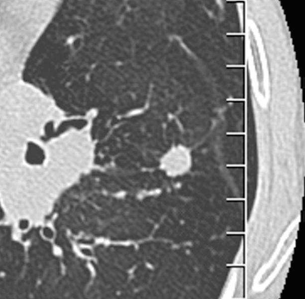
Spiculated Nodule in Amyloidosis
87 year old female with known nodular form of amyloidosis. The axial CT scan shows a spiculated nodule with surrounding ground glass changes
Ashley Davidoff Boston Medical Center TheCommonvein.net LV-006 mag spiculatedb
-
Links and References
- TCV
- TCV