
- Map of Size of Parts of the Lung
- Structures of the Path of the Air
- Right Lung
- Left Lung
- Embryology and Aging
- Blood Supply Pulmonary Artery
- Venous Drainage
- Lymphatic Drainage
- Pleura
42651c
keywords lung chest
Ashley Davidoff TheCommonVein.net
From the Macroscopic View
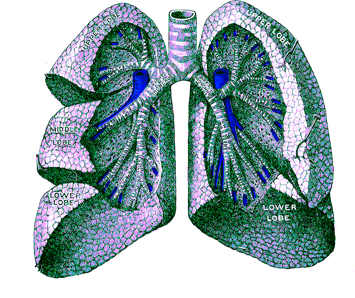
The right and left lung are asymmetric with the right having three lobes and the left two lobes. The lingula is part of the LUL.
The lung is divided into a right and left lung with the right lung being composed of an upper middle and lower lobe, and the left lung being composed of an upper lobe with the lingula as part of the upper lobe and the lower lower lobe.
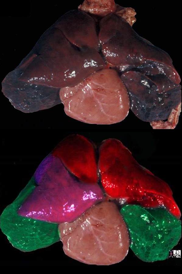
The post mortem specimen is viewed from the anterior aspect showing the upper lobes in red, the right middle lobe in pink and the lower lobes in green.
Ashley Davidoff MD TheCommonVein.net 32558c01
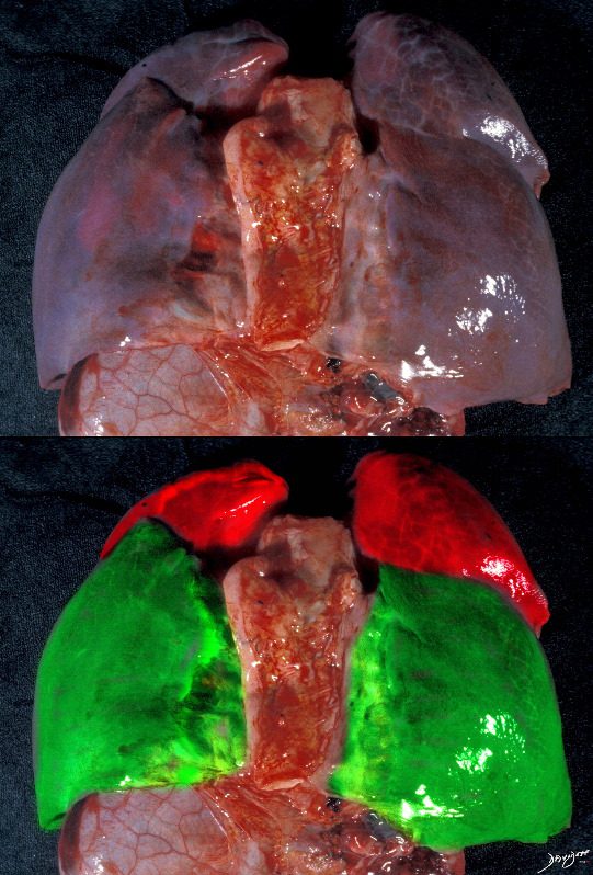
The same specimen as seen above is viewed from its posterior aspect showing the upper lobes in red and the lower lobes in green. Note that the lower lobes have a majority of their parenchyma posteriorly while the upper lobes are dominantly positioned anteriorly. Courtesy Ashley Davidoff MD 32557.8c
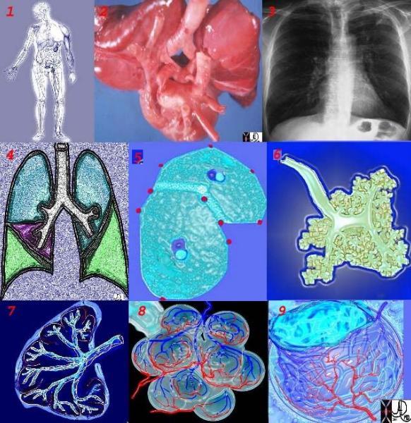
There are two lungs and they are made up of lobes which are divided into segments and these are discussed in the individual documents dedicated to each of the lungs.
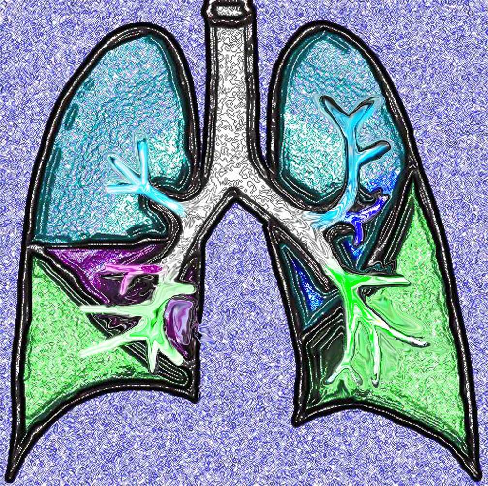
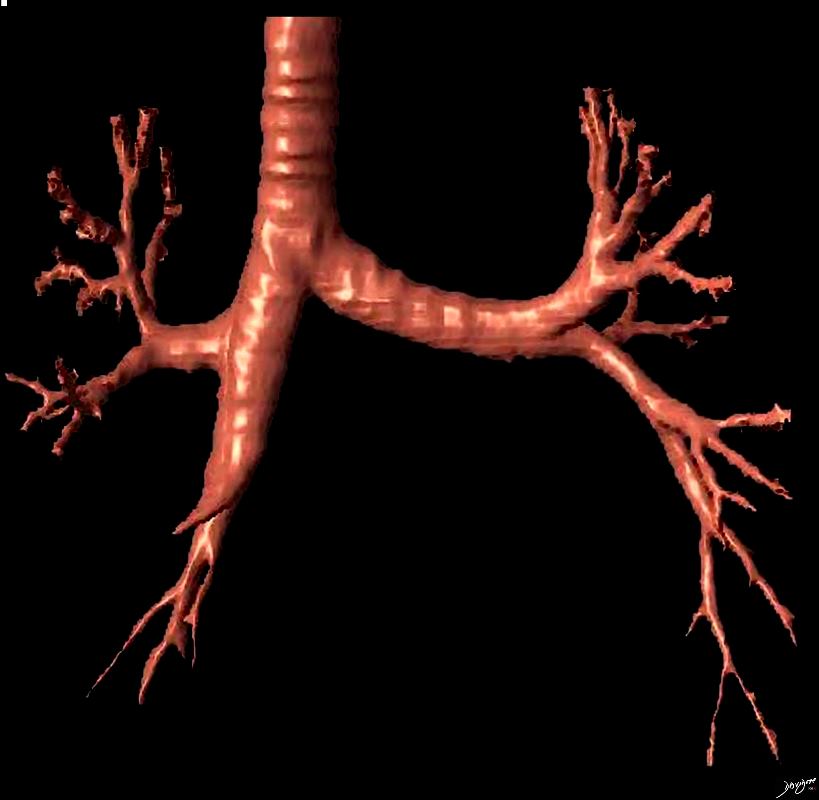
This diagram shows the basic division of the tracheobronchial tree into lobes. The right lung is divided into right upper (RUL) (teal) right middle, (RML pink) and right lower lobe (RLL green). The left lung is divided into left upper (LUL teal), which includes the lingula(dark blue), and left lower lobe (LLL= green). Note that the two mainstem bronchi are of unequal length and size. The right mainstem is short and fat while the left is long and thin. This irregular dichotomous branching pattern is characteristic of the branching pattern of all the conducting systems within the lungs.
Ashley Davidoff
TheCommonVein.net
32686b05
Ashley Davidoff
TheCommonVein.net
The segments are divided into the secondary lobules. The lobules are made up of the small airways including the terminal bronchioles, respiratory bronchioles, alveolar ducts alveolar sacs and the alveoli themselves.
Grays Anatomy
Right Lung Segments
- Upper lobe
- apical segment
- posterior segment
- anterior segment
- Middle lobe
- lateral segment
- medial segment
- Lower lobe
- superior segment
- medial-basal segment
- anterior-basal segment
- lateral-basal segment
- posterior-basal segment
Left Lung Segments
- Upper lobe
- apico-posterior segment (merger of “apical” and “posterior”)
- anterior segment
- Lingula
- inferior lingular segment
- superior lingular segment
- Lower lobe
- superior segment
- anteromedial basal segment (merger of “anterior basal” and “medial basal”)
- posterior basal segment
- lateral basal segment
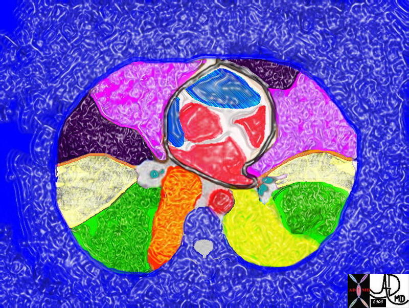
The axial CT through the level of the heart shows a few of the right and left pulmonary segments including parts of the middle lobe, lingula and of the lung bases
Ashley Davidoff MD TheCommonVein.net 42644.800
The Conducting System and Air Exchange
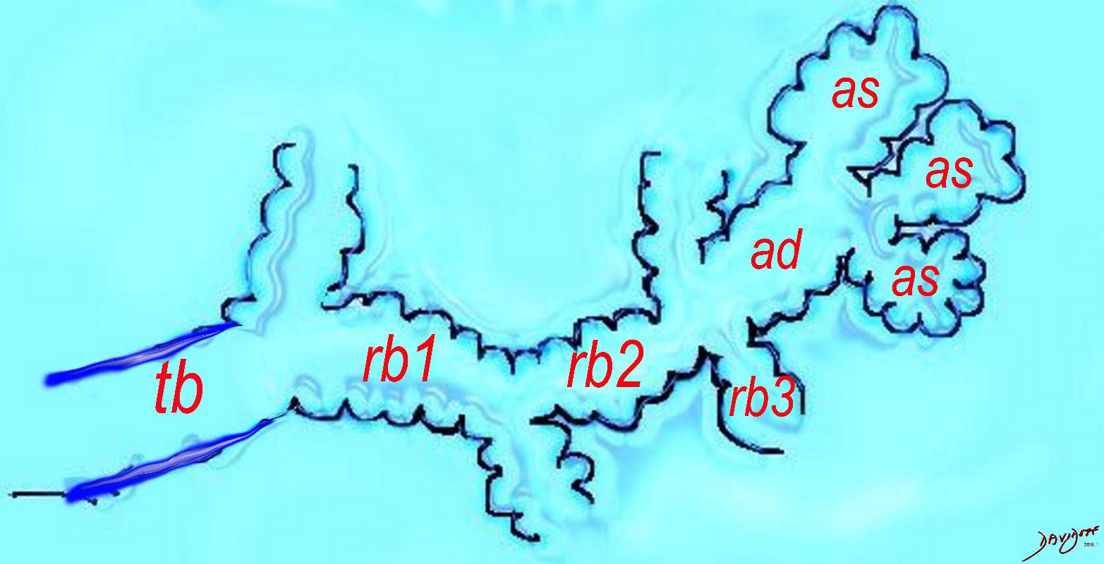
The diagram shows the ductal system of the acinus, starting with the tick walled terminal bronchiole (tb) that is the last duct of the conducting system of the airways. The respiratory bronchiole (rb) enters the secondary lobule and is the first duct to havediffusion capability. There can be 3 orders of rb’s, and they branch into alveolar ducts, which branch in turn to alveolar sacs until they reach the alveoli, which is the final destination.
Courtesy Ashley DAvidoff MD 2019
lungs-0032-low res
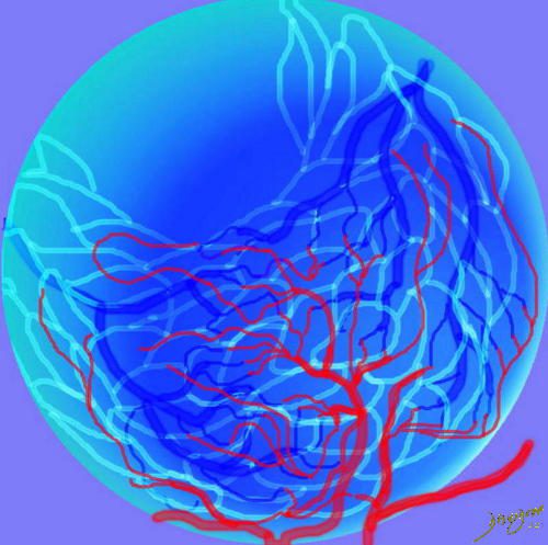
The pulmonary arteriole accompanies the airway as it carries oxygen from the trachea to the alveoli. They part ways at the alveoli where the pulmonary venule then takes the oxygenated blood from capillary network around the alveoli back to the left atrium.
The intimate relationship of the airways and the pulmonary artery and their close approximation in size, is helpful in radiology, firstly to identify these structures and secondly to define disease such as heart failure and bronchiectasis.
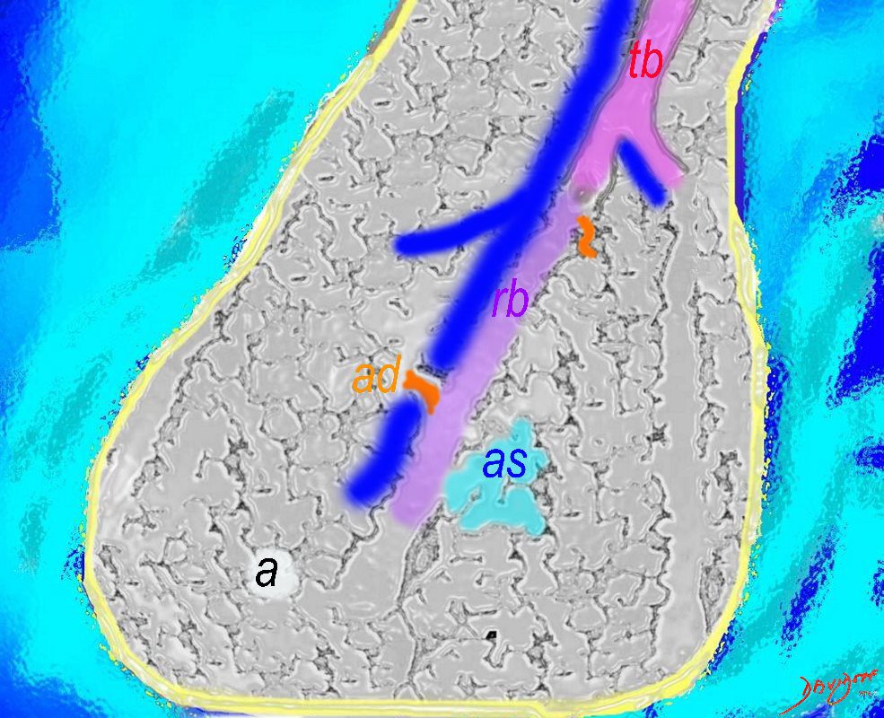
The acinus as shown in this image is defined as a unit of lung consisting of a single first order respiratory bronchiole that subtending a cluster of alveoli reminiscent of a bunch of grapes or berries (acinus in Latin means berry) . The lobular bronchiole (lb) branches into the terminal bronchiole (tb), which then branches into the first order respiratory bronchiole (rb). Subsequent branching after the respiratory bronchiole, includes in order, the alveolar duct (ad), alveolar sac (as), and then finally the berry like alveoli.
Courtesy Ashley Davidoff 2019
lungs-0033-low res
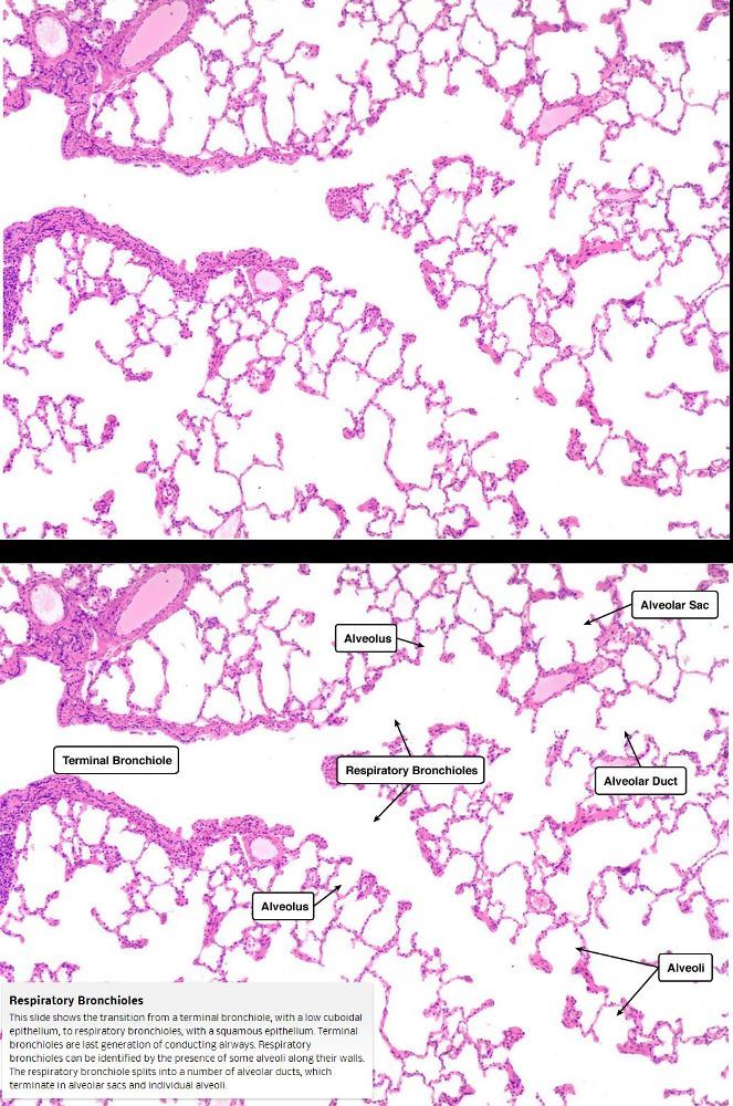
This slide shows the transition from a terminal bronchiole, with a low cuboidal epithelium, to respiratory bronchioles, with a squamous epithelium. Terminal bronchioles are last generation of conducting airways.
Respiratory bronchioles can be identified by the presence of some alveoli along their walls. The respiratory bronchiole splits into a number of alveolar ducts, which terminate in alveolar sacs and individual alveoli
Courtesy medcell.med.Yale.edu
The Secondary Lobule
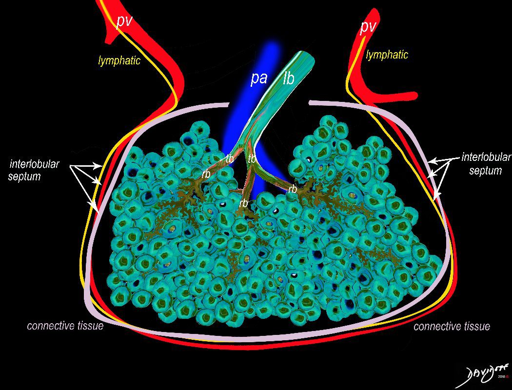
The secondary lobule is housed in a connective tissue framework in which run the lymphatic and venular tributaries . Together these 3 structures form the interlobular septum.
The lobar arteriole enters the framework, accompanied by the lobar bronchiole, and they all run together and form the interlobular septa. This structure measures between .5cms and 2cms and is visible on CT scan.
It is important in clinical radiology since many of the structures can be identified in health, and more particularly in disease, enabling the identification and characterization of many pathological processes.
Courtesy Ashley Davidoff MD
lungs-0036-low res
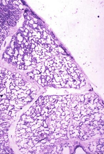
Normal lung histology
This image is a panoramic view of the lung showing secondary lobules and interlobular septa. Within the interalveolar septae, one sees small venules and lymphatics .Courtesy Armando Fraire MD. 32649b
code lung pulmonary alveoli alveolus secondary lobule interlobular septa vein lymphatic histology
interstitium interstitial
32649b
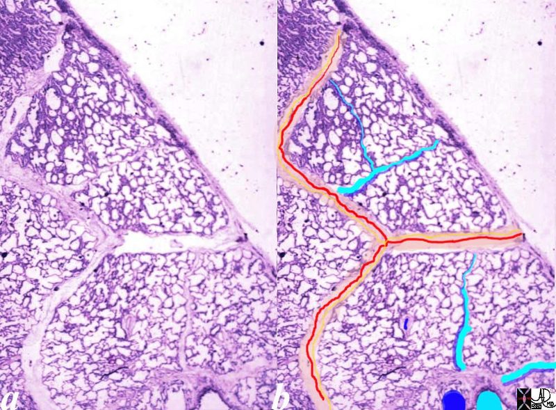
Normal lung histology
This image is a panoramic view of the lung showing secondary lobules and interlobular septa. Within the interalveolar septae, one sees small venules and lymphatics .
The side by side images show the interlobular septa within which reside the pulmonary venules (red) and lymphatics and within the center of the lobule run the respiratory bronchioles (teal) and pulmonary arterioles (blue)
Courtesy Armando Fraire MD. 32649b
key words
lung pulmonary alveoli alveolus secondary lobule interlobular septa vein lymphatic histology
interstitium interstitial
32649c06.8s
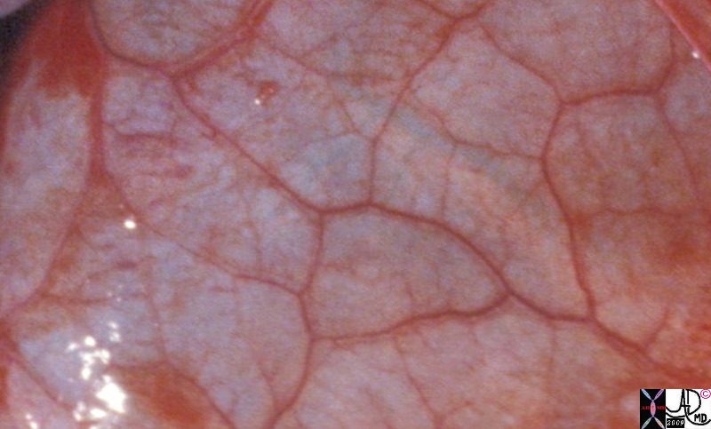
Gross anatomy specimen shows secondary lobules with interlobular septa containing pulmonary venules and the capillary components within the secondary lobules
Davidoff MD TheCommonVein.net 32557bb03.8s
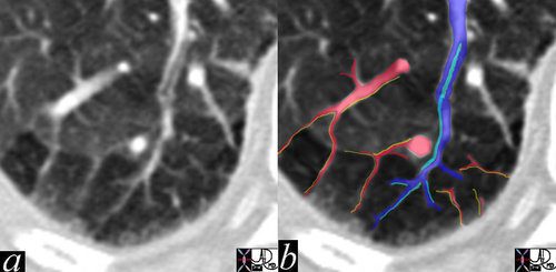
The high resolution CT shows multiple secondary lobules in the periphery of the right lung. A lobular bronchiole is overlaid in teal, while the accompanying lobular arteriole is seen in royal blue entering the lobule and then branching. The peripheral venules are identified in the interlobular septa (red) and joining to form the lobular vein.
In this remarkable CT we were able to identify a few secondary lobules at the periphery of the lung that have a rectangular shape in this instance. The branching structure that enters the lobule (blue in b), is characterised as an arteriole for two reasons. Firstly it is paired with a tubular airway seen in (a) in its most proximal portion as a lucent tubule, and subsequently interpolated in light blue in b. Secondly it branches in the centre of the lobule. It is distinct from the border forming interlobular septum which surrounds it. A second relatively large vessel colored in red receives a branch from the interlobular septum and by virtue of its size and position it has to be a pulmonary venule. We know that the lymphatic vessel accompanies the venule, and so the yellow lymphatic has been implied but not visualised. We also know that connective tissue surrounds these two structures. In this instance the matrix of the lobule that consists of the alveoli is less dense than it should be and is surrounded by normal alveoli. Lucency implies air trapping and air trapping implies small airway disease. Thus this image tells us that the criminal in this case of disorder is the small airway, We now can focus on the small airways with a pathological differential diagnosis, and from there plan the treatment.
key words
lung pulmonary lobule secondary lobule arteriole venule interlobular septa bronchovascular bundle mosaic pattern air trapping fx ground glass
CTscan
Ashley Davidoff MD
47152c01
The Secondary lobule is made of Acini
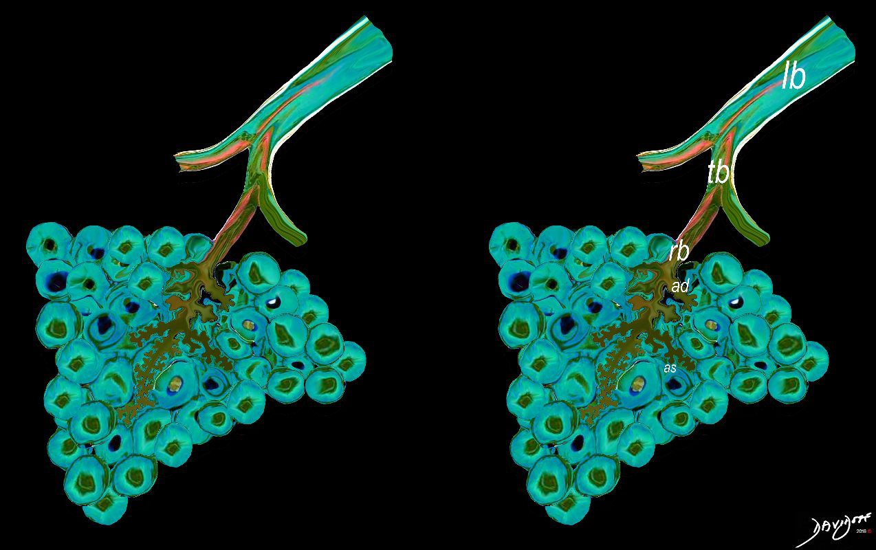
The acinus is defined as a unit of lung consisting of a single first order respiratory bronchiole that subtending a cluster of alveoli reminiscent of a bunch of grapes or berries (acinus in Latin means berry) . The lobular bronchiole (lb) branches into the terminal bronchiole (tb), which then branches into the first order respiratory bronchiole (rb). Subsequent branching after the respiratory bronchiole, includes in order, the alveolar duct (ad), alveolar sac (as), and then finally the berry like alveoli.
Ashley Davidoff MD TheCommonVein.net
lungs-0030-low res
key words
key words RS lung alveolus respiratory bronchiole artery vein pulmonary capillary normal anatomy histology drawing
Ashley Davidoff MD TheCommonVein.net 32164
This drawing demonstrates the open mouth view of the alveolus, which is surrounded by its capillary network. The lining single layer of squamous cells (pneumocytes) can be seen peaking through the vessels.
Ashley Davidoff MD. TheCommonVein.net 32166
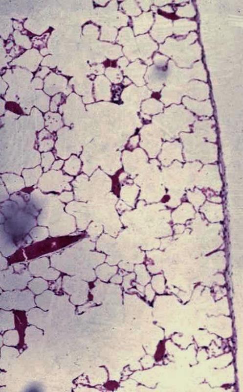
The low magnification at the periphery of the normal lung shows normal lining of the alveoli surrounding the air spaces (alveoli).
The layer of visceral pleura is shown to the right of the image
Small airways and accompanying blood vessels (with clotted blood)
Ashley Davidoff MD
lungs-0031-low-res
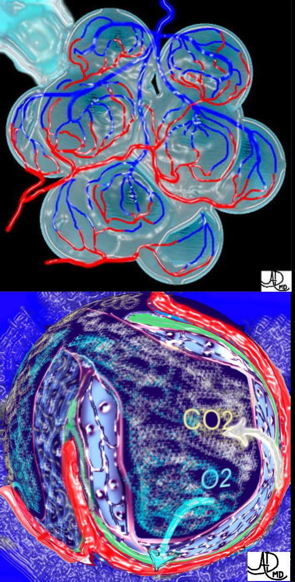
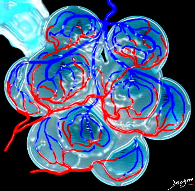
This is a drawing of a cluster of alveoli surrounded by the capillary network, fed by an arteriole in blue, and drained by a venule in red. The second image shows the exchange of life giving oxygen for the by product of metabolic activity – carbon dioxide
Ashley Davidoff MD TheCommonVein.net 32165c

Lower magnification of the lung with H and E stain shows cup-shaped alveolar spaces outlined by delicate thin alveolar capillary membrane.
key words
lung, pulmonary, normal alveolus, alveoli, histology, interstitium, interstitial
Courtesy Armando Fraire MD. 32819 TheCommonVein.net
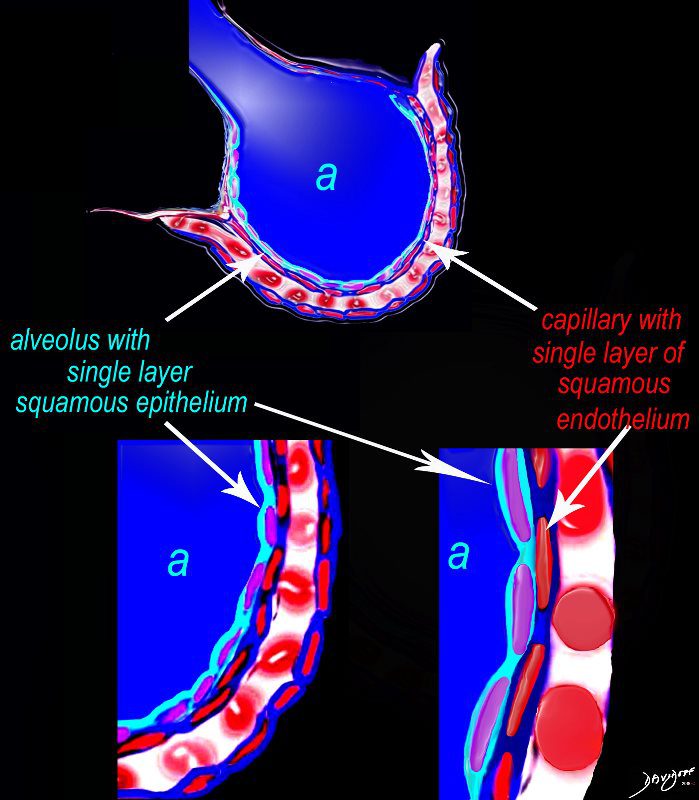
The diagram shows an alveolus (a) above, lined by a single layer of squamous cells, surrounded by a capillary with red cells which is also lined by a single layer of squamous endothelial cells . The images below show progressive magnification of the alveolar wall demonstrating the two thin layer of the alveolar membrane .
Courtesy Ashley Davidoff 2019
lungs-0027-low res
Links and References
- TCV
- Map of Size of Parts of the Lung
- Structures of the Path of the Air
- Trachea
- Carina
- Mainstem Bronchi
- Segmental Bronchi
- Small Airways
- Terminal Bronchiole
- Respiratory bronchiole
- Secondary Lobule
- Acinus
- Alveolus
- Cells
- Right Lung
- Left Lung
- Embryology and Aging
- Blood Supply Pulmonary Artery
- Venous Drainage
- Lymphatic Drainage
- Pleura
Findings
Diseases