
Pneumonia Etmology
- The word “pneumonia”
- from the Greek word
- “pneumon,” which means “lung.” and
- “-ia,” which is a suffix used to indicate a condition or state.
- from the Greek word
- Therefore, “pneumonia” translates to
- “lung condition” or
- “lung disease” in Greek. This is fitting given that pneumonia is a respiratory condition characterized by inflammation of the lungs, often caused by infection.
Infection
- Pneumonia
- general term
- filling the alveoli.
- usually purulent, (ie infection)
- generally caused by infecction but entities such as DIP (Desquamative Interstitial Pneumonia), LIP (Lymphocytic Interstitial Pneumonia), NSIP (Nonspecific Interstitial Pneumonia), UIP (Usual Interstitial Pneumonia), COP (Cryptogenic Organizing Pneumonia), and OP (Organizing Pneumonia)—refer to specific patterns of interstitial lung diseases and are called pneumonia but they fall under the broader category of pneumonitis, or inflammation of the lung tissue.
- general term
Pneumonias can be classified by:
- etiology
- infective agent
- bacterial (pyogenic) pneumonia
- cavitating bacterial pneumonia
- fungal pneumonia
pneumocystis pneumonia (PCP)
mycobacterial pneumonia
viral pneumonia
coronavirus
COVID-19
Middle East respiratory syndrome (MERS) infection
severe acute respiratory syndrome (SARS)
varicella pneumonia
setting of infection
community-acquired pneumonia
hospital-acquired pneumonia (HAP)
ventilator-associated pneumonia (VAP)
healthcare-acquired pneumonia (HCAP)
aspiration pneumonia
lipid: lipoid pneumonia
method of spread (a pathological description)
bronchopneumonia
lobar pneumonia
multilobar pneumonia
radiographic appearance
atypical pneumonia
round pneumonia
cavitating pneumonia
hemorrhagic pneumonia
- bacterial (pyogenic) pneumonia
- infective agent
TB
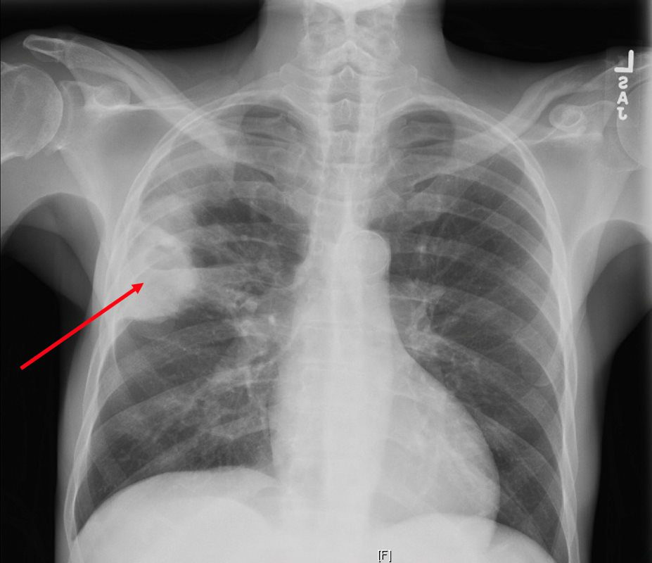
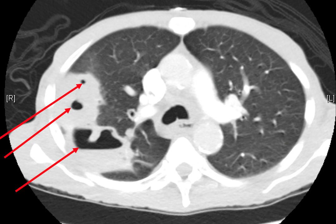
CXR reveals a dense consolidation in the right upper lobe (red arrow) with questionable air-fluid level. No pneumothorax. No pleural effusions. Differential includes right upper lobe pneumonia or tuberculosis. CT is recommended for further evaluation if there is concern for a cavity.
Courtesy Joseph Cannella,
Dr. Christina LeBedis, MD, MS
Courtesy Joseph Cannella,
Dr. Christina LeBedis, MD, MS
 Bronchopneumonia- Centrilobular
lung axial interstitium bronchioles connective tissue fx bronchial plugging peribronchial halo peribronchial thickening dx bronchopneumonia CTscan Davidoff MD 47614c01
Links and References
- Videos
- AUR
Title web link Infection in the Immuno-competent Patient AUR Cardiothoracic Imaging Infection in the Immuno-compromised Patient AUR Cardiothoracic Imaging
- AUR