
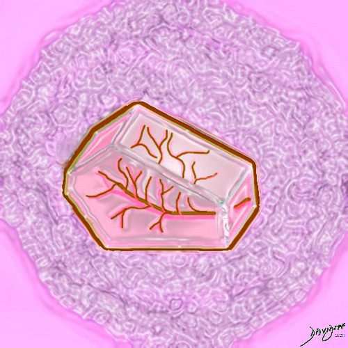
The secondary lobule is housed in a connective tissue framework in which run the lymphatic and venular tributaries . Together these 3 structures form the interlobular septum.
The lobular arteriole enters the framework, accompanied by the lobular bronchiole, and they all run together and form the interlobular septa. This structure measures between .5cms and 2cms and is visible on CT scan.
It is important in clinical radiology since many of the structures can be identified in health, and more particularly in disease, enabling the identification and characterization of many pathological processes.
Courtesy Ashley Davidoff MD TheCommonVein.net lungs-0036-low res
Keywords:
lung pulmonary alveoli alveolus secondary lobule interlobular septa vein lymphatic histology interstitium interstitial normal copyright 2009 all rights reserved
The segments form the secondary lobules.
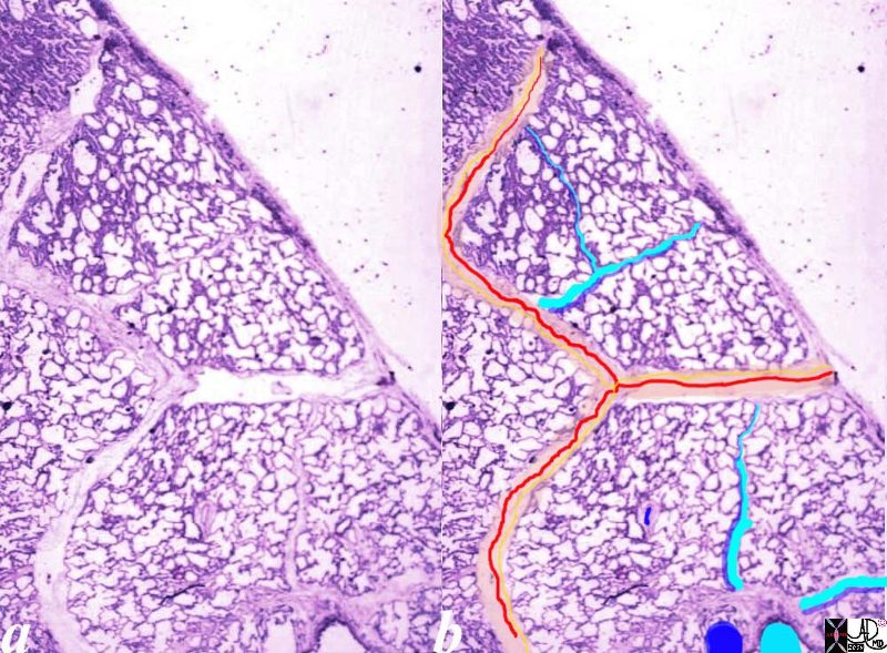
Normal lung histology. This image of the lung periphery shows secondary lobules and interlobular septa. Within the interlobular septae, one sees small venules and lymphatics. The matrix of the lobule contains alveoli.
Courtesy of: Armando Fraire, M.D. Ashley Davidoff TheCommonVein.net
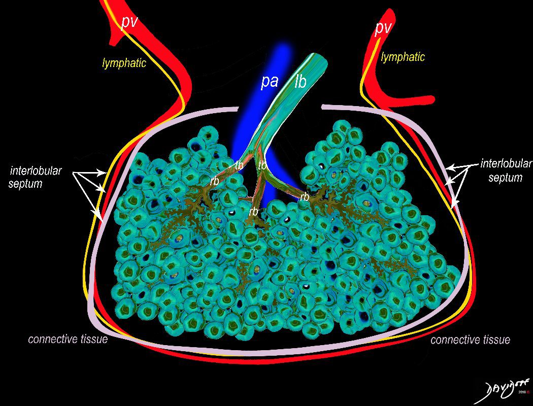
The arteries and airways pair up and travel together from the interlobular septa to the hilum. The pulmonary lobule, also called the secondary lobule is a structural unit surrounded by a membrane of connective tissue, and it is smaller than a subsegment of lung but larger than an acinus. This diagram shows two secondary lobules lying side by side. The pulmonary arteriole (royal blue) and bronchiole (pink) are shown together in the centre of the lobule (“centrilobular”), while the oxygenated pulmonary venules (red) and lymphatics (yellow) are peripheral and also form a formidable and almost inseparable pair.
42440b03
Ashley Davidoff MD
TheCommonVein.net
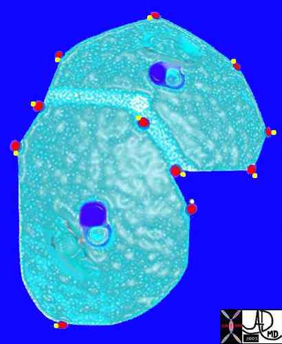
3D image of the polyhedral shapes secondary lobule with a centrilobular bronchovesicular bundle Ashley Davidoff MD. The Common Vein.net lungs-0003
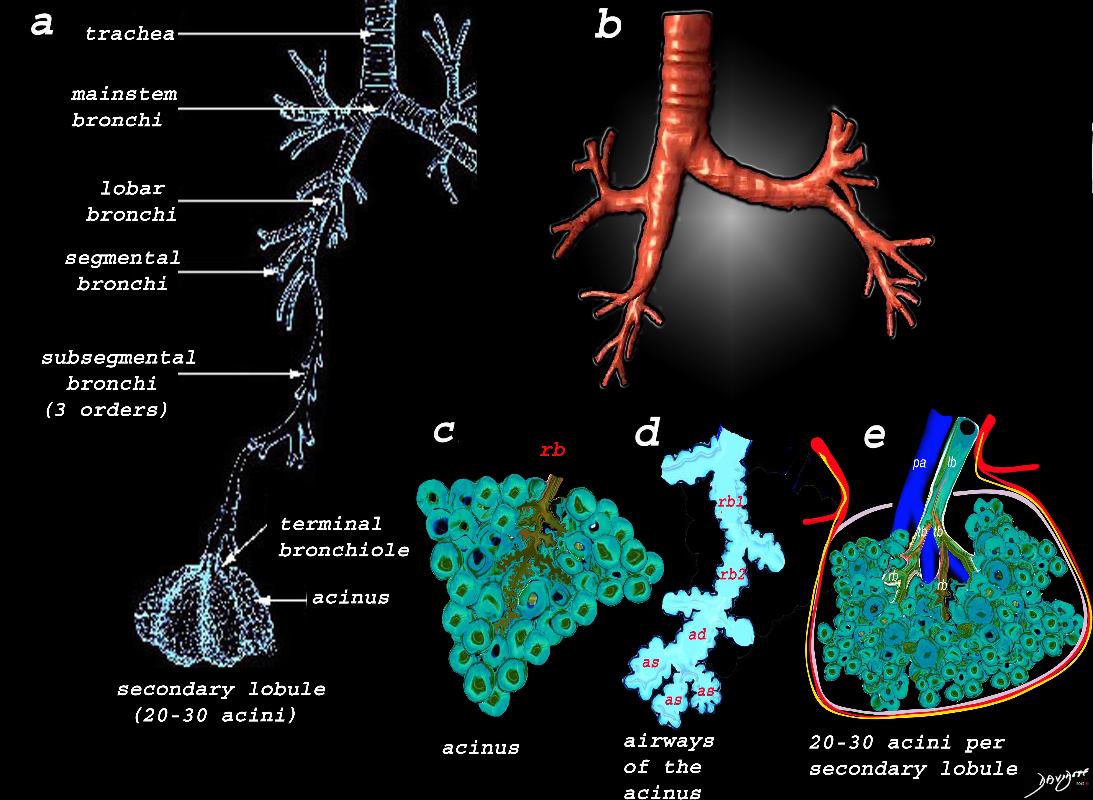
Image a shows the airways starting in the trachea and continuing to the mainstem bronchi, lobar bronchi, segmental bronchi, and subsegmental bronchi,. The subsegmental bronchi have 3 subsequent generations until the bronchiole is reached. The terminal bronchiole is the last of the transporting airways and is considered the most proximal small airway with a diameter of 2mm or less, and it gives rise to the respiratory bronchiole which is the feeding airway for the acinus . The acinus is the functional unit of the lung.
Image b is a 3D reconstruction of a CT scan showing the proximal airways from the trachea to the segmental airways.
Image c shows the structures that make up the acinus and the other parts of the small airways, starting with the respiratory bronchiole (rb) . The diagram in d, shows the detail of the small airways that participate in gas exchange, including the respiratory bronchiole, (rb) alveolar duct, (ad) and alveolar sac (as)
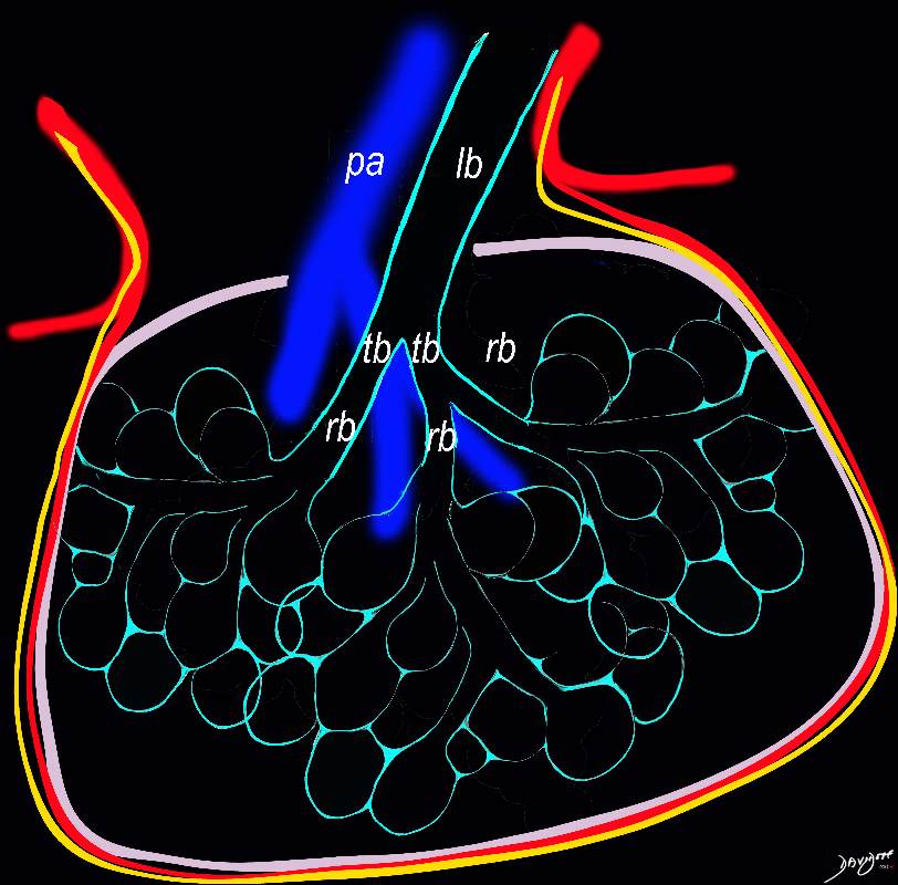
Image e shows the secondary lobule made from about 20-30 acini, arising from a single lobular bronchiole accompanied by a single pulmonary arteriole (pa).. Structure that surround and enclose the secondary lobule include the pulmonary venule, (red) lymphatics,(yellow) and a fibrous septum (pink).
Ashley Davidoff MD TheCommonVein.net
lungs-0739
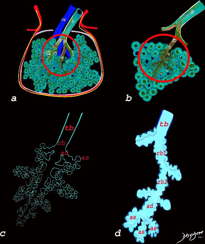
The diagram allows us to understand the the components and the position of the small airways starting in (a) which is a secondary lobule that is fed by a lobular bronchiole(lb) which enters into the secondary lobule and divides into terminal bronchioles (tb) which is the distal part of the conducting airways, and at a diameter of 2mm or less . It divides into the respiratory bronchiole (rb) a transitional airway which then advances into the alveolar ducts(ad) and alveolar sacs (as) Diseases isolated to the small airways do not affect the alveoli and hence there is peripheral sparing Ashley Davidoff MD TheCommonVein.net
The secondary lobule is housed in a connective tissue framework in which run the lymphatic and venular tributaries . Together these 3 structures form the interlobular septum.
The lobar arteriole enters the framework, accompanied by the lobar bronchiole, and they all run together and form the interlobular septa. This structure measures between .5cms and 2cms and is visible on CT scan.
It is important in clinical radiology since many of the structures can be identified in health, and more particularly in disease, enabling the identification and characterization of many pathological processes.
Courtesy Ashley Davidoff MD The CommonVein.net
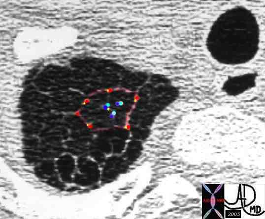
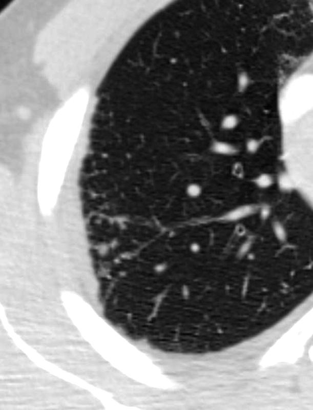
In this high resolution CTscan at the apex of the left lung the secondary lobule is seen with 3 sets of bronchovascular bundles at the door of the secondary lobule surrounded by the pulmonary venules and lymphatics in the surrounding septum. Note also that lymphatics are found both in the periphery of the lobule as well as in the centre of the lobule. This patient had mild emphysema and it was surprising to find this beautiful example of a pulmonary lobule in a patient who was almost normal. We suspect that with the higher resolution technology we will see the normal (or almost normal) pulmonary lobule with greater frequency.
Ashley Davidoff MD
TheCommonVein.net 31819b01
Sarcoidosis
SARCOIDOSIS, ACTIVE – ALVEOLAR FORM
Ashley Davidoff MD
SARCOIDOSIS, ACTIVE – ALVEOLAR FORM
Ashley Davidoff MD
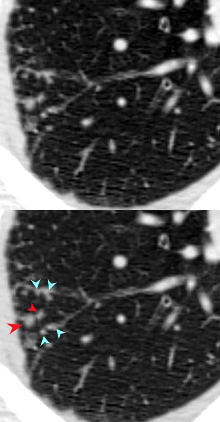
Miliary TB Along the Interlobular Septa – Venule Shown

68 year old female presented with malaise night sweats weight loss Quantiferon gold positive, with a past history of treated TB in her native country as a child. Axial CT images through the upper lobe shows a miliary pattern of disease affecting interlobular septa, centrilobular and tree in bud nodular patterns. Bronchoscopy isolated Mycobacterium complex. She was treated with good result
Ashley Davidoff MD TheCommonVein.net mycobacterium-complex-TB-68-001

68 year old female presented with malaise night sweats weight loss QuantiFeron gold positive, with a past history of treated TB in her native country as a child. Axial CT images through the upper lobe shows a miliary pattern of disease affecting interlobular septa, centrilobular and tree in bud nodular patterns. Bronchoscopy isolated Mycobacterium complex. She was treated with good result
Ashley Davidoff MD TheCommonVein.net mycobacterium-complex-TB-68-008