
- The respiratory bronchioles are the
- narrowest airways of the lungs,
- 0.5 mm across. the bronchioles
- transporting air to the exchange surfaces of the lungs including
- alveolar ducts
- alveolar sacs and finally the
- alveoli
-

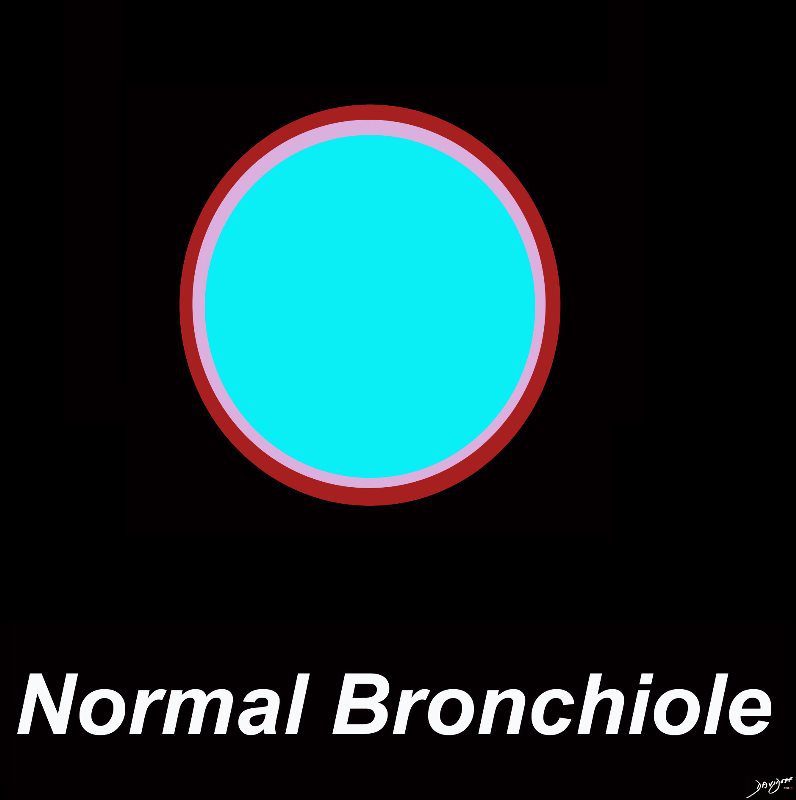
A normal bronchiole usually 1mm or less in diameter. The wall consists of ciliated cuboidal epithelium and a layer of smooth muscle. Bronchioles divide into even smaller bronchioles, called terminal bronchioles, which are 0.5 mm or less in diameter and are primarily lined by club cells, and accompanied by a small number of ciliated cuboidal cells.. Respiratory bronchioles are the final division of the bronchioles within the lung and they are .5mm or less in diameter and contain a simple non ciliated cuboidal epithelium and a thin layer of smooth muscle
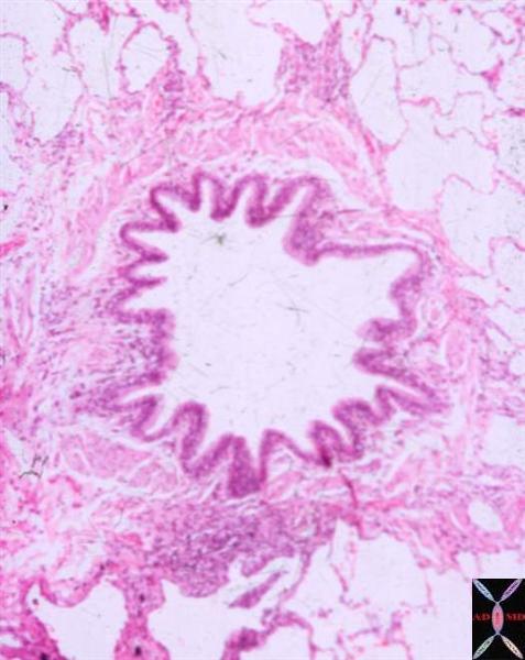
Ashley Davidoff MD TheCommonVein.net lungs-0721Membranous Bronchiole Note folded respiratory folded epithelium resting upon connective tissue. Mucous glands and cartilage are not evident.
Courtesy Dr. Armando Fraire
TheCommonVein.net
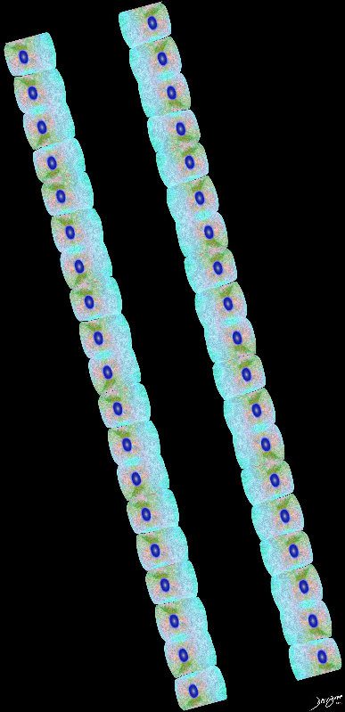
32508As the terminal bronchial transitions to the respiratory bronchial the mucosa becomes non ciliated and cuboidal
Ashley Davidoff
TheCommonVein.net lungs-00676
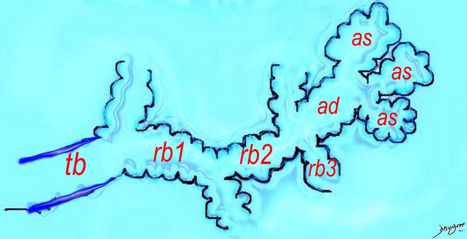
The diagram shows the ductal system of the acinus, starting with the tick walled terminal bronchiole (tb) that is the last duct of the conducting system of the airways. The respiratory bronchiole (rb) enters the secondary lobule and is the first duct to have diffusion capability. There can be 3 orders of rb’s, and they branch into alveolar ducts, which branch in turn to alveolar sacs until they reach the alveoli, which is the final destination.
Courtesy Ashley DAvidoff MD 2019
lungs-0032-low res
The Duct, and the Artery
The pulmonary arteriole accompanies the airway as it carries oxygen from the trachea to the alveoli. They part ways at the alveoli where the pulmonary venule then takes the oxygenated blood from capillary network around the alveoli back to the left atrium.
The intimate relationship of the airways and the pulmonary artery and their close approximation in size, is helpful in radiology, firstly to identify these structures and secondly to define disease such as heart failure and bronchiectasis.
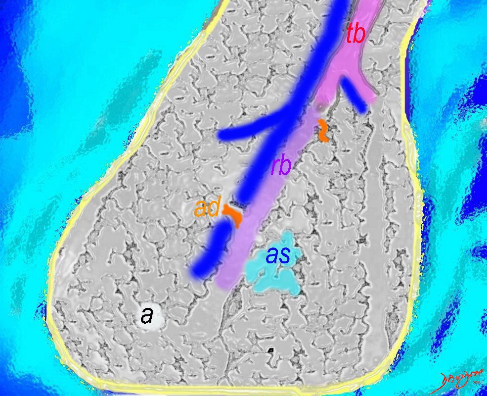
The acinus as shown in this image is defined as a unit of lung consisting of a single first order respiratory bronchiole that subtending a cluster of alveoli reminiscent of a bunch of grapes or berries (acinus in Latin means berry) . The lobular bronchiole (lb) branches into the terminal bronchiole (tb), which then branches into the first order respiratory bronchiole (rb). Subsequent branching after the respiratory bronchiole, includes in order, the alveolar duct (ad), alveolar sac (as), and then finally the berry like alveoli.
Courtesy Ashley Davidoff 2019
lungs-0033-low res
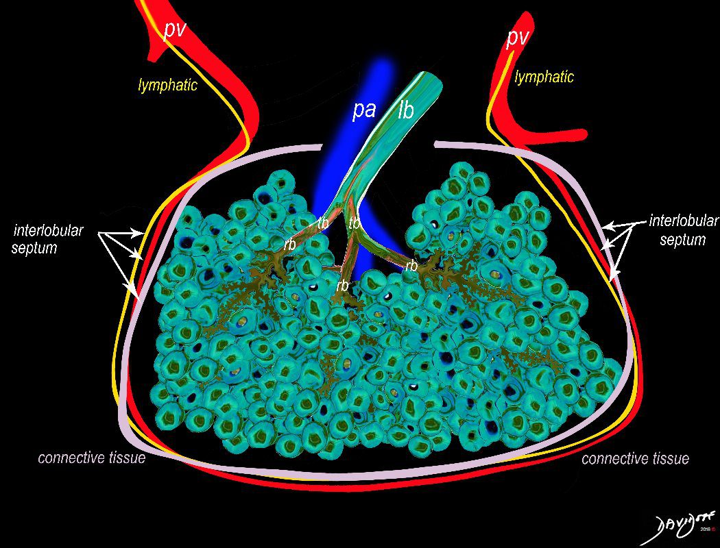
The secondary lobule is housed in a connective tissue framework in which run the lymphatic and venular tributaries . Together these 3 structures form the interlobular septum.
The lobar arteriole enters the framework, accompanied by the lobar bronchiole, and they all run together and form the interlobular septa. This structure measures between .5cms and 2cms and is visible on CT scan.
It is important in clinical radiology since many of the structures can be identified in health, and more particularly in disease, enabling the identification and characterization of many pathological processes.
Courtesy Ashley Davidoff MD
lungs-0036-low res
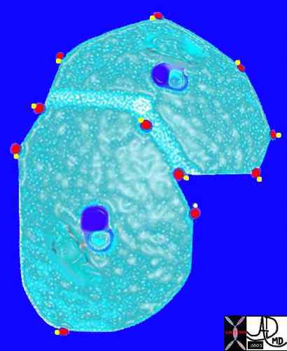
The arteriole and bronchiole lie in the center of the lobule.
Pulmonary venules (red) and lymphatics (yellow). lie in the periphery of the lobule
42440b03
Davidoff Art Courtesy Ashley Davidoff MD
Ashley Davidoff MD

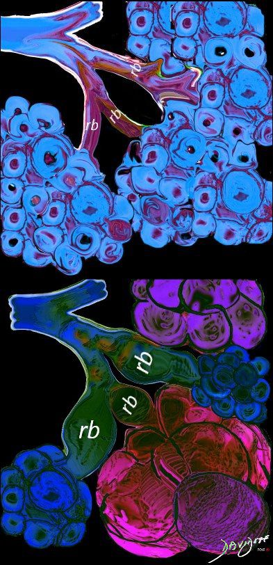
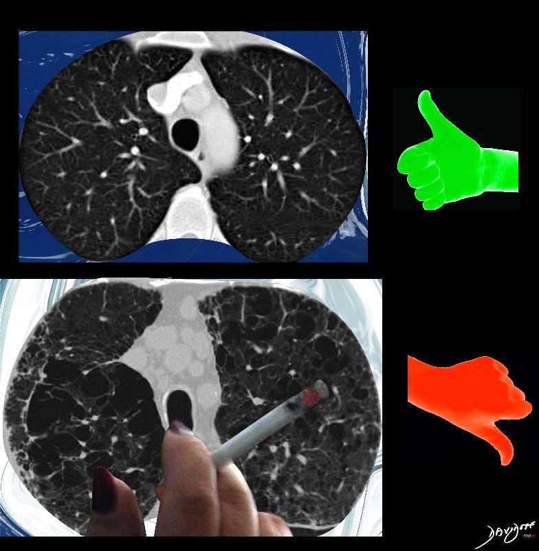
Image on the left shows normal size and appearance of terminal bronchioles and alveoli. On the right the image shows the effects on the respiratory bronchioles and when severe, on the alveoli as well
Ashley Davidoff MD
TheCommonVein.net
Ashley Davidoff Art TheCommonVein.net

Ashley Davidoff MD TheCommonvein.net
References and Links
see Bronchiole Wiki