
- Normal
- Adenopathy
- Non Calcified
- Calcified
- Stippled
- Egg Shell
Normal
Adenopathy
Bilateral hilar adenopathy is most common and usually symmetric (50 percent of cases) or the right may be slightly more prominent . Unilateral adenopathy is uncommon (<5 percent of cases).
See Garland Triad, Pawnbrokers Sign and 1,2,3 Sign
Non Calcified
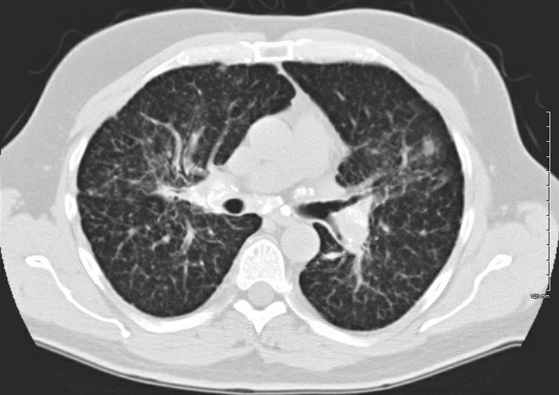
SARCOIDOSIS, ACTIVE – ALVEOLAR FORM
Ashley Davidoff MD
Solid
Solid Calcifications
TB
INACTIVE SECONDARY TB WITH EXTENSIVE PARENCHYMAL AND LYMPHOVASCULAR INVOLVEMENT
48-year-old male with history of TB
Ashley Davidoff MD
INACTIVE SECONDARY TB WITH EXTENSIVE PARENCHYMAL AND LYMPHOVASCULAR INVOLVEMENT
48-year-old male with history of TB presents with back pain
AP view of the spine shows complex lesion in the right apex characterized by fibronodular opacities. There are scattered calcifications throughout the lungs but some are centered around the lymphatics, including the interlobular septa and centrilobular region
Ashley Davidoff MD
Histoplasmosis
PULMONARY HISTOPLASMOSIS
77-year-old male presents for preop CABG and admitting CXR shows multiple large pulmonary nodules
Chest CT shows innumerable pulmonary nodules ranging from 5mm to 17mms. A few of these nodules are calcified.
CT guided biopsy of the largest irregular nodules in the right lower lobe showed granulomatous pneumonitis with intracellular fungal spores, positive PAS and GMS most compatible with histoplasmosis
Ashley Davidoff MD
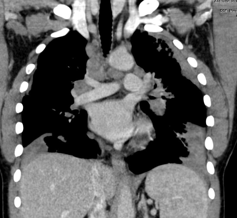
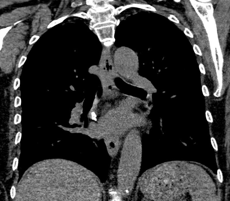
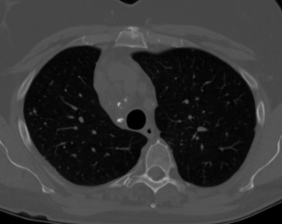
Amyloid

CALCIFIED NODES OF AMYLOIDOSIS
72-year-old male with history of an amyloidoma removed with right middle lobectomy
The CXR shows left ventricular enlargement
The current CT is characterized by stable small hilar nodal calcifications that likely represent amyloidosis
There are calcifications on right side of the left atrium and by the left heart border with associated focal regions of pericardial thickening. Involvement of the pericardium may be due to amyloidosis. However the LS calcification could also be post op and the calcification along on the left heart border could also be branch of circumflex with unusually chunky appearance which would be out of proportion to the degree of calcification elsewhere in the coronaries.
Fat in the LV apex indicates previous LAD territory infarction and likely account for the LVE noted on CXR
Ashley Davidoff MD
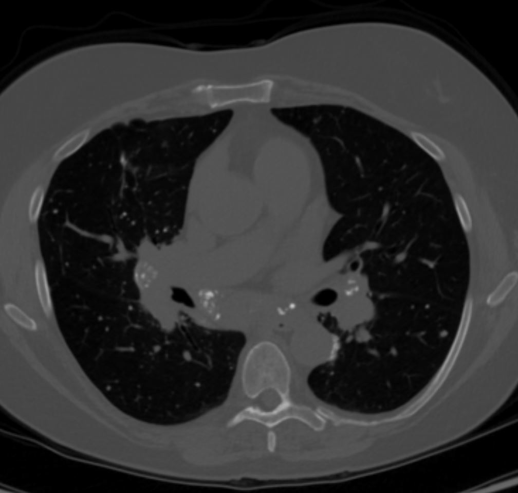
Amyloid
58 F Heterogeneous calcifications in mediastinal and hilar adenpathy
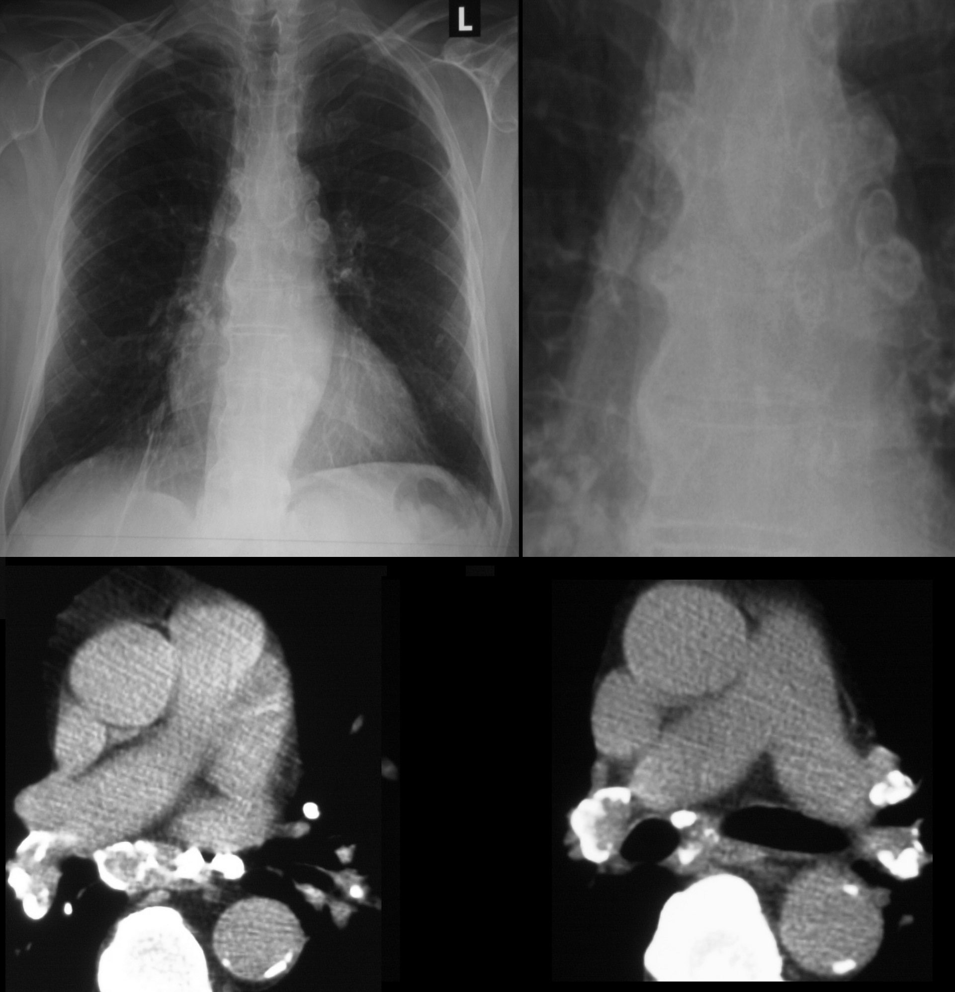
Egg Shell
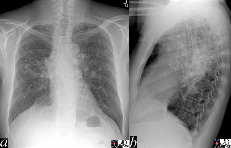
The A-P and lateral view of the chest is from a patient with sarcoidosis showing classical egg shell calcification of the mediastinal nodes and hilar nodes.
Ashley Davidoff MD TheCommonVein.net 42195c01
SARCOIDOSIS AND EGG SHELL CALCIFICATION OF THE LYMPH NODES
51-year-old male with Stage 3 Sarcoidosis and egg shell calcification of lymph nodes
Ashley Davidoff MD
51-year-old male with Stage 3 Sarcoidosis and egg shell calcification of lymph nodes
Ashley Davidoff MD
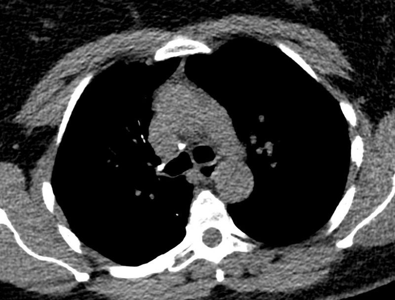
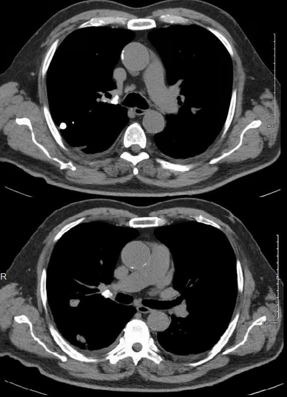
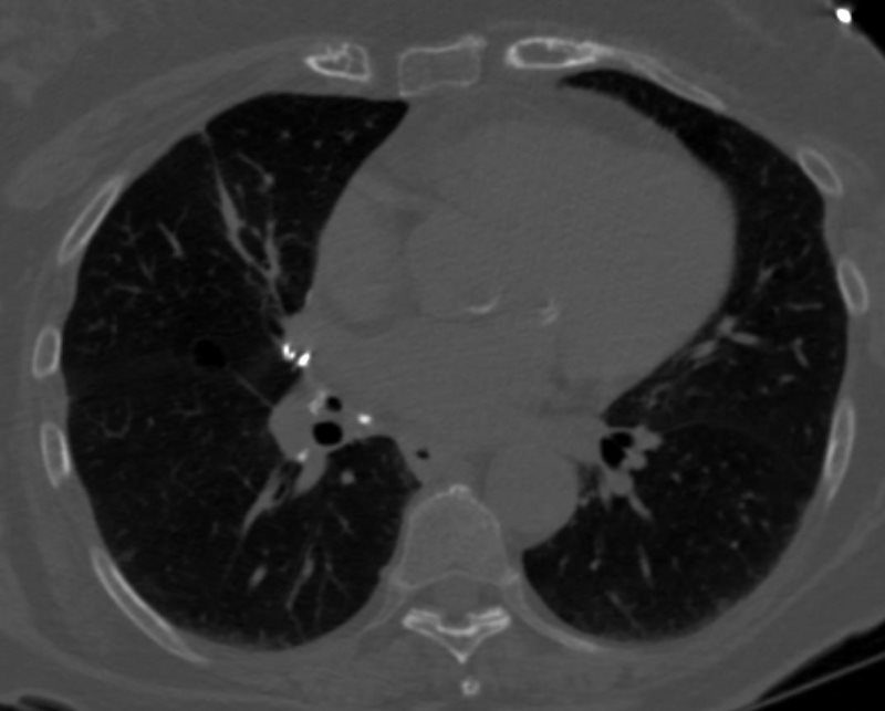
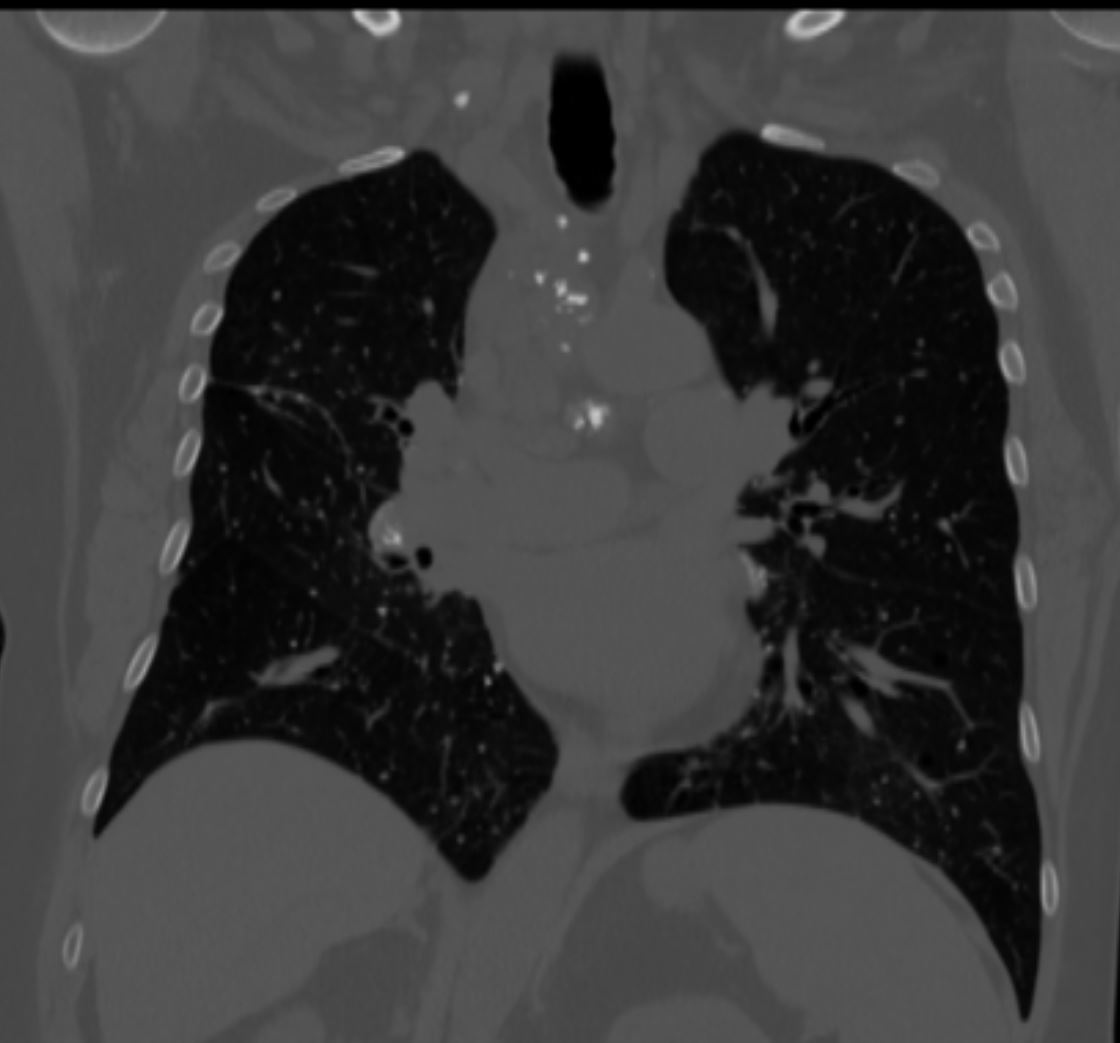
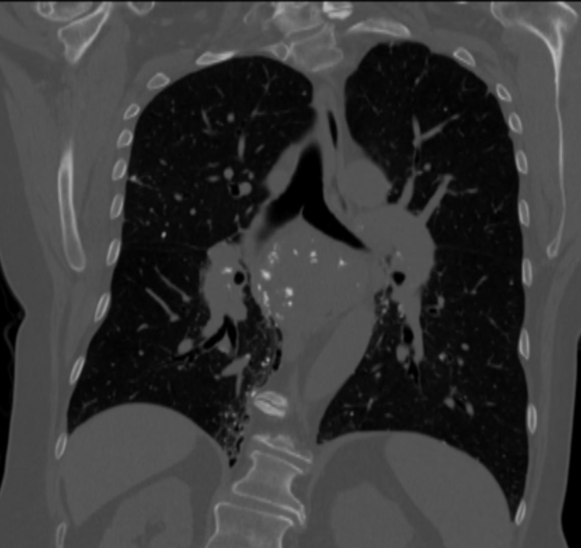
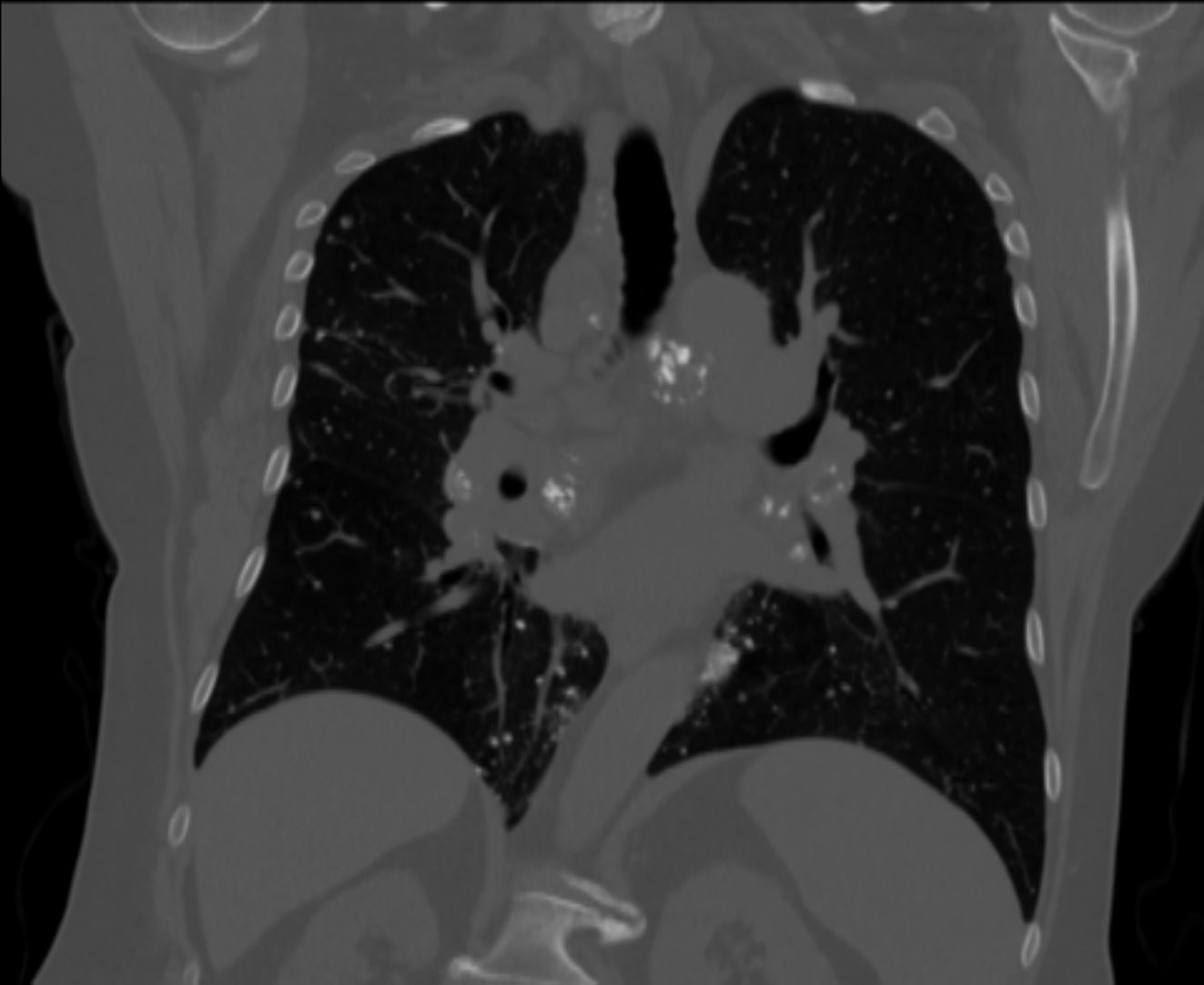
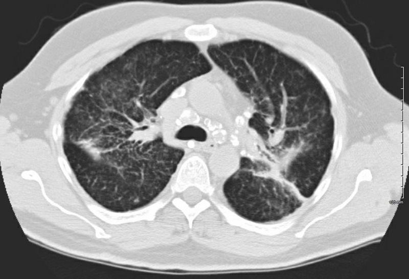
Stippled
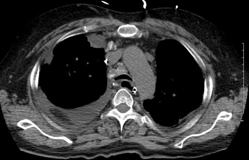
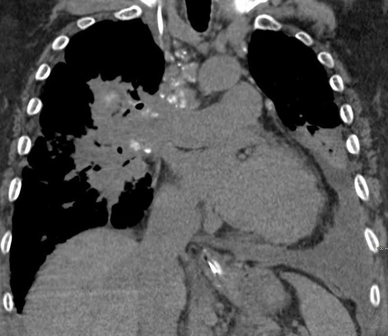
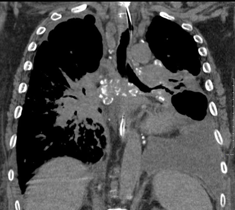
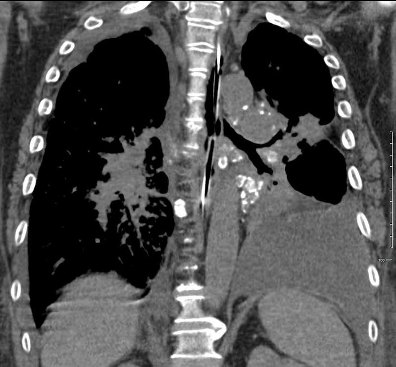
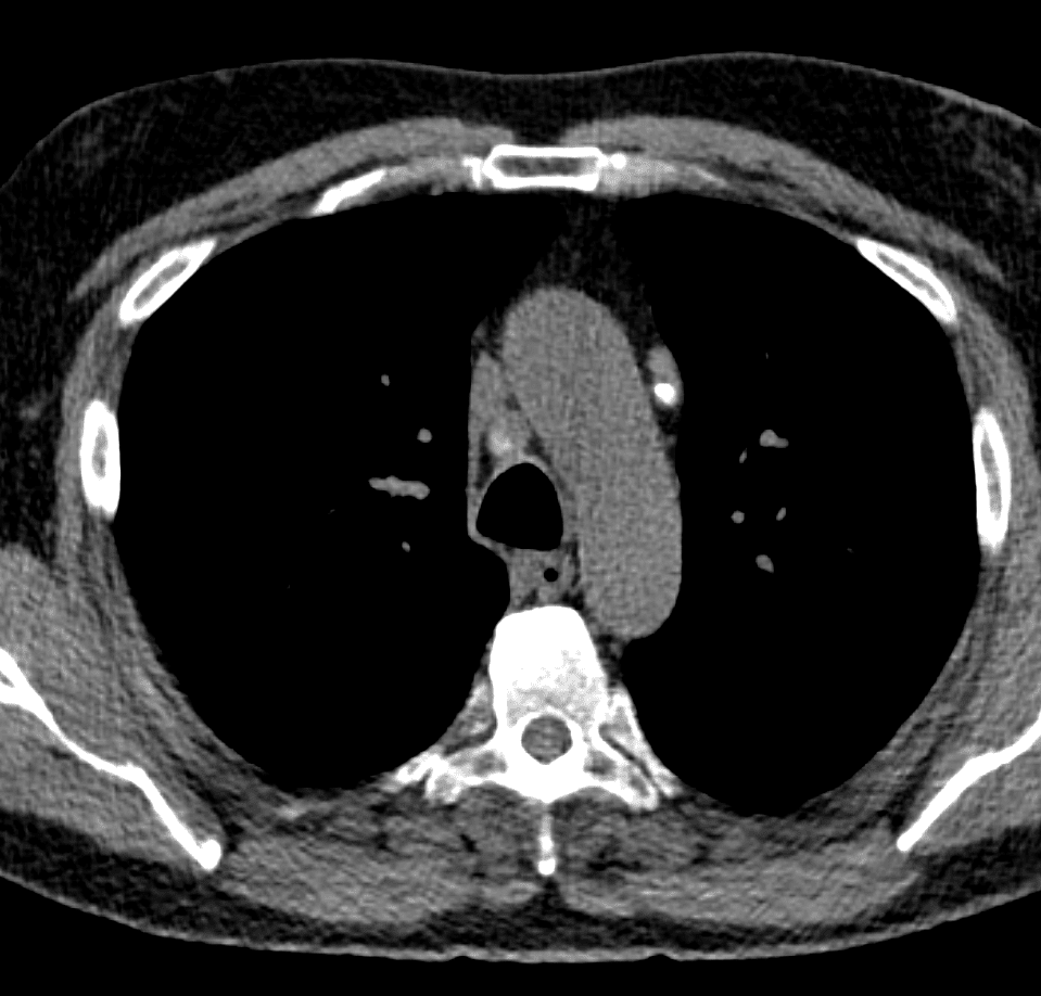
SARCOIDOSIS, STAGE IV, PTX, ENCASEMENT
Ashley Davidoff MD
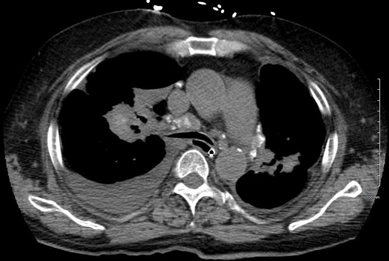
SARCOIDOSIS, STAGE IV, PTX, ENCASEMENT
Ashley Davidoff MD
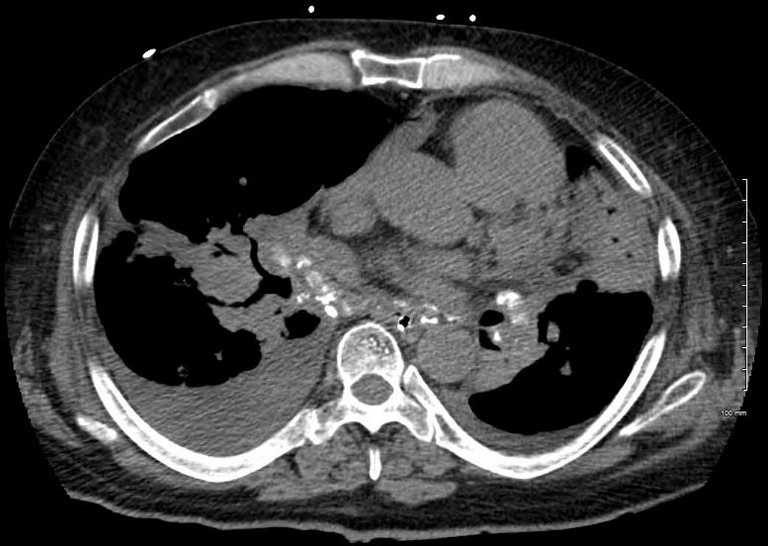
SARCOIDOSIS, STAGE IV, PTX, ENCASEMENT
Ashley Davidoff MD
SARCOIDOSIS, STAGE IV, PTX, ENCASEMENT
Ashley Davidoff MD

SARCOIDOSIS, STAGE IV, PTX, ENCASEMENT
Ashley Davidoff MD
SARCOIDOSIS, STAGE IV, PTX, ENCASEMENT
Ashley Davidoff MD
SARCOIDOSIS, STAGE IV, PTX, ENCASEMENT
50-year-old male presents with history of Stage 4 sarcoidosis acute chest pain and dyspnea
Ashley Davidoff MD
Egg Shell
The A-P and lateral view of the chest is from a patient with sarcoidosis showing classical egg shell calcification of the mediastinal nodes and hilar nodes.
Ashley Davidoff MD TheCommonVein.net 42195c01
79 year old male with sarcoidosis and egg shell calcification of the hilar and mediastinal nodes
Ashley Davidoff MD