
The Secondary Lobule (aka pulmonary lobule)
We will now move upstream toward the terminal bronchiole to gain an understanding of the secondary lobule. From a radiological point of view, it is structurally a more important unit than the acinus. It is a conglomerate of 3 to 5 acini enclosed in a membrane with a polyhedral shape. From a structural and radiological point of view it is important to understand this entity, since it has implications in the understanding of the CXR, conventional CT, high resolution CT and in the structural changes that occur in some of the lung diseases.
We have already described the “buddy system” that exists in the lung, with the branches of arterioles and bronchi traveling together, and the venules and lymphatics going together. Lymphatics sometimes also accompany the bronchovascular bundle. A story called “Grapes of Exchange” about two friends on a journey will describe the structures within the secondary lobule.
In the story there are two pairs of structures that travel together as they branch through the lungs. The first pair is an artery and an airway and the second is a vein and a lymphatic. The story is described by the airway – a female — who travels with the artery – the male. They travel for many divisions together, dividing at the same time. In the early phases they are both tubular in their form and equal in size. As they enter close to the chambers of gas exchange the airway develops little sacs off her body and she becomes rounder and fatter, while the artery continues to be sleek thin and tubular. While she becomes the chamber through which gas exchange takes place he becomes at one with the vein after traversing the capillaries. Once revived by the oxygen, he becomes a vein and he picks up his new partner, the skinny lymphatic, and continues on his travels accompanied by the lymphatic. The airway feels at this stage like a ditched lover but there is hope for her as she continues on her return journey.
Grapes of Exchange – A Romantic Story
The following audio is a story that describes the relationship between the pulmonary artery and the bronchiole as they branch through the parenchyma and eventually reach the secondary lobule. It is told in the form of a story to promote understanding . If you do not have an audio system the story is in written form below.
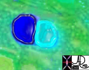
This image shows the arteriole (royal blue) and the bronchiole (teal) travelling side by side bith of equal size. Ashley Davidoff MD TheCommonVein.net 42440b05
From the minute I saw him I loved him and I knew that no matter what evolved, we would spend our lives together… little did I know what the working relationship would be.
Here follows a true story about me, the bronchiole, and my lifelong buddy the pulmonary arteriole.
He and I were on a mission. I had my origins in the atmosphere and he from the heart. We came from very different backgrounds and had come a long way on this trip on the highways and byways of the airways and circulation. My mission was to take products from the air in the atmosphere and deliver them to the grapes of exchange and his was to deliver blue blood to the same grapes. In this communication we will speak mostly of the journey to the grapes in the house of the pulmonary lobule. The grapes of exchange in my imagination had something to do with bonding and marriage – perhaps the exchange of vows. The story turns out quite differently. We had great travels together and took a lot of pictures!!!
I think by this time you know what we had already been through. He had started out as a large elastic vessel off the right heart called the main pulmonary artery (nickname MPA) and I had started out as the trachea. (nickname “trach”). We met at the doorway of the lung called the hilum, and took a fancy to each other right away and so we decided to travel together. We had traveled a long way by the time our story begins both experiencing many divisions and were right in the middle of an inspiration. We both looked quite different at this point having given birth to many offspring. In the new language – we had both “morphed” quite a bit but this was a necessary part of the mission. Of course we were much smaller than we had been. He had lost some of his elasticity and developed a bit of muscle. I had lost my entire cartilagenous skeleton and had developed some muscle as well. We were told that from now on we were both going to lose muscle and I in particular was going to change drastically. I could not wait?!!!! I secretly hoped that this change would make me more attractive and bring me closer to a happy union since I was promised a happy union in the end. My name at this stage was “terminal bronchiole”. A foreboding and deathly chill rattled down my muscularis as I said the word “terminal” knowing that I was going to lose the small amount of muscle that I had.
We reached the doorway surrounding the secondary lobule and faced the polyhedral entrance. It was quite beautiful I thought in my teal blue outfit.
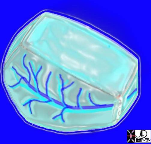
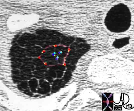
Here is a picture of the outside of the polyhedral pulmonary lobule from the side. It looked quite futuristic. Through the transparent side window we saw a couple similar to ourselves. From this vantage point the morphing did not look too different from what we had already been through – division after division – leaner and meaner. Ashley Davidoff MD. The Common Vein.net 42449b02
We took a quick walk around the polyhedral structure.
We returned to the entrance and took a closer look at the goings on inside through the large front entrance windows of the pulmonary lobule. Below is a picture of what we saw.
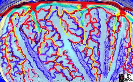
The arteries and airways pair up and travel together from the interlobular septa to the hilum. The pulmonary lobule, also called the secondary lobule is a structural unit surrounded by a membrane of connective tissue, and it is smaller than a subsegment of lung but larger than an acinus. This diagram shows two secondary lobules lying side by side. The pulmonary arteriole (royal blue) and bronchiole (pink) are shown together in the centre of the lobule (“centrilobular”), while the oxygenated pulmonary venules (red) and lymphatics (yellow) are peripheral and also form a formidable and almost inseparable pair.
42440b03
Ashley Davidoff MD
TheCommonVein.net
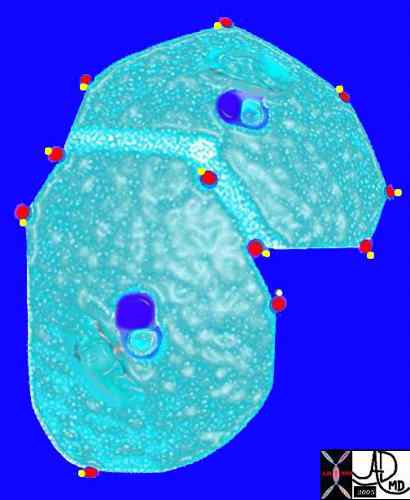
As we were ushered into the lobule, we were faced by a refreshing atmosphere of comforting air and a hub of activity. In addition to the swishing sounds of air movement we heard a hubbub of clinking and clanking. We were told that the sounds were coming from the grapes of exchange (wedding bells?). We also noticed that there were at least three other couples who looked like the people of our tribe – three other bronchovascular bundles. We called our tribe the “bronchovascular bundle” with the one part of the bundle being the progeny of the bronchus and the other, the progeny of the pulmonary artery. A group picture inside the lobule is shown below. Fortunately the flash was working because it was a little dark inside.
This picture shows us on the left with a white ring around us (we were the tallest) and the other couples who looked so much like us (also ringed). We called our tribe the “bronchovascular bundle” with the one part of the bundle being the progeny of the bronchus and the other the progeny of the pulmonary artery. In the distance at the periphery we could see the pairs from the other friendly tribe – the red pulmonary vein with its smaller yellow buddy the lymphatic. Behind them we could see the transparent window membrane through which we had peaked earlier. Oh my goodness!!! Look what has happened to my body!!!!!!!…… Ashley Davidoff MD. The Common Vein.net 42447b03b01
I did not realize that my body had already started to morph at the time of the picture – little sac-like love handles spreading all along my formerly sleek and beautiful body Yech! I did not look good at all. My buddy the arteriole on the other hand as well as members of the other tribe maintained their sleek tubular looks. “Why me?” I shouted. “All for the good of the nation and good gas exchange!!” they shouted above the swishing of air and clanging of the factories. “Easy for you to say,” I replied. “Some of us have to suffer some bad morphs for the good of the whole” they said, “but those love handles kinda suit you,” they exclaimed with a wry smile. I was so sad.
Well the divisions started coming rapidly and so we all became smaller very quickly. I was getting rounder and rounder while they were getting thinner and sleeker. Things seemed to be rushing at an accelerated pace and I must say that there was some excitement in the air as we all got closer and closer to each other. In the big picture we seemed to be coming together as a team, and the whole landscape seemed more colorful and more promising. We reached out to the other tribe and they too us.
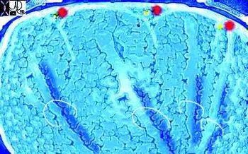
This picture was taken just before the real drama started. The image gives a sense of what was to come. You can see here in the house of the lobule that we were all dividing into smaller parts and were getting smaller and the picture was quite colorful and rosy. I fully expected to have intimate contact with the arteriole… but it did not happen as I expected…… Ashley Davidoff MD. The Common Vein.net 42447b05b02
I remained on the plump side as my love handles grew more pronounced and rounder while the clanging noises grew louder and louder. I was dividing into alveolar ducts and alveolar sacs and finally into alveolus which of course is a complete transformation from my former tube like shape to the spherical shape of the grape. In the mean time my buddy the artery grew as small as an 8 micron red cell and remained true to his tubular form. He started to surround me completely but was more in concert with that beastly red vein as he ran headstrong in blind passion to join her in capillary union. For a moment they each lost their identity becoming the tiniest of tubes neither arteriole nor venule. I on the other hand became at one with the fruit of the vine and in stuporose state I think I became the grape itself. Through my waist I felt the pleasant freshness of gusts of air going back and forth through my skin. My delivery of fresh air moved very quickly into the capillary. Little did I know that it was the oxygen that I carried which gave my competition the beautiful red glow of health. The odious carbon dioxide moved the other way. This toxic waste came directly from the man I had known and trusted for so long. Thanks for nothing.
I do not like to show the next picture much since for me it is a sad one. There I am a ditched lover, fat as a grape, in the middle of the capillary union between my blue “ex” and his new partner in crime, the red beast. Yes of course it was for the better – they all say that – but what about me?
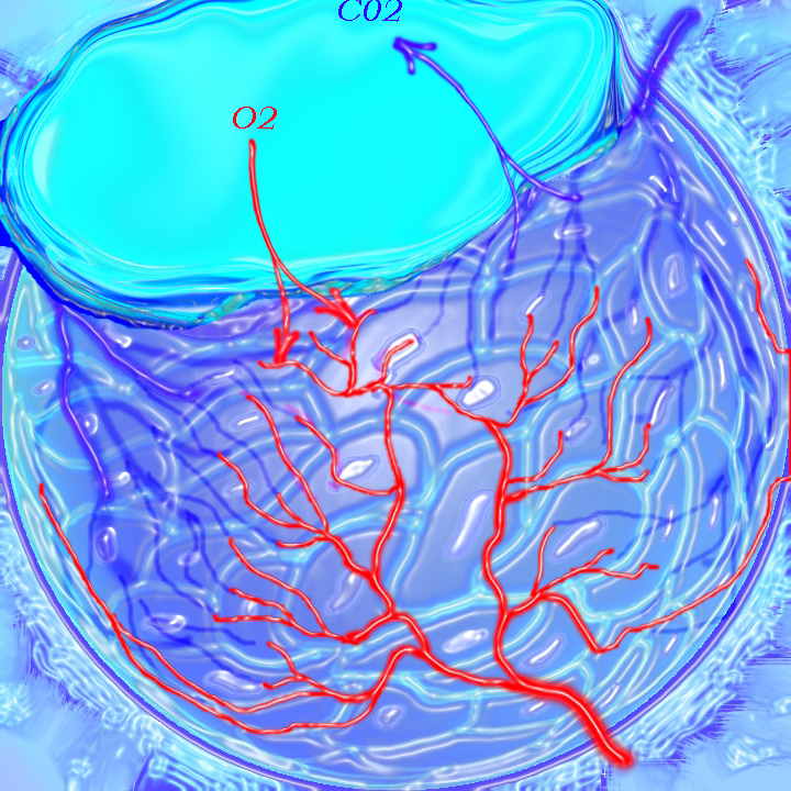
There I am the fat alveolus surrounded by the gracile capillary network, with breezes of oxygen and carbon dioxide whiffing through my waist. The authors of this module seem to like this picture – but for me I an in my worst physical shape.
Ashley Davidoff MD. The Common Vein.net 42438B03
As you have learned already, the blood circulation needs two types of vessels; one to carry the blood to the lungs and a second to carry it from the lungs. I on the other hand do it all by myself. On inspiration I take air to the lungs and on expiration I take it away through the same vessel to the atmosphere. This is the lot of all women. We do the double the amount of work shlepping here and there and everywhere and get no respect for it.
So at this junction we are in the middle of an inspiration (for me – what kind of inspiration could I feel in my morphed format) and we were just about to start the expiration. While I was traveling to the exit of the lobule during this phase of my life, my “ex” was going in the same direction – now transformed into a beautiful vessel with that healthy glow. Despite my odious load and my downtrodden feeling I moved with a sense of optimism.
What do you know? As I left the chamber of the lobule I shed my love handles one by one and my saccular form started to take on the sleek and tubular look again. I looked and felt brand new. “Hmm… I thought – perhaps he will fall in love with me again. I was hoping for another inspiration, just to show him once again how beautiful I was both on the outside and the inside. And that my friends was my nightmare in the Grapes of Exchange.
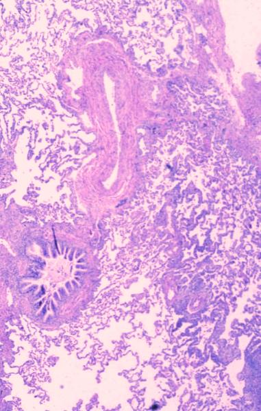
This histological section shows the bronchovascular bundle in their true form. We are still fairly proximal since you can see the much smaller alveoli and alveolar ducts in the background. The mucosa of the bronchiole is thrown into a series of folds while the endothelium of the arteriole smooth and the lumen is collapsed.
Courtesy Armando Fraire MD 32699b
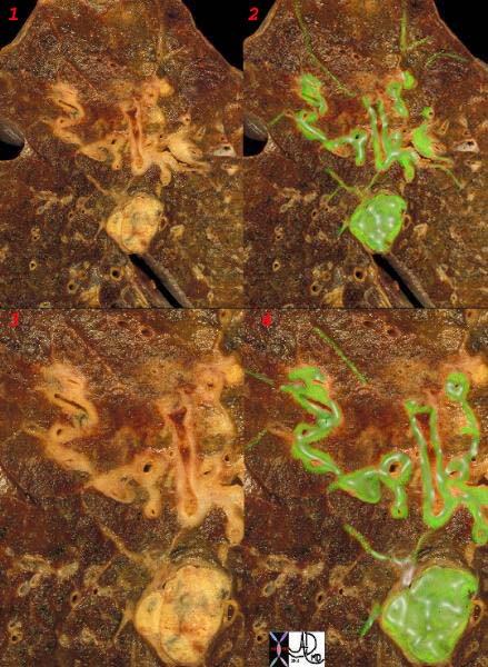
The secondary lobule contains all the structural elements of the lung compacted into a small space and it is a recognizable structural entity that measures about 1-2.5cms. Prior to entering the secondary lobule the bronchovascular bundle branches at approximately 1cm intervals. However, once they enter the confines of the lobule the branching becomes fast furious and a new branch originates every 1-3mms. A lot of structure subsequently packs into a small space. The lobules are well formed in the apices and the peripheral aspects of the lungs and poorly formed posteriorly. In the image below the lymphatic system has taken up carbon particles and the blackened lymphatics outline the interlobular septa, which in turn outline the lobule.
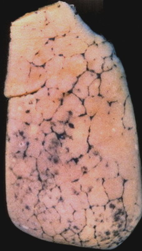
Anthracosis – Note the accumulation of carbon particles within the lymphatics along the interlobular septa, outlining the secondary lobules. The carbon particles are inhaled from an anthracotic urban environment. Courtesy Ashley Davidoff MD. TheCommonvein.net 32291 keywords lung interlobular septum septa secondary lobule pulmonary lobule intertstitium interstitial grosspathology carbon
The interlobular septa and the secondary lobules are not normally visualized. However any disease of the lung, particularly if the lymphatics are involved, will expose the lobules mostly by providing better visualization of the interlobular septa. If more than 3 contiguous secondary lobules are visualized then the lung is abnormal. At the level of the terminal bronchiole the arteriole and bronchiole measure about 0.2 mms each and it is the challenge of our CT technology to resolve these structures. At this level we are starting to push the limits of resolution. Thin collimation allows us to optimally visualize the lobule. We are thus better able to visualize the septa and the lobules with 1.5mm collimation than with 3mm collimation and our chances diminish as collimation gets thicker. The evolution of high resolution CT scan has allowed us insight into the lobule and provides a better understanding of the diseases that affect the lobule.
In this high resolution CTscan at the apex of the right lung the secondary lobule is seen with 3 sets of bronchovascular bundles at the door of the secondary lobule surrounded by the pulmonary venules and lymphatics in the surrounding septum. Note also that lymphatics are found both in the periphery of the lobule as well as in the centre of the lobule. This patient had mild emphysema and it was surprising to find this beautiful example of a pulmonary lobule in a patient who was almost normal. We suspect that with the higher resolution technology we will see the normal (or almost normal) pulmonary lobule with greater frequency.
Ashley Davidoff MD TheCommonVein.net 31819b01
Congestive Heart Failure
We are never able to visualize the normal lobule on a plain film but in diseases such as congestive cardiac failure when the lymphatics become overwhelmed they distend and become visible. In congestive cardiac failure the heart fails as a pump. In left ventricular failure the resulting congestion is reflected in the left ventricle (LV), left atrium (LA), pulmonary veins and capillaries as increased pressure. It is reflected structurally as dilatation and of the chambers and vessels. The increased fluid and pressure not only cause dilatation but also cause the fluid to leak out into the interstitium. The lymphatics are able remove and mop up some of the excess fluid, but there is a point when the volume overwhelms the lymphatics and the excess fluid remains in the interstitium. On CXR the findings include dilatation and cephalization of the arterioles, peribronchial cuffing, Kerley B lines (thickening of the interlobular septa), pleural effusion and subpleural edema.

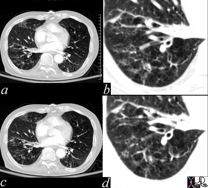
In these images. and c are normal and b and d represent thickened interlobular septa in a patient with congestive heart failure. These are the well known Kerley lines, often spoken about but rarely seen. They are identified as thin horizontal lines usually seen in the costophrenic angles, not being longer than 2 cms in length and touching the pleural surface.
Ashley Davidoff MD TheCommonVein.net
42545c01
lung axial interstitium bronchioles connective tissue fx bronchial plugging peribronchial halo peribronchial thickening dx bronchopneumonia CTscan
Ashley Davidoff MD. The Common Vein.net 47614c01
Other Diseases Carcinoma Sarcoidosis
Ashley Davidoff MD
TheCommonVein.net
32199cw
keywords
lungs pulmonary parenchymal mass neoplasm primary lymphatics distended metastases lymphangitis gross pathology
Sarcoidosis
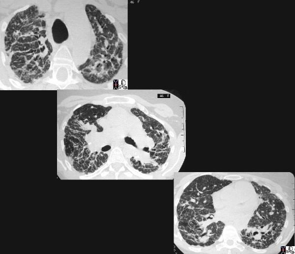
This cross sectional series of 3 CT images shows end stage sarcoidosis characterised by marked thickening along the lymphovascular bundles. The first image in the upper left shows marked thickening of the interlobular septa caused by granulomatous changes along the lymphatics. Courtesy Priscilla Slanetz MD. 31866c
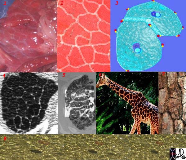
This is a series of images demonstrating the shape of the secondary lobule. The first image (1) is a post mortem specimen with congested lungs showing the interlobular septa, while the next (2), is an overlay of the septa in white showing their polygonal shape. The next drawing reveals side-by-side secondary lobules with central bronchovascular bundles and peripheral lympho-vascular bundles. Image 4 is a CT image through the apex of the lung showing thickened secondary lobules in a patient with mild emphysema, and 5 shows marked thickening of the interlobular septa in a patient with end stage sarcoidosis. 6,7,8 show the shape of the secondary lobules in the skin of a giraffe, the bark of a pine, and the ripples of the water respectively.
Ashley Davidoff MD TheCommonVein.net 31866collage