
Benign Hamartoma
Ashley Davidoff MD TheCommonVein.net hamartoma 006v
Ashley Davidoff MD TheCommonVein.net hamartoma 003c
Cancer
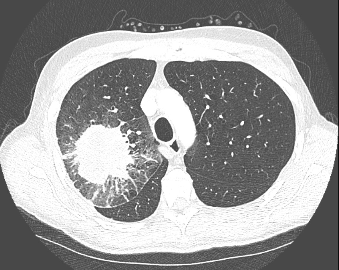
CT in the axial plane demonstrates a large, spiculated mass in the right upper lobe with surrounding halo likely reflecting hemorrhage or lymphatic edema around the mass. In addition, there is evidence of irregular interlobular septal thickening likely reflecting lymphatic invasion and indicating lymphangitis carcinomatosa. There is irregular thickening of the major fissure suggesting involvement.
Ashley Davidoff MD TheCommonVein.net 135865
Cavitation and Malignancy
Squamous Cell Carcinoma

Ashley Davidoff MD TheCommonVein.net squamous cell carcinoma cavitating 003
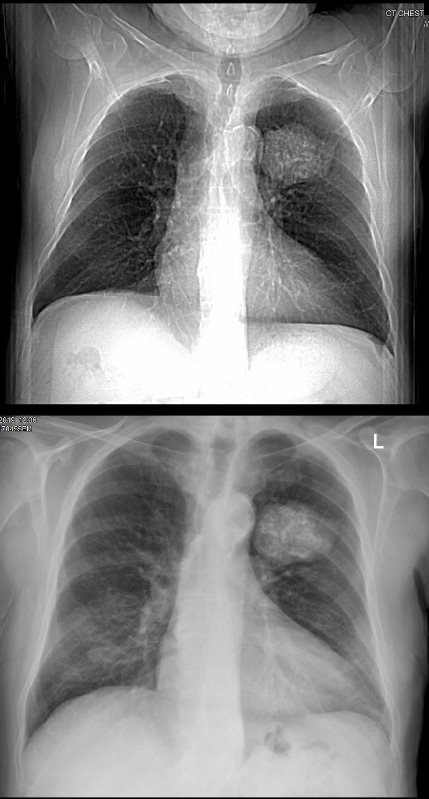
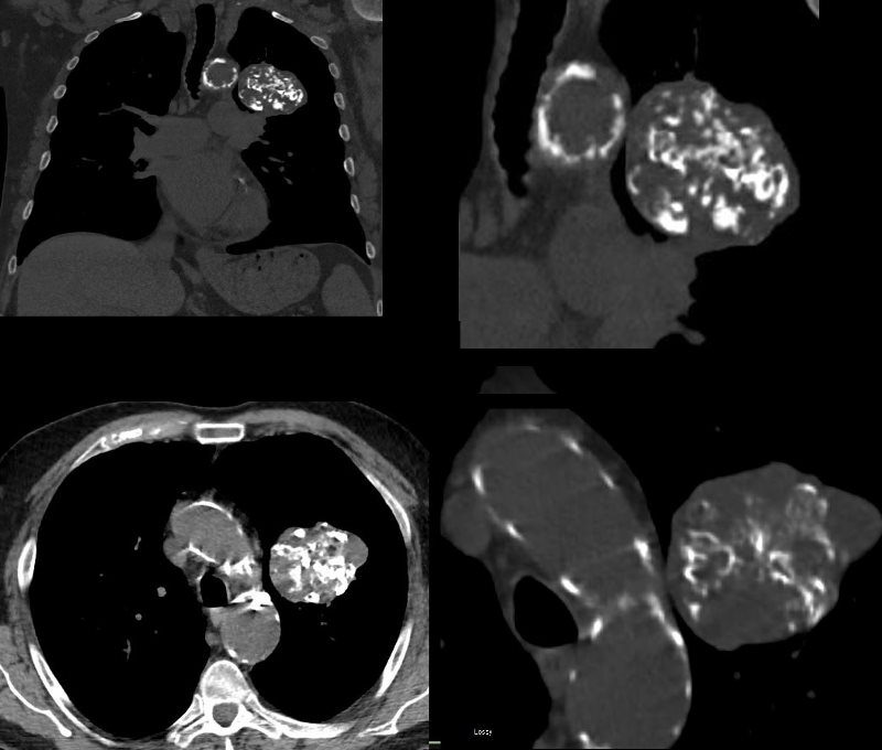
Calcification in Malignancy
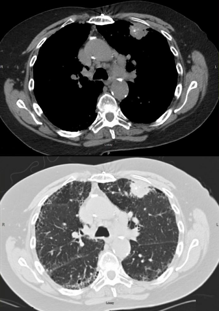
73 year old female with left mastectomy and IPF with new onset bilateral upper lobe masses. The LUL mass has scattered calcifications, and the right shows early cavitation.
Both lesions are PET positive
The LUL lesion was biopsied revealing squamous cell carcinoma with calcifications showing progressive growth and enlarging ipsilateral lymphadenopathy.
Ashley Davidoff MD TheCommonVein.net