
In a Nutshell
- Size
-

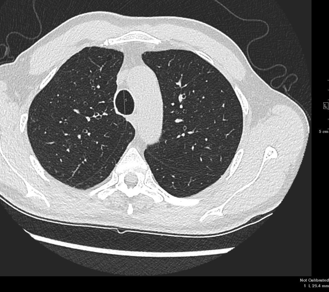
66 year old male - 1.3- 2.5cms in the male and from 1-2.1cms in the female
- Tracheomalacia
-
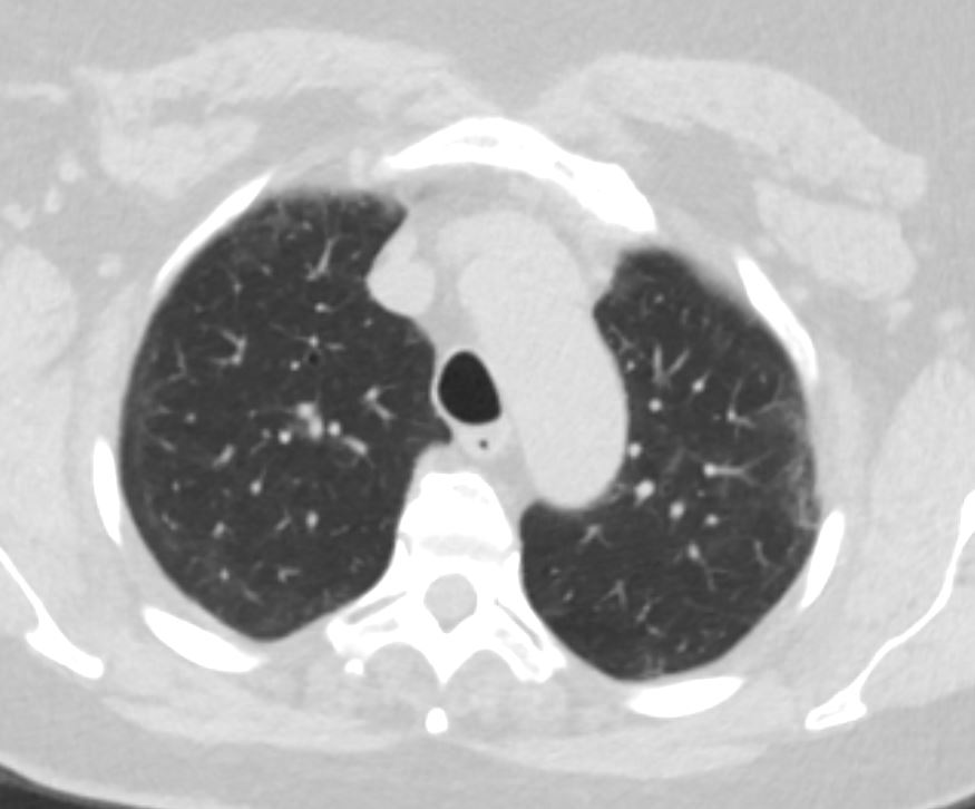
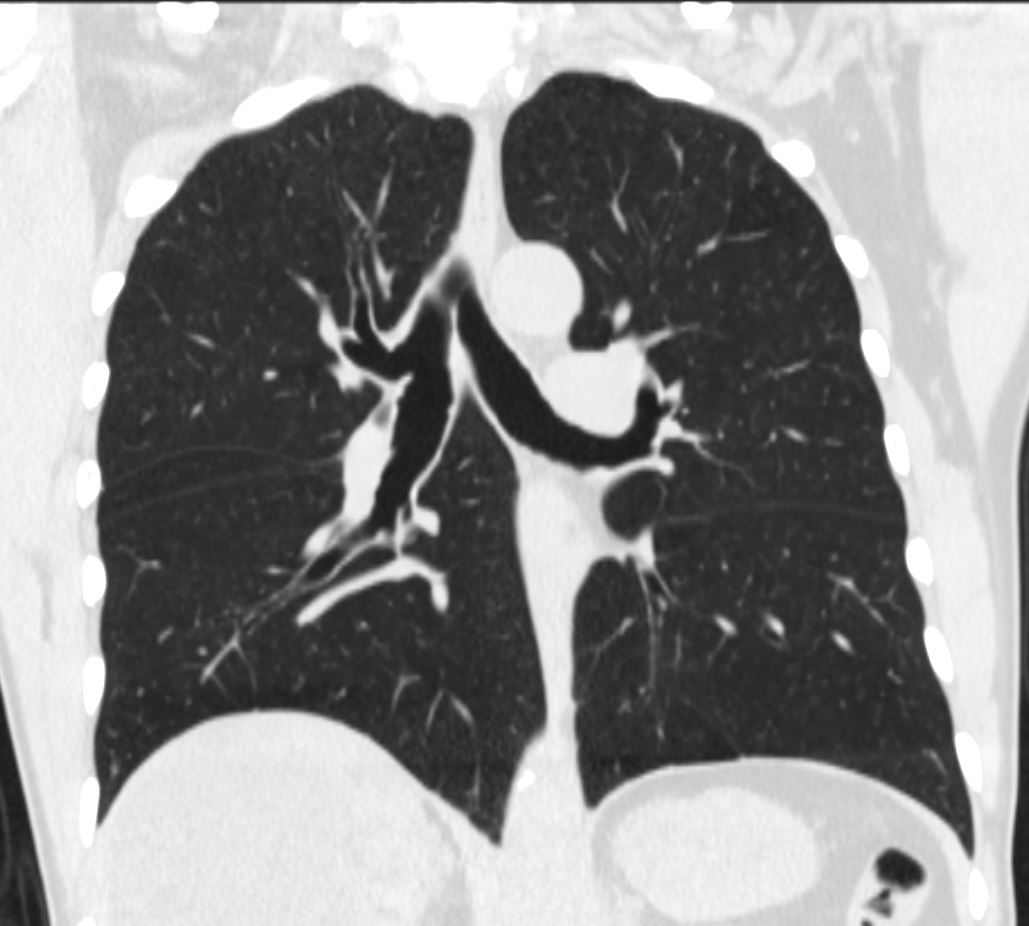
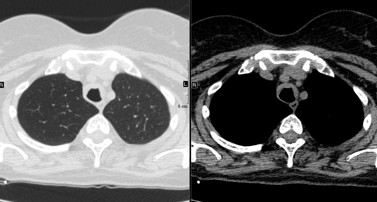
INSPIRATION
68 year female with tracheomalacia
On inspiration the trachea is normal and widely patent and on expiration the trachea collapses to greater than 50% of its original diameter
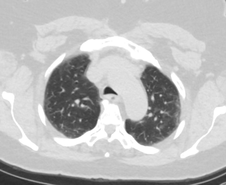
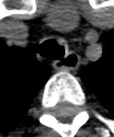
Ashley Davidoff MD TheCommonVein.netEXPIRATION
68 year female with tracheomalacia
On inspiration the trachea is normal and widely patent and on expiratation the trachea collapses to greater than 50% of its original diameter
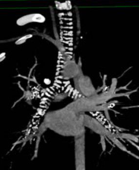
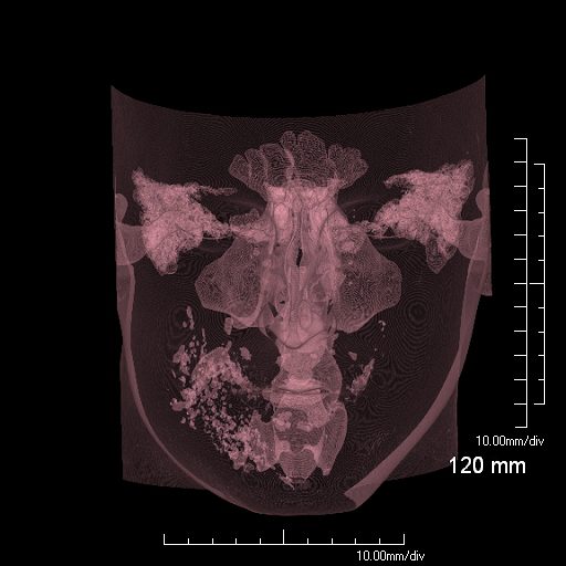
Ashley Davidoff MD TheCommonVein.netGoitre displacing the trachea to the left
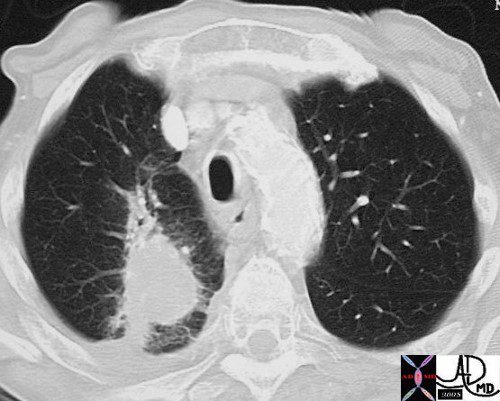
In the CT above a retrosternal thyroid goitre is compressing and displacing the trachea from its normal position to a more leftward and anterior location.
45796c key words thyroid trachea enlarged compression surface rendering normal anatomy
Ashley Davidoff TheCommonVein.net
-
- Tracheomalacia
-
- Shape
-
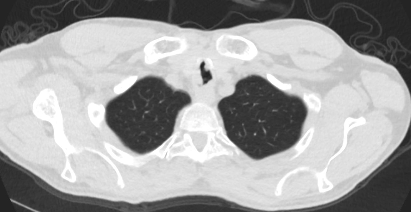
Trachea – shape
Normal chest CT of the upper lobes of both lungs. The trachea is horse shoe shaped.
Ashley Davidoff TheCommonVein.net 32158 42636c01Saber Sheath Trachea in COPD
Ashley Davidoff TheCommonVein.net
-
- Position
-
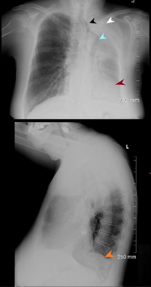
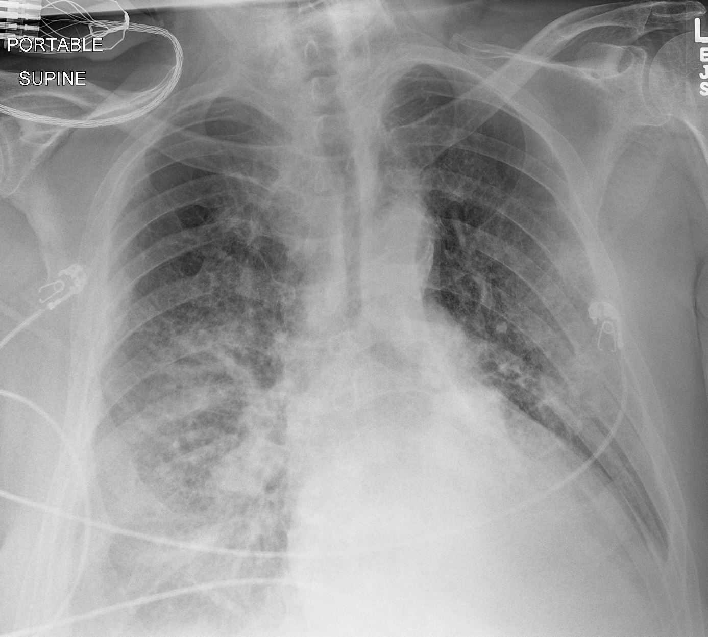
76-year old female presents with dyspnea. PA CXR shows airless consolidation of the left upper lobe (white arrowhead), volume loss of the left hemithorax, with leftward shift of the trachea (black arrowhead) and cardio mediastinal structures (maroon arrowhead). The right lung is hyperinflated with herniation across the midline (light blue arrowhead). The lateral examination shows an anterior airless consolidation, and elevation of the left hemidiaphragm orange arrowhead). Granulomatous calcifications are noted in the left upper lobe atelectatic segment
Ashley Davidoff MD TheCommonVein.net - Character
- Amyloidosis of the Trachea
-
Amyloid in the Trachea
Ashley Davidoff
TheCommonVein.net 44f Amyloid airways 001Tracheobronchial Papillomatosis caused by Infection with Human Papillomavirus
44M Tracheobronchial Papillomatosis caused by infection with human papillomavirus
Ashley Davidoff TheCommonVein.net - Time
-
Tracheobronchial calcifications occur as a normal aging process
Courtesy CTisUS
-
The trachea is a tubular portion of the respiratory system that connects the larynx to the bronchi. It is relatively thin walled, about 6 inches long in the adult, consists of a series of c-shaped cartilagenous rings anteriorly and a membrane posteriorly. At the level of the carina it divides into left and right bronchi.
Parts
Principles
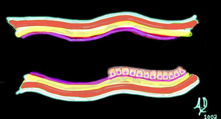
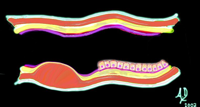
key words art mucosa submucosa muscularis adventitia serosa histology 32347 tube colon small bowel lung bronchus bronchi esophagus stomach large bowel bile duct ureter tube principles
Ashley Davidoff TheCommonVein.net
32347d01 key words mucosa submucosa muscularis adventitia serosa mucosal mass polyp neoplasm carcinoma acute angles with the lumen histopathology imaging diagnosis Ashley Davidoff TheCommonVein.net
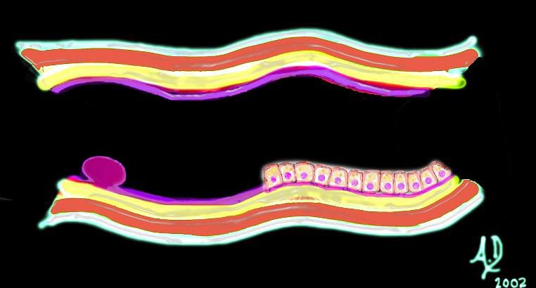
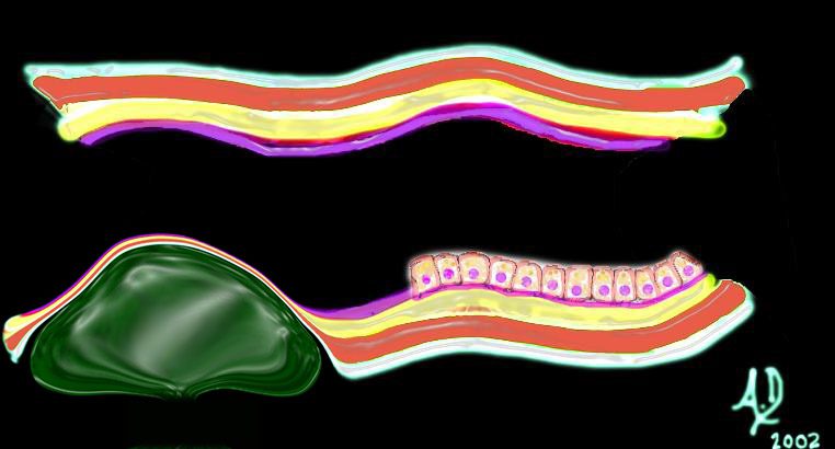
32347d02 key words mucosa submucosa muscularis adventitia serosa submucosal mass edema hemorrhage obtuse angles or right angle 90 degree ninety degree angle with the lumen histopathology imaging diagnosis
Ashley Davidoff TheCommonVein.net
32347d03 key words mucosa submucosa muscularis adventitia serosa submucosal mass edema hemorrhage obtuse angles or right with the lumen histopathology imaging diagnosis Ashley Davidoff TheCommonVein.net
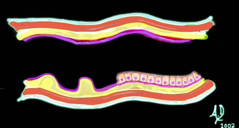
32347d04 key words mucosa submucosa muscularis adventitia serosa submucosal mass edema hemorrhage neoplasm malignancy benign obtuse angles with the lumen histopathology imaging diagnosis
Ashley Davidoff TheCommonVein.net
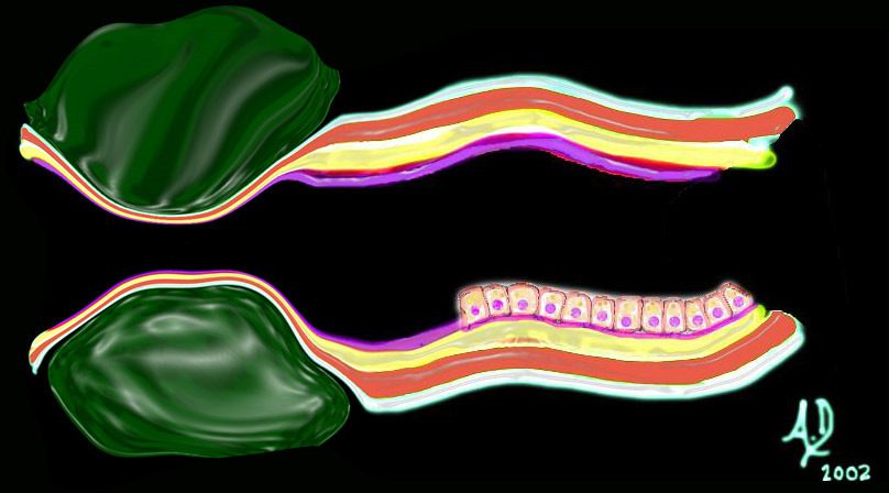
32347d06
key words mucosa submucosa muscularis adventitia serosa submucosal mass edema hemorrhage neoplasm malignancy benign obtuse angles with the lumen circumferential narrowing constriction obstruction histopathology imaging diagnosis Davidoff art Davidoff MD
Ashley Davidoff MD TheCommonvein.net
Diameter
The mean transverse dimension of the trachea is 1.6cm, ranging from 1.3- 2.5cms in the male and from 1-2.1cms in the female. The mean A-P dimension is slightly less, measuring 1.4cm.
Consideration of the size of the conducting system as a tubular and transporting system where diameter and length are functionally relevant is far different from the considerations given to the gas exchange system where surface area is the prime functional consideration. Measurements of the larger airways have practical significance for the bronchoscopist who has to know how far it is possible to advance the bronchoscope into the airways, for the anesthesiologist who needs to intubate the patient, and for the interventionalist who may need to stent a stenotic segment. The smaller airways are more difficult to measure but in general they should approximate the size of the accompanying arteries.

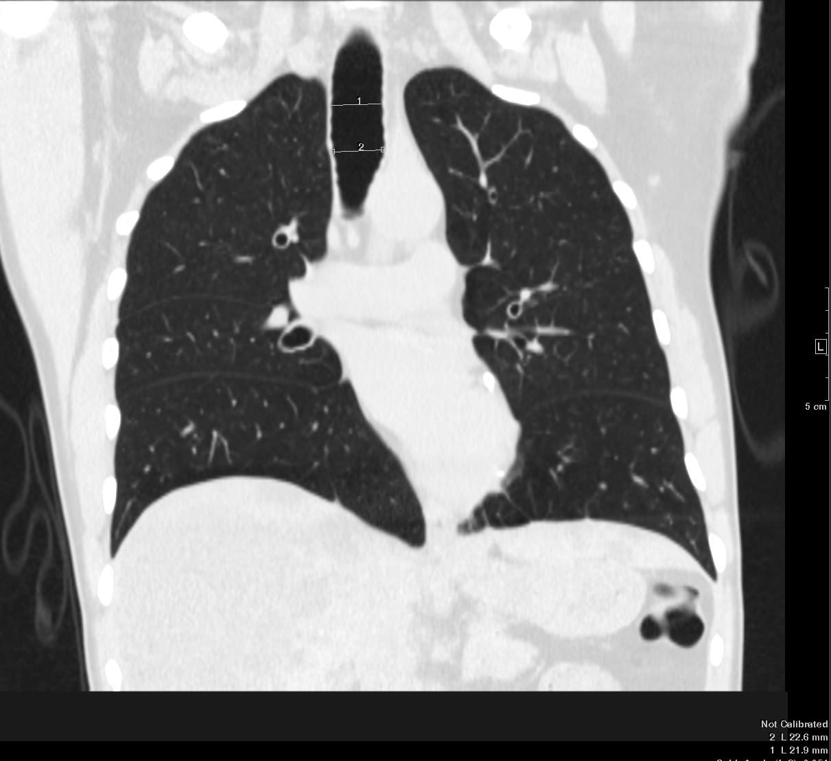
Normal chest CT of the upper lobes of both lungs showing the trachea. The transverse dimension of the trachea at this level is slightly larger than its A-P dimension, and both are slightly smaller than the diameter of the aortic arch.
Ashley Davidoff TheCommonVein.net 32158
The trachea, although it is a tube, is made up of 15-20 C-shaped cartilaginous rings with a posterior membranous surface, giving it a horseshoe shape in cross section. Its AP dimension is thus longer than its transverse dimensions.
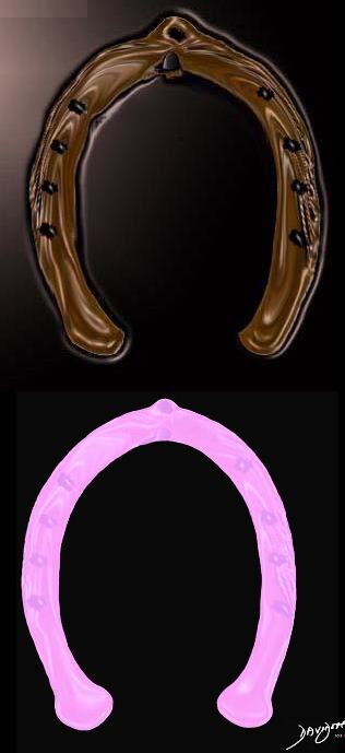
Normal chest CT of the upper lobes of both lungs. The trachea is horse shoe shaped.
Ashley Davidoff TheCommonVein.net 32158 42636c01
The shape of the tracheobronchial tree at large is very similar to a branching tree. If you took an image of the tracheobronchial tree and turned it upside down you would see the following;
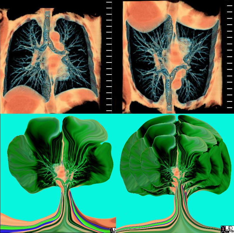
The tracheobronchial tree turned upside down shows it’s similarity to the branching pattern of a tree.
keywords lung bronchus tracheobronchial tree airway tree the common vein applied biology
Ashley Davidoff MD TheCommonVein.net
32620c02.800
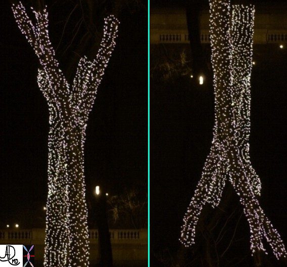
This is a photo taken near Lake Michigan at the end of the Fall in Chicago showing the illuminated base of the naked trees. The dichotomous branching is reminiscent of the tracheal branching into the two mainstem bronchi. Turn them upside down and the similarity is remarkable.
Ashley Davidoff TheCommonVein.net22153e
It has an irregular dichotomous branching pattern meaning it divides into paired branches of unequal length and diameter. The dichotomous branching pattern mirrors the branches of a tree. Similarly if you took the illuminated bare trees on Michigan Avenue in Chicago at RSNA, and turned them upside down you would see the following
Applied Anatomy
Once we move beyond the trachea into the rest of the tubular system, the rounded cross sectional shape is at first maintained by full circle cartilage and then by muscle. The irregular dichotomous branching pattern is maintained.
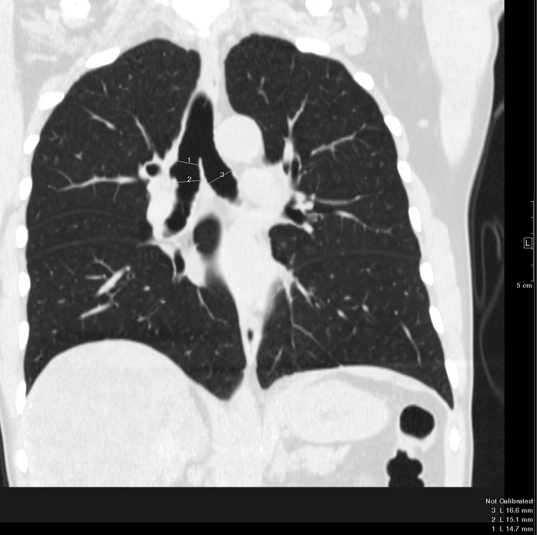
Trachea and Bronchi Upper Limits Normal in Size
Ashley Davidoff TheCommonVein.net
Trachea and Bronchi Upper Limits Normal in Size
Ashley DAvidoff TheCommonVein.net
Trachea and Bronchi Upper Limits Normal in Size
Ashley DAvidoff TheCommonVein.net
Trachea and Bronchi Upper Limits Normal in Size
Ashley DAvidoff TheCommonVein.net
Trachea and Bronchi Upper Limits Normal in Size
Ashley DAvidoff TheCommonVein.net
Trachea and Bronchi Upper Limits Normal in Size
Ashley DAvidoff TheCommonVein.net
Trachea and Bronchi Upper Limits Normal in Size
Ashley DAvidoff TheCommonVein.net
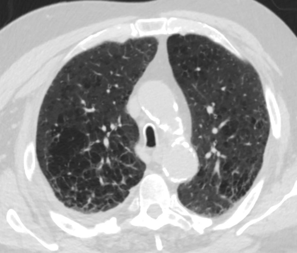
We have noted the normal horseshoe shape of the trachea. In COPD the tracheal A-P dimension increases relative to its transverse dimension, and the trachea assumes the shape of a “sabre” and is known as a “sabre trachea .
This is a patient with COPD and who has a cancer in the RUL. Note the shape of this trachea which has a AP diameter more than twice the transverse diameter. This is known as a sabre trachea or a sabre sheath trachea. The second image shows the sabre sword (a) the sabre sheath (b) a view from above the sheath, (c) and then a view from above the sheath, magnified and oriented in line with the CT orientation.
Ashley Davidoff TheCommonVein.net 28417
Position of the Trachea
The trachea is central in its position, and lies anterior to the esophagus. It originates below the pharynx and ends at the carina where it bifurcates into left and right main stem bronchi.
In the CT above a retrosternal thyroid goiter is compressing and displacing the trachea from its normal position to a more leftward and anterior location.
45796c key words thyroid trachea enlarged compression surface rendering normal anatomy
Ashley Davidoff TheCommonVein.ne
The tracheobronchial tree changes its morphology (and so its nature) as it progresses from a relatively rigid cartilaginous structure strengthened by C-shaped cartilages. The cartilage accompanies and supports the bronchi but disappears at the level of the bronchioles. The structure of the walls progresses to muscular, elastic, and mucus secreting bronchioles and then to delicate one cell layered airways at the alveolar level. The bronchi contain cartilage the bronchioles do not
Applied Anatomy
Tracheomalacia is softening of the tracheal cartilages which results in relative collapse of the trachea during inspiration and thus affecting airflow. The cause can be due to congenital weakening of the cartilage, due to extrinsic disease such as vascular rings and slings, or acquired from prolonged intubation or chronic infection. (eMedicine).
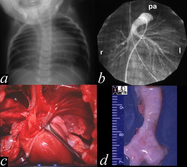
This autopsy is from a neonate who had respiratory difficulty at birth and showed a hyperinflated right lung with secondary leftward mediastinal shift. (a) A pulmonary angiogram showed the right pulmonary artery arising from the left PA rather than from the MPA (b) The gross in situ pathology shows the RPA coursing behind the trachea (c), while the last image (d) shows the narrowing of the left distal trachea. Histologically there was tracheomalacia secondary to the pressure effects of the LPA n the right side of the trachea causing right died bronchial obstruction with air trapping in the right lung. 01533c02
Ashley Davidoff MD TheCommonVein.net
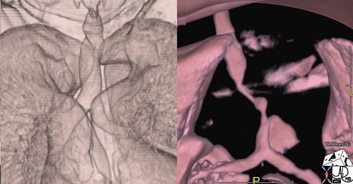
Normal on the right
Abnormal on the left is a 3D CT scan from a 50 year old female with respiratory difficulty trachea bronchi rectus abdominis muscle compression fractures kyphosis dwarf dwarfism right aortic arch tracheomalacia tracheal stenosis rectus abdominis muscle hypertrophy
Ashley Davidoff TheCommonVein.net
key words CTscan shape size position character growth
49838d01
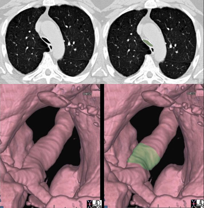
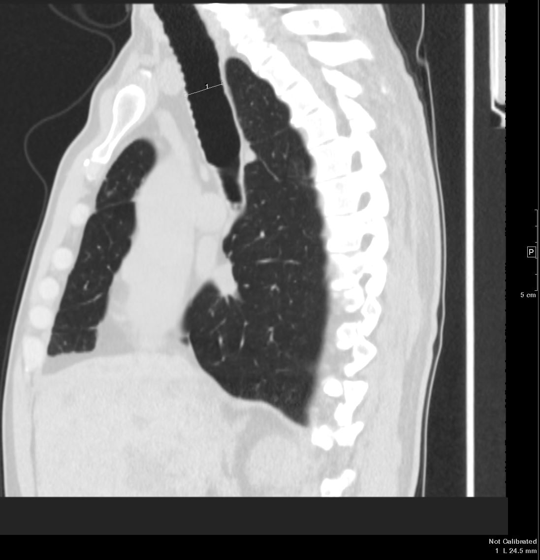
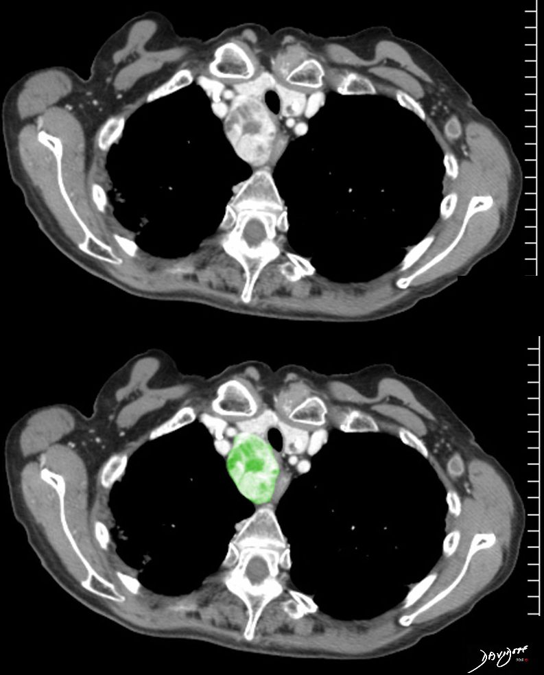
The trachea narrows significantly in its A-P dimension above the carina as seen in the cross sectional CT (a,b) with the narrowing noted in green overlay, and the change in diameter is better appreciated in the surface rendering of the trachea in c and d.
46114c04 Ashley Davidoff TheCommonVein.net
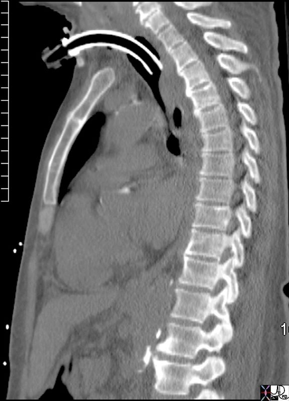
72333 keywords airway tracheostomy tubes treatment airways trachea ventilation Ashley Davidoff TheCommonVein.net
Amyloid Involvement
Ashley DavidoffTheCommonVein.net 44f Amyloid airways 001
Ashley Davidoff amyloid airways 001c
TheCommonVein.net
Ashley Davidoff amyloid airways 001b
Ashley Davidoff amyloid airways 001