
- Definition:
- bronchial dilation
- resulting from
- fibrosis and scarring in the surrounding lung tissue,
- due to
- chronic inflammatory or
- fibrotic lung diseases.
- Imaging Appearances:
- dilated bronchi
- “tram-track” or “railroad-track”
- absence of normal tapering of the bronchi toward the lung periphery.
- reticulations
- linear or irregular branching opacities
- honeycombing, and
- ground-glass opacities.
- Contributing Lung Diseases
- Traction bronchiectasis is often seen in association with various chronic lung diseases, including:
- Idiopathic pulmonary fibrosis (IPF)
- Connective tissue diseases such as rheumatoid arthritis and systemic sclerosis
- Chronic hypersensitivity pneumonitis
- Asbestosis
- Radiation-induced lung fibrosis
- Non-specific interstitial pneumonia (NSIP)
- NSIP
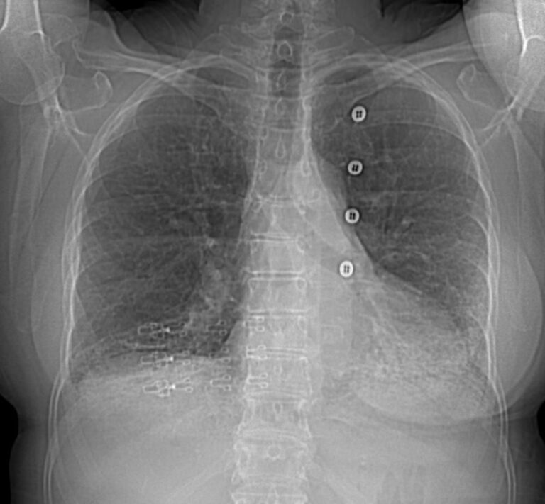
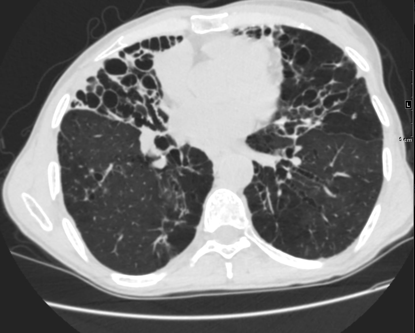
71-year-old female presents with a history of scleroderma, ILD, hypothyroidism and dcSSc
Scout film of the CT shows bibasilar reticular changes
Ashley Davidoff MD TheCommonVein.net 196Lu 136604
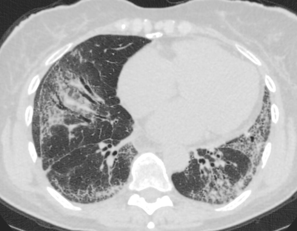
71-year-old female presents with a history of scleroderma, ILD, hypothyroidism and dcSSc
CT in the axial plane, shows extensive ground glass changes in the inferior lingula middle lobe and traction bronchiectasis in the middle lobe. The left lower lobe shows bronchovascular changes, and bronchiolectasis and there is bilateral subpleural sparing
The fissures show irregular thickening
Ashley Davidoff MD TheCommonVein.net 196Lu 136610
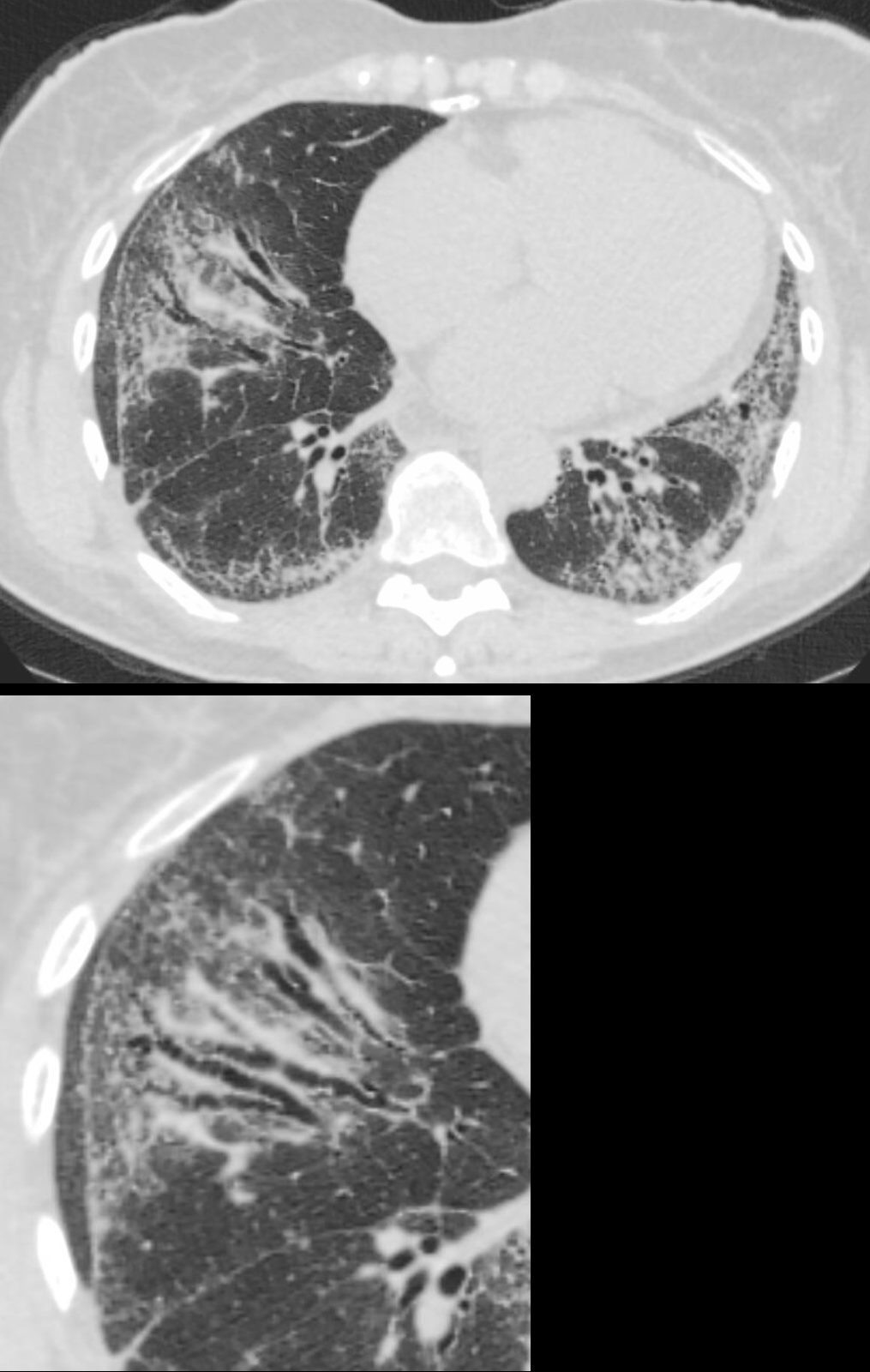
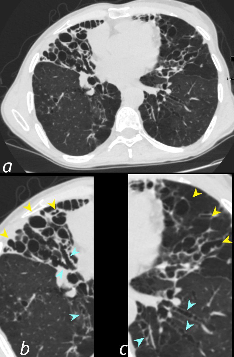
71-year-old female presents with a history of scleroderma, ILD, hypothyroidism and dcSSc
CT in the axial plane, shows extensive ground glass changes in the inferior lingula middle lobe and traction bronchiectasis in the middle lobe. The left lower lobe shows bronchovascular changes, and bronchiolectasis and there is bilateral subpleural sparing
The fissures show irregular thickening.
The lower image exemplifies the traction bronchiectasis
Ashley Davidoff MD TheCommonVein.net 196Lu 136610c
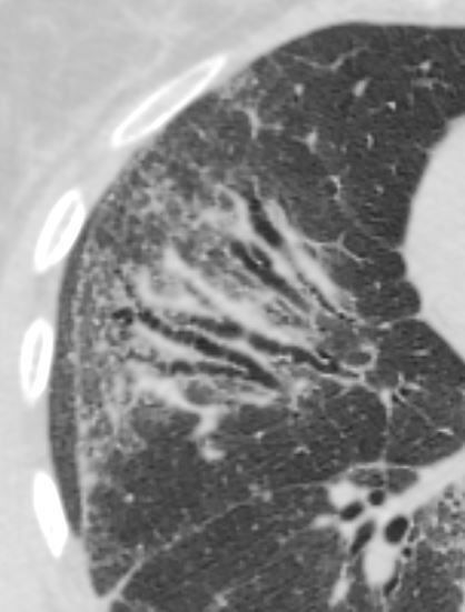
71-year-old female presents with a history of scleroderma, ILD, hypothyroidism and dcSSc
CT of the middle lobe in the axial plane, shows extensive traction bronchiectasis in a background of ground glass changes and reticulations caused by thickening of the interlobular septa.
The fissures show irregular thickening.
Ashley Davidoff MD TheCommonVein.net 196Lu 136611
Mounier Kuhn
61-year-old male with a history of treated mycobacterial infections including MAC and chronic cough.
Axial CT at the level of the mid to lower chest shows mildly ectatic segmental airways to the lower, and middle lobe bronchi but significant bronchiectasis to the middle lobe and lingula involving the subsegmental airways. There is a relative paucity of mucus in the ectatic airways. The history of MAC and the distribution of the bronchiectasis in the middle lobe and lingula are reminiscent of the diagnosis of Lady Windermere syndrome
Ashley Davidoff MD TheCommonVein.net 250Lu 135876
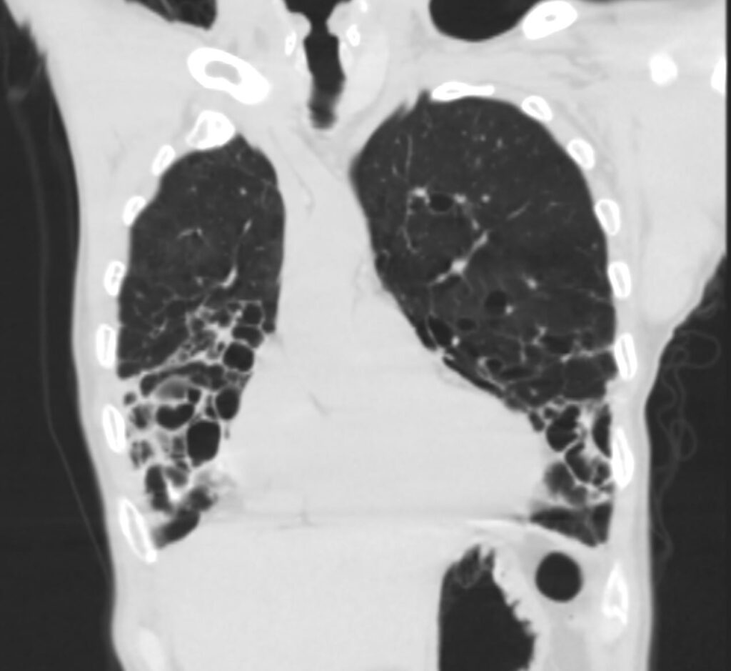
61-year-old male with a history of treated mycobacterial infections including MAC and chronic cough.
Coronal CT at the level of the heart shows significant bronchiectasis to the middle lobe and lingula and as a result abut the right and left heart border accounting for the CXR findings of a “shaggy heart border”. There is a relative paucity of mucus in the ectatic airways. The history of MAC and the distribution of the bronchiectasis in the middle lobe and lingula are reminiscent of the diagnosis of Lady Windermere syndrome
Ashley Davidoff MD TheCommonVein.net 250Lu 135879
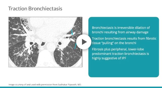
Bronchiectasis is irreversible dilation of the bronchi resulting from airway damage due to a variety of causes, including infection, airway obstruction, or fibrosis.7 By definition, traction bronchiectasis results from fibrotic tissue “pulling” on the bronchi, while freestanding bronchiectasis is unrelated to fibrosis.7 Traction bronchiectasis is often seen at the periphery of the lung where there is less supportive connective tissue, leaving the bronchi prone to distortion.10 Peripheral, lower-lobe predominant traction bronchiectasis in the setting of changes consistent with fibrosis is highly suggestive of IPF.545
Radiology Rounds