
Ashley Davidoff
TheCommonVein.net
Ashley Davidoff
TheCommonVein.net

Ashley Davidoff
TheCommonVein.net
Ashley Davidoff MD The CommonVein.net
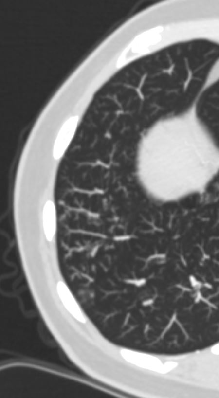
- Is a
- radiologic finding on chest CT
- Characterised by
- small, peripheral nodules,
- originating from a centrilobular location extending into
- distal smaller airways as
- soft tissue nodules
- connected to
- multiple contiguous,
- linear branching opacities.
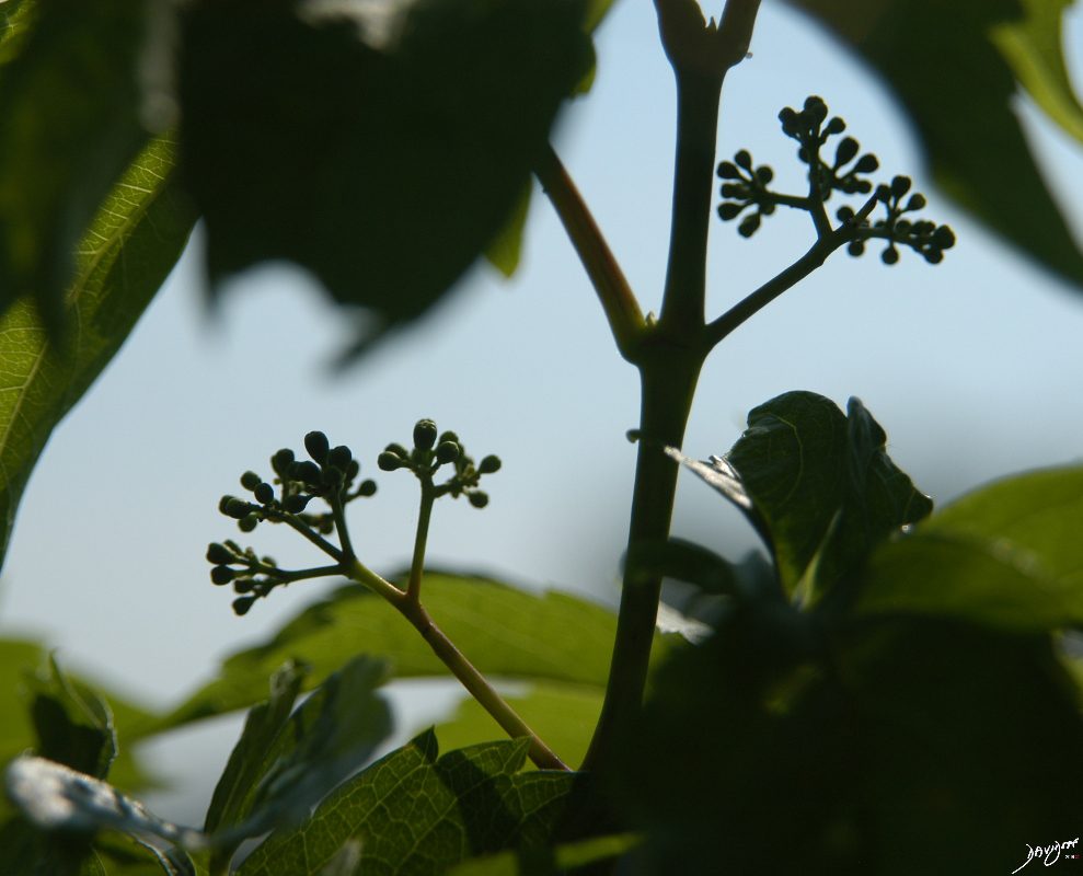
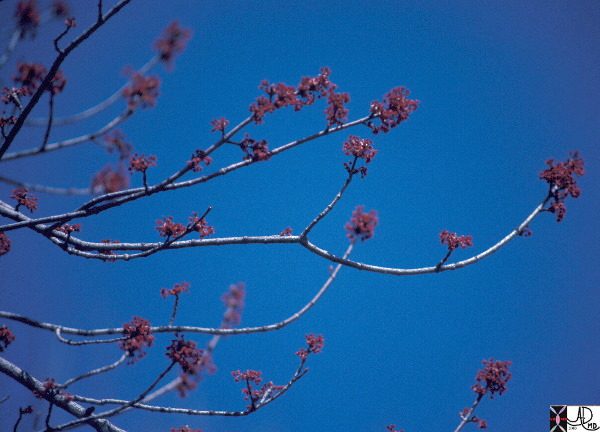
- reminiscent of a tree in bud in the spring
- Representing
- mucous plugging,
- bronchial dilatation, and
- wall thickening of bronchiolitis.
- histopathological correlate
- small airway plugging with
- mucus,
- pus, or
- fluid, with
- dilated intralobular bronchioles,
- peribronchiolar inflammation, and
- wall thickening.
- small airway plugging with
- Cause
-
- commonly associated with
- infectious etiologies
- aspiration
- endobronchial spread of Mycobacterium tuberculosis,
- Pneumocystis jiroveci (Pneumocystis carinii) pneumonia
- Also other infections
- bacterial,
- fungal,
- viral, or
- parasitic
- invasive pulmonary aspergillosis.
- various
- immunodeficiency states,
- congenital disorders (cystic fibrosis),
- malignancy (lymphoma),
- panbronchiolitis,
- various
-
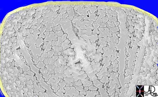 Respiratory bronchioles, alveolar sacs, and alveoli
This drawing shows about 3-4 respiratory bronchioles that serve to make a secondary lobule. Alveolar sacs and individual alveoli are also seen. The yellow border represents the visceral pleura on the surface.
Courtesy of: Ashley Davidoff, M.D.
Infection
- small (2–4-mm) centrilobular nodules and
- branching linear opacities of similar caliber
- originating from a single stalk
- bronchial wall thickening with or without
- bronchiectasis.
- Active TB
-

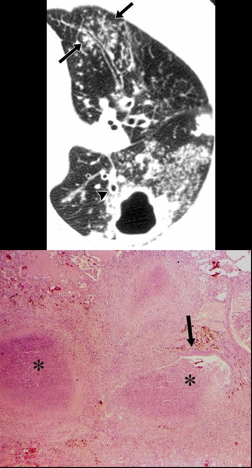
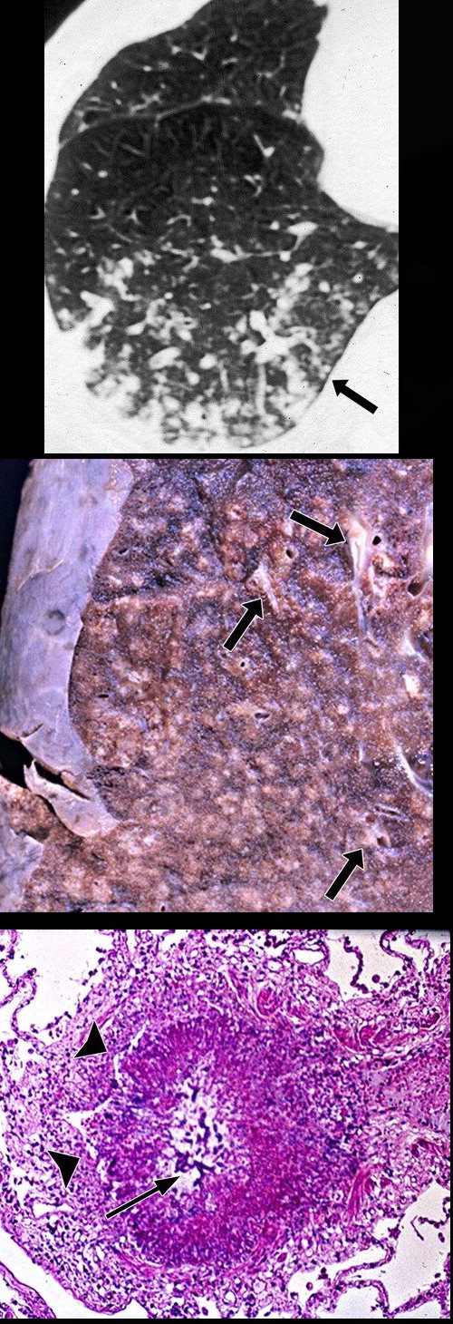
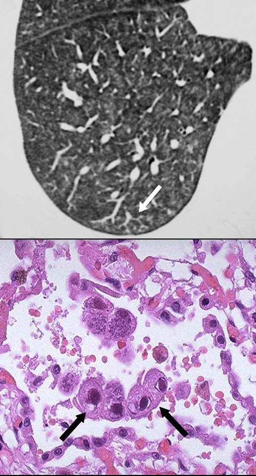
Postprimary active tuberculosis in a 34-year-old man with weight loss and a chronic cough. (a) High-resolution CT scan of the left lung shows a thick-walled cavity and multiple peripheral small nodules and branching linear structures (arrows). Note the thickening of the bronchial walls (arrowhead). (b) Photomicrograph (original magnification, ×400; hematoxylin-eosin stain) shows impacted caseous material (*) in small peripheral airways (arrow).
Rossi, SE et al Tree-in-Bud Pattern at Thin-Section CT of the Lungs: Radiologic-Pathologic Overview RadioGraphics Vol. 25, No. 3 2005 - TB
-
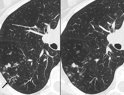
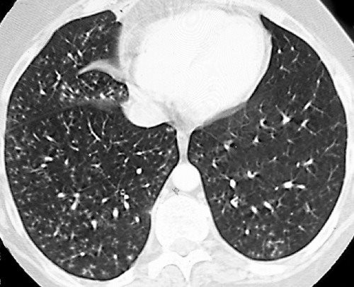
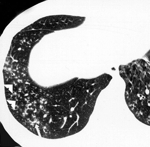
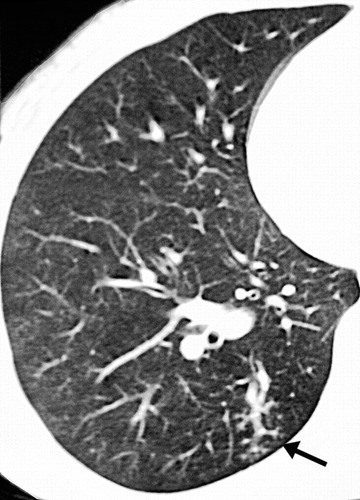
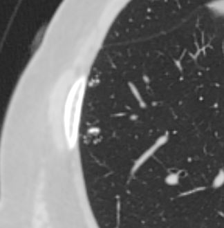
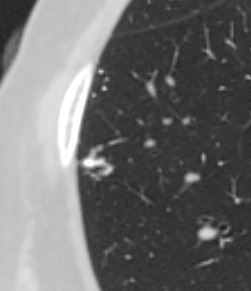
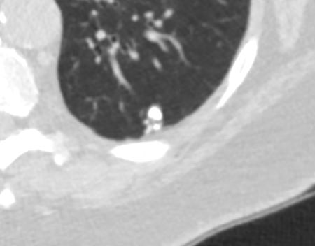
Post primary active tuberculosis in a 66-year-old woman with a chronic cough. High-resolution CT scans of the right lung show peripheral, poorly defined, small (2–4-mm-diameter) centrilobular nodules and branching linear opacities of similar caliber originating from a single stalk (the tree-in-bud pattern) in the lower lobe (arrow). These findings represent endobronchial spread of tuberculosis.
Rossi, SE et al Tree-in-Bud Pattern at Thin-Section CT of the Lungs: Radiologic-Pathologic Overview RadioGraphics Vol. 25, No. 3 2005Postprimary active tuberculosis in a 66-year-old woman with a chronic cough. High-resolution CT scans of the right lung show peripheral, poorly defined, small (2–4-mm-diameter) centrilobular nodules and branching linear opacities of similar caliber originating from a single stalk (the tree-in-bud pattern) in the lower lobe (arrow). These findings represent endobronchial spread of tuberculosis.
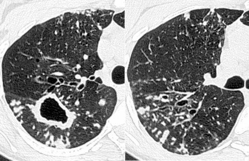
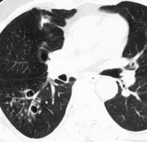
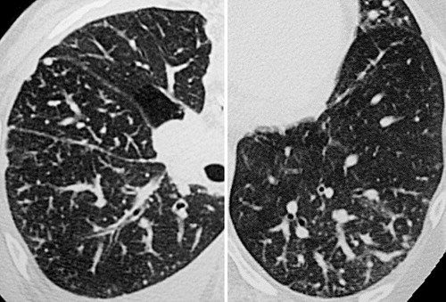
Rossi, SE et al Tree-in-Bud Pattern at Thin-Section CT of the Lungs: Radiologic-Pathologic Overview RadioGraphics Vol. 25, No. 3 2005Infection with M avium-intracellulare complex in a 44-year-old woman with malaise and a chronic cough. High-resolution CT scans of the right lung show multiple peripheral small nodules connected to branching linear opacities and a thick-walled cavity in the superior segment of the lower lobe. Note the thickening of the bronchial walls, bronchial dilatation, and mucus impaction. The diagnosis was confirmed with bronchoalveolar lavage.
Rossi, SE et al Tree-in-Bud Pattern at Thin-Section CT of the Lungs: Radiologic-Pathologic Overview RadioGraphics Vol. 25, No. 3 2005 - Aspergillus and Tree in Bud Sign

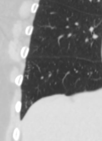
CT scan through the chest shows medium sized bronchi, bronchioles and small airways impacted with fluid. This collage is presented to reveal tree in bud changes resulting from impaction in the smaller terminal bronchioles and respiratory units. The tree-in-bud pattern also results in small centrilobular nodules connected to multiple branching linear structures of similar caliber from a single stalk. Originally it was felt to result from endobronchial spread of Mycobacterium tuberculosis, but is is now recognized in diverse entities including peripheral airway diseases caused by infection (bacterial, fungal, viral, or parasitic), congenital disorders, idiopathic disorders (obliterative bronchiolitis, pan bronchiolitis), aspiration or inhalation of foreign substances, immunologic disorders, connective tissue disorders and peripheral pulmonary vascular diseases such as neoplastic pulmonary emboli.
In this case there are also dilated medium sized airways, impacted with soft tissue characteristic of the finger in glove sign and most likely due to allergic bronchopulmonary aspergillosis (ABPA)
Ashley Davidoff MD Ashley Davidoff MD TheCommonVein.net
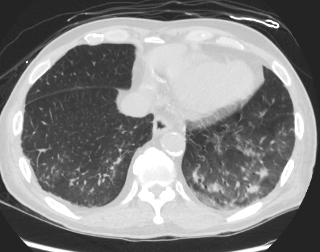
47113c01
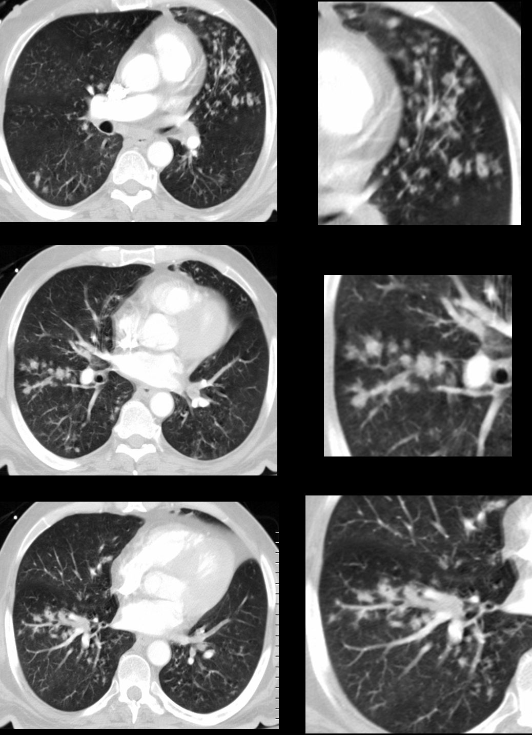
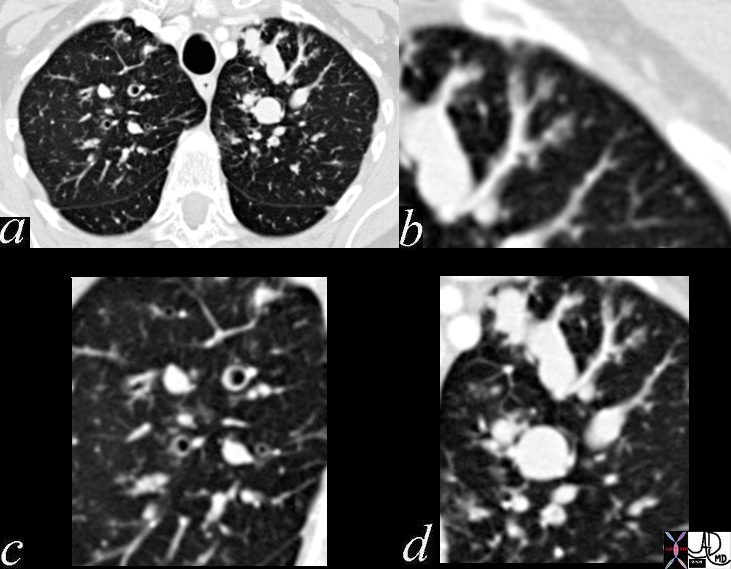
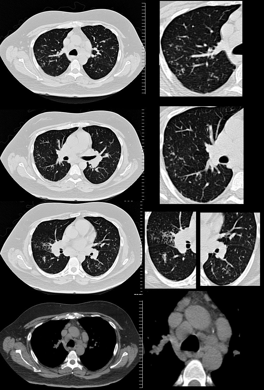
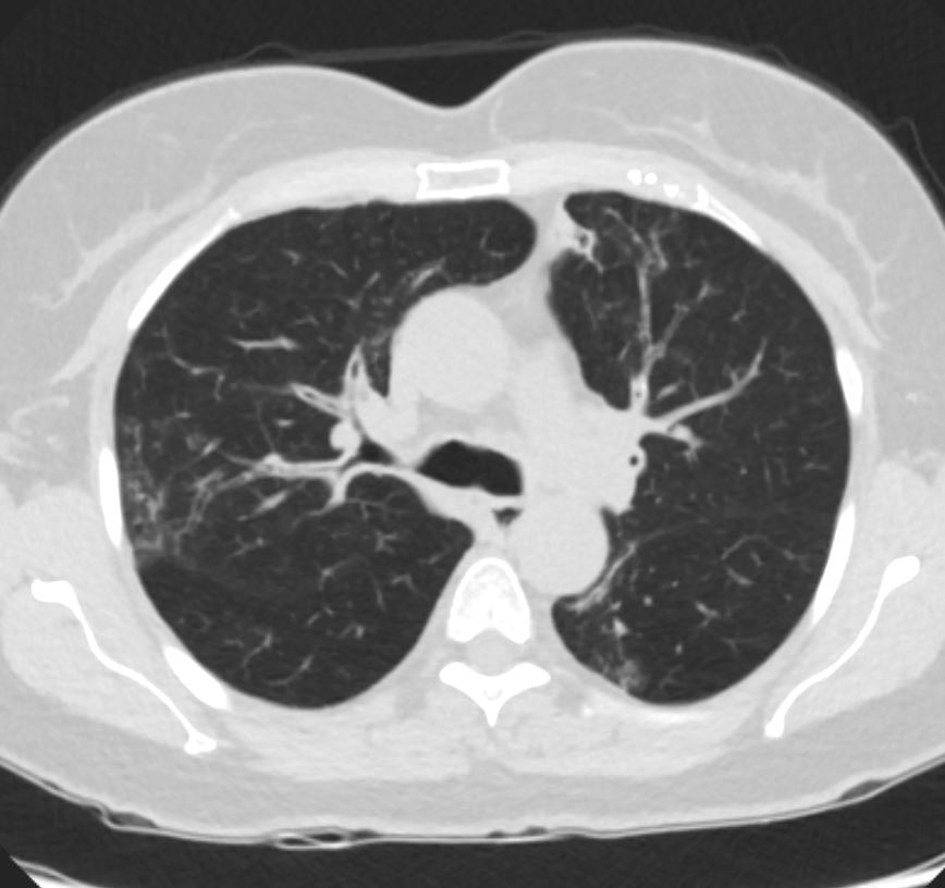
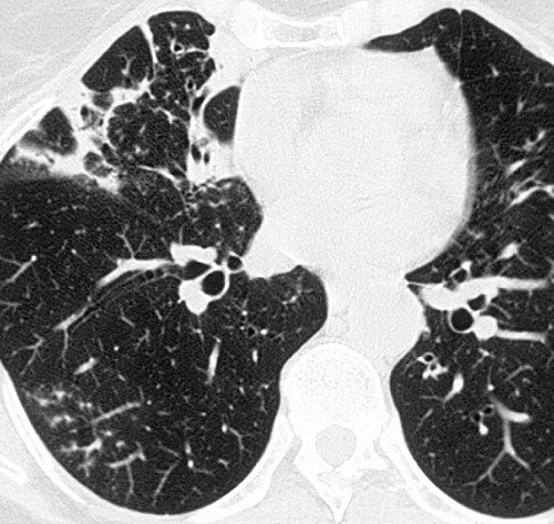
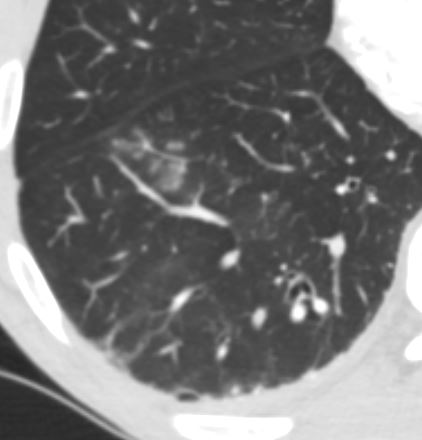
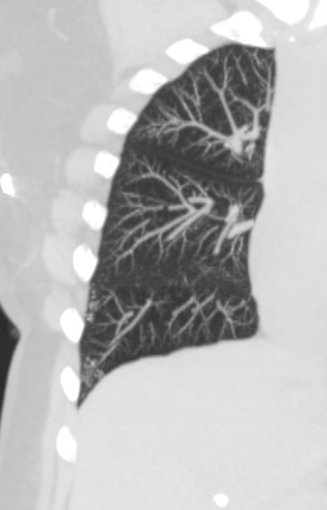
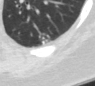
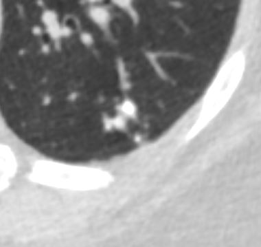
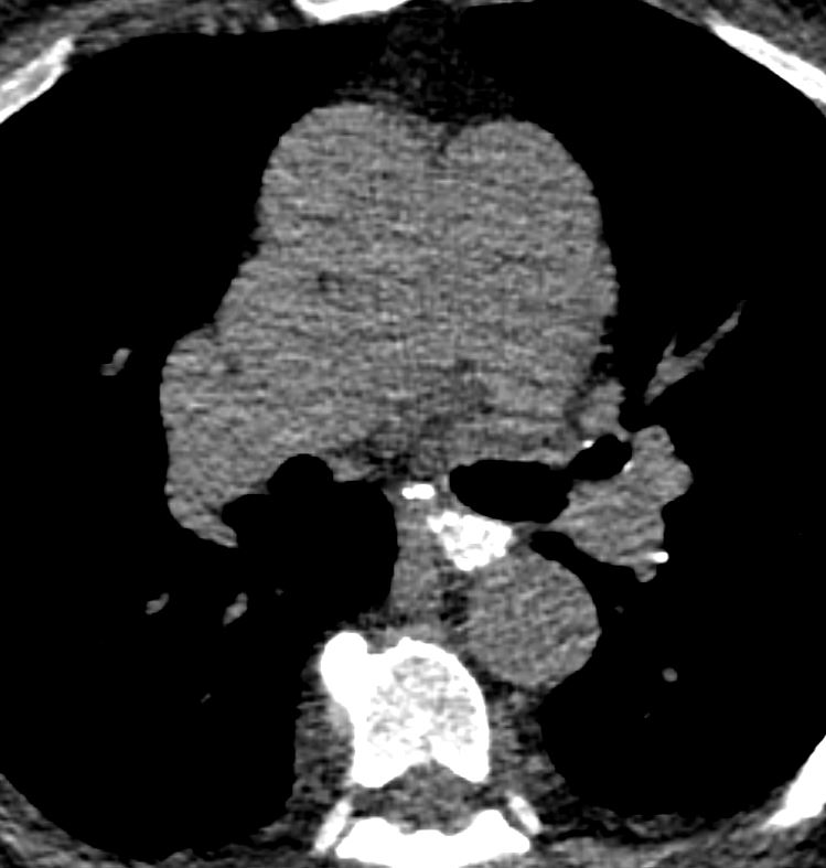
43 year old man with known aspergillus infection. Note the thickening of the walls of the segmental subsegmental and small airways with extensive tree in bud changes and bronchial wall thickening. There are centrilobular nodules indicating the small airway disease
Ashley Davidoff MD TheCommonVein.net
117816c
Rossi, SE et al Tree-in-Bud Pattern at Thin-Section CT of the Lungs: Radiologic-Pathologic Overview RadioGraphics Vol. 25, No. 3 2005
Other Infections
Staphylococcus Aureus
Rossi, SE et al Tree-in-Bud Pattern at Thin-Section CT of the Lungs: Radiologic-Pathologic Overview RadioGraphics Vol. 25, No. 3 2005
Hemophilus Influenza
Rossi, SE et al Tree-in-Bud Pattern at Thin-Section CT of the Lungs: Radiologic-Pathologic Overview RadioGraphics Vol. 25, No. 3 2005
CMV
Rossi, SE et al Tree-in-Bud Pattern at Thin-Section CT of the Lungs: Radiologic-Pathologic Overview RadioGraphics Vol. 25, No. 3 2005
Respiratory Syncytial Virus

Rossi, SE et al Tree-in-Bud Pattern at Thin-Section CT of the Lungs: Radiologic-Pathologic Overview RadioGraphics Vol. 25, No. 3 2005
87 year old male with history of cough and suspicion of aspiration shows barium aspiration into the proximal trachea (upper right) The scout view ( upper right) shows an infiltrate at the right base, Thickened airways in the right lower lobe (2nd row left ) is associated with a pneumonic infiltrate in the right lower lobe (lower right) consistent with aspiration. There are thickened airways to the lingula (3rd and 4th row) with magnified view showing tree in bud changes (right sided images 3rd and 4th row)
These findings likely relate to infection, often fungal or granulomatous
Ashley Davidoff MD Ashley Davidoff MD TheCommonVein.net
Inflammatory Diseases
Aspiration and Tree in Bud
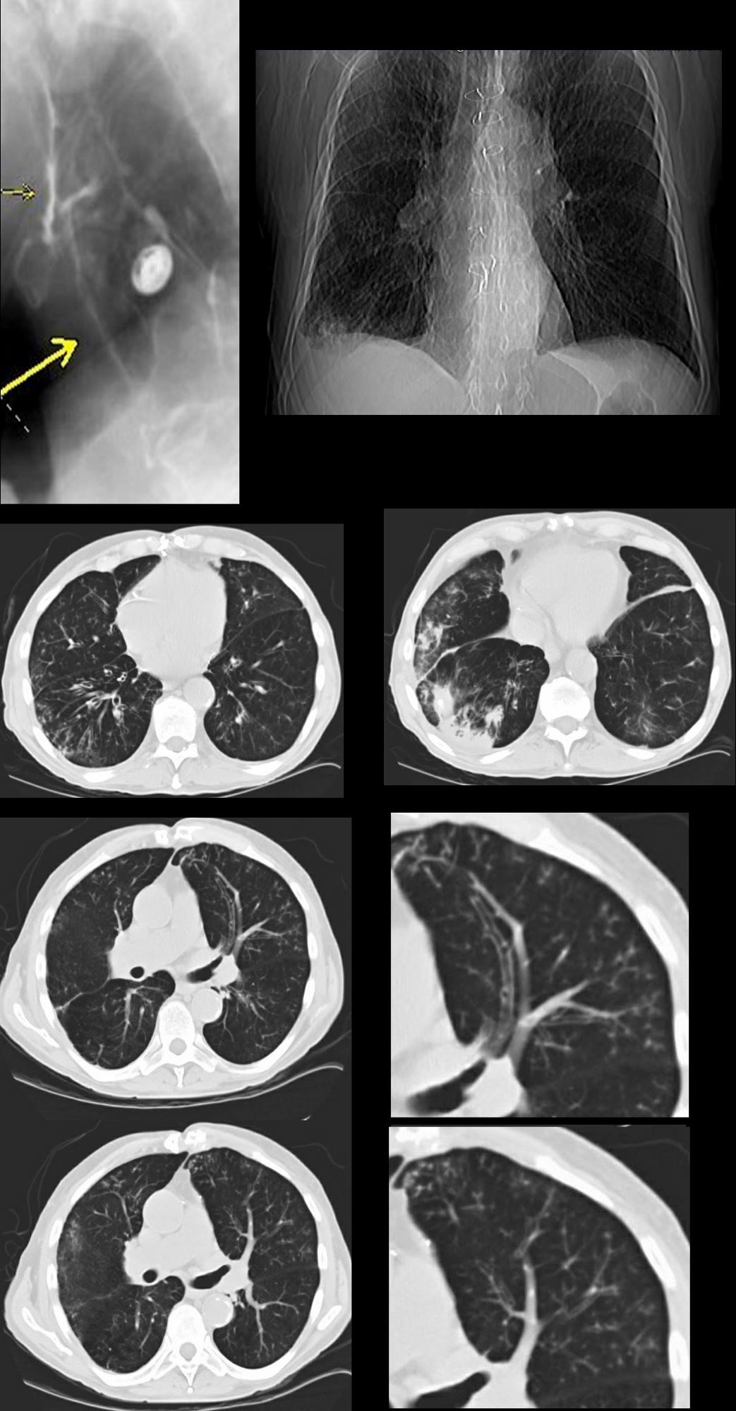
87 year old male with history of cough and suspicion of aspiration shows barium aspiration into the proximal trachea (upper right) The scout view ( upper right) shows an infiltrate at the right base, Thickened airways in the right lower lobe (2nd row left ) is associated with a pneumonic infiltrate in the right lower lobe (lower right) consistent with aspiration. There are thickened airways to the lingula (3rd and 4th row) with magnified view showing tree in bud changes (right sided images 3rd and 4th row)
All these finding likely relate to aspiration though lingula involvement is not usual
Ashley Davidoff MD Ashley Davidoff MD TheCommonVein.net
Rossi, SE et al Tree-in-Bud Pattern at Thin-Section CT of the Lungs: Radiologic-Pathologic Overview RadioGraphics Vol. 25, No. 3 2005
Asthma
55F-asthma-involving medium and small sized airways focal consolidations and chronic sinusitis with tree in bud
Ashley Davidoff TheCommonVein.net
Inhalation Bronchiolitis
Rossi, SE et al Tree-in-Bud Pattern at Thin-Section CT of the Lungs: Radiologic-Pathologic Overview RadioGraphics Vol. 25, No. 3 2005
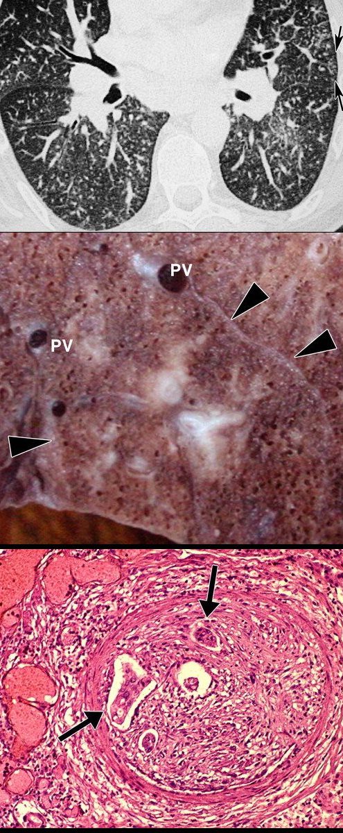
Obliterative Bronchiolitis aka (bronchiolitis obliterans is also known and constrictive bronchiolitis) association to lung transplant and bone marrow transplant
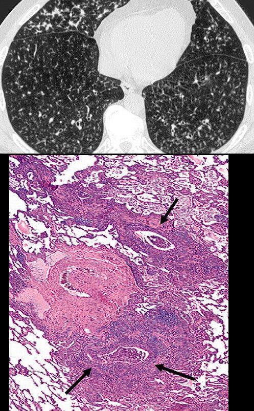
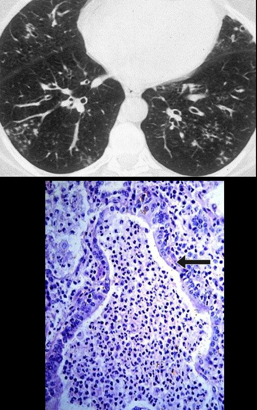
Obliterative bronchiolitis after bone marrow transplantation in a 47-year-old man with myeloma. (a) Expiratory high-resolution CT scan shows diffuse centrilobular nodules connected to branching linear opacities bilaterally. Note the air trapping in the right lower lobe. (b) Photomicrograph (original magnification, ×200; hematoxylin-eosin stain) of a specimen from open lung biopsy shows the bronchiolar walls surrounded by concentric chronic inflammatory infiltrates (arrows).
Rossi, SE et al Tree-in-Bud Pattern at Thin-Section CT of the Lungs: Radiologic-Pathologic Overview RadioGraphics Vol. 25, No. 3 2005
Rossi, SE et al Tree-in-Bud Pattern at Thin-Section CT of the Lungs: Radiologic-Pathologic Overview RadioGraphics Vol. 25, No. 3 2005
Rossi, SE et al Tree-in-Bud Pattern at Thin-Section CT of the Lungs: Radiologic-Pathologic Overview RadioGraphics Vol. 25, No. 3 2005
Circulatory
Rossi, SE et al Tree-in-Bud Pattern at Thin-Section CT of the Lungs: Radiologic-Pathologic Overview RadioGraphics Vol. 25, No. 3 2005
Rossi, SE et al Tree-in-Bud Pattern at Thin-Section CT of the Lungs: Radiologic-Pathologic Overview RadioGraphics Vol. 25, No. 3 2005
Rossi, SE et al Tree-in-Bud Pattern at Thin-Section CT of the Lungs: Radiologic-Pathologic Overview RadioGraphics Vol. 25, No. 3 2005
Tree in Bud with
Ashley Davidoff MD TheCommonVein.net
Source
Signs in Thoracic Imaging
Journal of Thoracic Imaging 21(1):76-90, March 2006.
https://pubs.rsna.org/doi/full/10.1148/radiol.13120908
Ashley Davidoff
TheCommonVein.net
Ashley Davidoff
TheCommonVein.net
Ashley Davidoff
TheCommonVein.net
Ashley Davidoff
TheCommonVein.net
Ashley Davidoff
TheCommonVein.net
Ashley Davidoff
TheCommonVein.net
Ashley Davidoff
TheCommonVein.net
Ashley Davidoff
TheCommonVein.net
Ashley Davidoff MD TheCommonvein.net
Ashley Davidoff MD TheCommonvein.net
References and Links
Rossi, SE et al Tree-in-Bud Pattern at Thin-Section CT of the Lungs: Radiologic-Pathologic Overview RadioGraphicsVol. 25, No. 3 2005